Statistical Validation of Pairwise and High-Order Brain Connectivity: Methods, Applications, and Clinical Translation
This article provides a comprehensive resource for researchers and drug development professionals on the statistical validation of brain functional connectivity.
Statistical Validation of Pairwise and High-Order Brain Connectivity: Methods, Applications, and Clinical Translation
Abstract
This article provides a comprehensive resource for researchers and drug development professionals on the statistical validation of brain functional connectivity. It covers the foundational shift from traditional pairwise analysis to high-order interaction models that capture complex, synergistic brain dynamics. The content details rigorous methodological frameworks, including surrogate data and bootstrap analyses for single-subject significance testing, essential for clinical applications. It addresses common pitfalls in connectivity analysis and offers optimization strategies to enhance robustness. Furthermore, the article explores the translation of these validated connectivity biomarkers into drug development pipelines, discussing their growing role in pharmacodynamic assessments and clinical trials for neurological and psychiatric disorders.
From Pairwise Links to High-Order Networks: Uncovering the Brain's Complex Dialogue
The Fundamental Limitation of Pairwise Connectivity
Functional connectivity (FC) mapping has become a fundamental tool in network neuroscience for investigating the brain's functional organization. Traditional models predominantly represent brain activity as a network of pairwise interactions between brain regions, typically using measures like Pearson's correlation or partial correlation [1] [2]. While these approaches have revealed fundamental insights into brain organization, they operate under a limiting constraint: the assumption that all interactions between brain regions can be decomposed into dyadic relationships [1] [3]. This perspective inherently neglects the possibility of higher-order interactions (HOIs) that simultaneously involve three or more brain regions [1] [3].
Mounting theoretical and empirical evidence suggests that the brain's complex functional architecture cannot be fully captured by pairwise statistics alone. Higher-order interactions appear to be fundamental components of complexity and functional integration in brain networks, potentially linked to emergent mental phenomena and consciousness [3]. At both micro- and macro-scales, studies indicate that significant information may be detectable only in joint probability distributions and not in pairwise marginals, meaning pairwise approaches may fail to identify crucial higher-order behaviors [1].
This application note examines the fundamental limitations of pairwise connectivity approaches and demonstrates how emerging higher-order methodologies provide a more comprehensive framework for understanding brain function in both basic research and drug development contexts.
Theoretical Limitations of Pairwise Approaches
The Incomplete Picture of Brain Dynamics
Pairwise connectivity models suffer from several theoretical shortcomings that limit their ability to fully characterize brain dynamics:
Simplified Representation: By reducing complex multivariate interactions to a set of dyadic connections, pairwise approaches potentially miss collective dynamics that emerge only when three or more regions interact simultaneously [1] [3].
Synergy Blindness: Information-theoretic research reveals two distinct modes of information sharing: redundancy and synergy. Synergistic information occurs when the joint state of three or more variables is necessary to resolve uncertainty arising from statistical interactions that exist collectively but not in separately considered subsystems [3]. Pairwise measures are inherently limited in detecting these synergistic relationships.
Network Context Neglect: Approaches like Pearson correlation do not account for the broader network context in which pairwise connections occur. While partial correlation methods attempt to address this by removing shared influences, they can be overly conservative and may eliminate meaningful higher-order dependencies [4] [2].
Empirical Evidence of Limitations
Comparative studies demonstrate concrete scenarios where pairwise methods fall short:
Consciousness Detection: Research differentiating patients in different states of consciousness found that higher-order dependencies reconstructed from fMRI data encoded meaningful biomarkers that pairwise methods failed to detect [1].
Task Performance Prediction: Higher-order approaches have demonstrated significantly stronger associations between brain activity and behavior compared to traditional pairwise methods [1].
Sensitivity to Intervention Effects: In clinical applications, investigating brain connectivity developments at a high-order level has proven essential to fully capture the complexity and modalities of recovery following treatment [3].
Table 1: Comparative Performance of Connectivity Approaches
| Analysis Domain | Pairwise Methods Performance | Higher-Order Methods Performance | Key Findings |
|---|---|---|---|
| Task Decoding | Moderate dynamic differentiation between tasks | Greatly enhanced dynamic task decoding [1] | HOIs improve characterization of dynamic group dependencies in rest and tasks |
| Individual Identification | Limited fingerprinting capability | Improved functional brain fingerprinting based on local topological structures [1] | HOIs provide more distinctive individual signatures |
| Brain-Behavior Association | Moderate associations | Significantly stronger associations with behavior [1] | HOIs capture more behaviorally-relevant neural signatures |
| Clinical Differentiation | Limited ability to differentiate patient states | Effective differentiation of states of consciousness [1] | HOIs encode meaningful clinical biomarkers |
Higher-Order Connectivity Frameworks
Topological Data Analysis Approach
One innovative method for capturing higher-order interactions leverages topological data analysis to reveal instantaneous higher-order patterns in fMRI data [1]. This approach involves four key steps:
Signal Standardization: Original fMRI signals are z-scored to normalize the data [1].
Higher-Order Time Series Computation: All possible k-order time series are computed as element-wise products of k+1 z-scored time series, representing instantaneous co-fluctuation magnitude of associated (k+1)-node interactions [1].
Simplicial Complex Encoding: Instantaneous k-order time series are encoded into weighted simplicial complexes at each timepoint [1].
Topological Indicator Extraction: Computational topology tools analyze simplicial complex weights to extract global and local indicators of higher-order organization [1].
This framework generates several key metrics beyond pairwise correlation:
Hyper-coherence: Quantifies the fraction of higher-order triplets that co-fluctuate more than expected from corresponding pairwise co-fluctuations [1].
Violating Triangles: Identify higher-order coherent co-fluctuations that cannot be described in terms of pairwise connections [1].
Homological Scaffold: Assesses edge relevance toward mesoscopic topological structures within the higher-order co-fluctuation landscape [1].
Information-Theoretic Framework
Multivariate information theory provides another framework for capturing HOIs through measures like O-information (OI), which evaluates whether a system is dominated by redundancy or synergy [3]. This approach distinguishes between:
Redundancy: Group interactions explainable by communication of subgroups of variables, representing information replicated across system elements [3].
Synergy: Information that emerges only from the joint interaction of three or more variables, reflecting the brain's ability to generate new information by combining anatomically distinct areas [3].
Table 2: Information-Theoretic Measures for Brain Connectivity
| Measure | Formula | Interpretation | Interaction Type Captured |
|---|---|---|---|
| Mutual Information (Pairwise) | I(Si;Sj) = H(Si) - H(Si∣Sj) | Information shared between two variables | Pairwise interactions only |
| O-Information | O(X) = TC(X) - DTC(X) | Overall evaluation of redundancy vs. synergy dominance | Higher-order interactions |
| Redundancy | Not directly computed; inferred when O(X) > 0 | Information replicated across system elements | Duplicative interactions |
| Synergy | Not directly computed; inferred when O(X) < 0 | Novel information from joint interactions | Emergent, integrative interactions |
Experimental Protocols and Validation
Protocol: Topological Analysis of Higher-Order fMRI Connectivity
Purpose: To detect and quantify higher-order interactions in resting-state or task-based fMRI data that are missed by pairwise correlation methods.
Materials:
- fMRI data (preprocessed and denoised)
- Brain parcellation atlas (e.g., Schaefer 100x7, HCP 119-region)
- Computational tools: Python with NumPy, SciPy, Topological data analysis libraries
- High-performance computing resources (for large-scale simplicial complex computation)
Procedure:
Data Preparation:
- Extract time series from N brain regions according to your chosen atlas
- Apply quality control and remove artifacts
- Standardize each regional time series using z-scoring: z(t) = (x(t) - μ)/σ
Compute k-Order Time Series:
- For each timepoint t, compute all possible simplex weights:
- For edges (1-simplices): w{ij}(t) = zi(t) × z_j(t)
- For triangles (2-simplices): w{ijk}(t) = zi(t) × zj(t) × zk(t)
- For higher-order simplices (as needed)
- Apply sign remapping based on parity of contributing signals
- For each timepoint t, compute all possible simplex weights:
Construct Weighted Simplicial Complexes:
- At each timepoint t, construct a simplicial complex K_t
- Assign weights to each simplex based on computed k-order time series values
Extract Topological Indicators:
- Compute hyper-coherence: fraction of "violating triangles" where triangle weight exceeds constituent edge weights
- Identify and record all violating triangles Δv
- Calculate homological scaffolds to assess edge importance in mesoscopic structures
- Analyze contributions to topological complexity from coherent vs. decoherent signals
Statistical Analysis:
- Compare higher-order indicators across experimental conditions or groups
- Assess relationship between higher-order features and behavioral measures
- Evaluate individual identification capacity using higher-order fingerprints
Validation:
- Benchmark against traditional pairwise methods (correlation, partial correlation)
- Test retest reliability in longitudinal data
- Verify biological plausibility through structure-function coupling analysis
Protocol: Single-Subject Statistical Validation of High-Order Interactions
Purpose: To statistically validate subject-specific pairwise and high-order connectivity patterns using surrogate and bootstrap analyses.
Materials:
- Single-subject multivariate fMRI time series
- Surrogate data generation algorithms (e.g., IAAFT, phase randomization)
- Bootstrap resampling implementation
- Information-theoretic computation tools
Procedure:
Connectivity Estimation:
- Compute pairwise connectivity using Mutual Information (MI) for all region pairs
- Compute high-order connectivity using O-information (OI) for all region triplets (or higher combinations)
Surrogate Data Analysis:
- Generate multiple surrogate datasets (typically 100-1000) that preserve individual signal properties but remove coupling
- Compute MI and OI for each surrogate dataset
- Construct null distributions for both pairwise and high-order measures
- Determine significance thresholds (e.g., 95th percentile of null distribution)
Bootstrap Validation:
- Generate bootstrap samples by resampling original time series with replacement
- Compute confidence intervals for MI and OI estimates
- Assess stability and reliability of connectivity patterns
Single-Subject Inference:
- Identify statistically significant pairwise and high-order connections for the individual
- Compare connectivity patterns across different conditions (e.g., pre-/post-treatment)
- Calculate significance rate as proportion of connections exceeding surrogate thresholds
Validation Metrics:
- False positive rate assessment using null data
- Effect size quantification for significant connections
- Intra-subject reliability across bootstrap samples
Table 3: Essential Resources for Higher-Order Connectivity Research
| Resource Category | Specific Tools/Resources | Function/Purpose | Key Considerations |
|---|---|---|---|
| Neuroimaging Data | HCP 1200 Subject Release [1] | Gold-standard public dataset for method development | Includes resting-state and task fMRI; extensive phenotypic data |
| Brain Parcellations | Schaefer 100x7, HCP 119-region [1] [2] | Define regions of interest for time series extraction | Choice affects sensitivity to detect HOIs; multiple resolutions recommended |
| Pairwise Statistics | PySPI package (239 statistics) [2] | Comprehensive benchmarking against pairwise methods | Includes covariance, precision, spectral, information-theoretic families |
| Topological Analysis | Simplicial complex algorithms [1] | Detect and quantify higher-order interactions | Computationally intensive; requires HPC resources for full brain |
| Information-Theoretic Measures | O-information, Mutual Information [3] | Quantify redundancy and synergy in multivariate systems | Sensitive to data length; requires adequate statistical power |
| Statistical Validation | Surrogate data methods, Bootstrap resampling [3] | Establish significance of connectivity patterns | Critical for single-subject analysis; controls for multiple comparisons |
| Computational Frameworks | Python (NumPy, SciPy, scikit-learn) | Implement analysis pipelines | Open-source ecosystem facilitates reproducibility |
Implications for Drug Development and Clinical Applications
The shift from pairwise to higher-order connectivity analysis has significant implications for pharmaceutical research and clinical applications:
Improved Biomarker Sensitivity: Higher-order interactions may provide more sensitive biomarkers for tracking treatment response, particularly for neuropsychiatric disorders and neurological conditions [3]. The enhanced individual fingerprinting capacity of HOIs enables more precise monitoring of intervention effects.
Target Engagement Assessment: HOI analysis could provide novel metrics for assessing how pharmacological agents engage distributed brain networks rather than isolated regions, potentially revealing mechanisms that transcend single neurotransmitter systems.
Personalized Treatment Approaches: Single-subject statistical validation of both pairwise and high-order connectivity enables subject-specific investigation of network pathology and recovery patterns, supporting personalized treatment planning [3].
Clinical Trial Optimization: The enhanced brain-behavior relationships demonstrated by higher-order approaches may improve patient stratification and endpoint selection in clinical trials, potentially reducing required sample sizes.
Future methodological developments should focus on optimizing the balance between computational complexity and biological interpretability, particularly for large-scale clinical studies where practical constraints remain significant.
Conceptual Framework and Quantitative Definitions
In the analysis of multivariate brain connectivity, higher-order interactions (HOIs) describe statistical dependencies that cannot be explained by pairwise relationships alone. These interactions are qualitatively categorized into two fundamental modes: redundancy and synergy [5] [3].
- Redundancy refers to information that is duplicated across multiple variables. This common information can be learned by observing any single variable or proper subset of variables within the system [5]. It represents a failure to reduce uncertainty by measuring additional components.
- Synergy constitutes information that is exclusively present in the joint state of three or more variables. It cannot be accessed by observing any proper subset and is only revealed when all components are considered together [5] [3]. Synergy is a marker of emergent information and is mathematically irreducible.
The O-information (Ω), a key metric from multivariate information theory, provides a scalar value to quantify the net balance between these two modes within a system [3]. A negative O-information indicates a system dominated by synergy, whereas a positive value signifies a redundancy-dominated system [3].
Table 1: Core Information-Theoretic Measures for HOIs
| Measure | Formula | Interpretation | Application in Brain Connectivity |
|---|---|---|---|
| Total Correlation (TC) | ( TC(\mathbf{X}) = \left[\sum{i=1}^{N} H(Xi)\right] - H(\mathbf{X}) ) | Quantifies the total shared information or collective constraints in the system; reduces to mutual information for two variables [5]. | Measures overall integration and deviation from statistical independence among brain regions [5]. |
| Dual Total Correlation (DTC) | ( DTC(\mathbf{X}) = H(\mathbf{X}) - \sum{i=1}^{N} H(Xi \mid \mathbf{X}^{-i}) ) | Quantifies the total information shared by two or more variables; captures the complex, multipartite dependencies in a system [5]. | Popular for identifying genuine HOIs; sensitive to shared information that is distributed across multiple nodes [5]. |
| O-Information (Ω) | ( \Omega(\mathbf{X}) = TC(\mathbf{X}) - DTC(\mathbf{X}) ) | A metric of the overall informational character of the system. Ω < 0 indicates synergy-dominance; Ω > 0 indicates redundancy-dominance [3]. | Used to characterize whether a brain network or subsystem operates in a synergistic or redundant mode [3]. |
Experimental Protocols for HOI Analysis in fMRI
The following protocol outlines a robust methodology for the statistical validation of higher-order functional connectivity on a single-subject basis, leveraging resting-state fMRI (rest-fMRI) data [3].
Protocol: Statistically Validated Single-Subject HOI Analysis
I. Objective To identify and validate significant pairwise and higher-order functional connectivity patterns from an individual's multivariate fMRI recordings, enabling subject-specific investigations across different physiopathological states [3].
II. Materials and Reagents
- Data Acquisition: Resting-state fMRI BOLD signal time series [3].
- Software/Packages: Tools for linear parametric regression modeling, surrogate data generation, and bootstrap resampling [3].
- Computational Environment: A high-performance computing cluster is recommended for processing-intensive steps like surrogate and bootstrap analyses [3].
III. Procedure
Data Preprocessing and Parcellation
- Acquire rest-fMRI data and preprocess using a standard pipeline (e.g., motion correction, normalization, denoising with ICA-AROMA and CompCor) [6].
- Apply a brain atlas to parcellate the data into Q regions of interest (ROIs), resulting in a set of random variables ( S = {S1, …, SQ} ) [3] [1].
- Extract the mean BOLD time series for each ROI. For subsequent static connectivity analysis, temporal correlations are disregarded, focusing only on zero-lag effects [3].
Connectivity Estimation
- Pairwise Connectivity: For all pairs of ROIs ( (Si, Sj) ), compute the Mutual Information (MI), ( I(Si; Sj) ), to quantify dyadic statistical dependencies [3].
- High-Order Connectivity: For groups of N ROIs (where ( N = 3, ..., Q )), compute the O-information ( \Omega ) to assess the net redundant or synergistic informational character of the subsystem [3].
Statistical Validation via Surrogate Data (for MI)
- Objective: Test the null hypothesis that two observed ROI time series are independent [3].
- Method: Generate an ensemble of surrogate time series (e.g., Iterative Amplitude Adjusted Fourier Transform - IAAFT). These surrogates preserve the individual linear properties (e.g., power spectrum) of the original signals but destroy any nonlinear or phase-based coupling between them [3].
- Significance Testing: Calculate the MI for each surrogate pair. The MI value from the original data is considered statistically significant if it exceeds the ( (1-\alpha) )-percentile (e.g., 95th for α=0.05) of the surrogate MI distribution [3].
Statistical Validation via Bootstrap (for O-Information)
- Objective: Establish confidence intervals for HOI estimates and enable cross-condition comparisons at the individual level [3].
- Method: Apply a bootstrap resampling technique. Generate a large number of bootstrap samples (e.g., 1000+ ) by randomly resampling the original multivariate time series with replacement [3].
- Confidence Intervals & Comparison: Calculate the O-information for each bootstrap sample. Use the distribution of these values to construct confidence intervals (e.g., 95% CI). For comparing two conditions (e.g., pre- vs. post-treatment), the difference in O-information is deemed significant if the confidence intervals do not overlap or if the bootstrap-derived p-value is below the significance threshold [3].
IV. Expected Results and Analysis
- This approach yields a statistical map of an individual's significant pairwise (MI) and higher-order (Ω) connections [3].
- It allows for the robust identification of "shadow structures"—synergistic subsystems that are missed by standard pairwise functional connectivity analyses but are crucial for capturing the full statistical structure of the brain network [3].
- The method has demonstrated clinical relevance, showing subject-specific changes in high-order connectivity associated with treatment, such as in a pediatric patient with hepatic encephalopathy following liver vascular shunt correction [3].
The Researcher's Toolkit for HOI Analysis
Table 2: Essential Research Reagents and Resources
| Category | Item / Metric | Function / Explanation |
|---|---|---|
| Theoretical Framework | Multivariate Information Theory [5] [3] | Provides the mathematical foundation for defining and disentangling redundancy and synergy using concepts from Shannon entropy. |
| Core Metrics | O-Information (Ω) [3] | Serves as the key scalar metric to determine if a system or subsystem is redundancy-dominated (Ω > 0) or synergy-dominated (Ω < 0). |
| Statistical Validation | Surrogate Data Analysis [3] | Used to test the significance of pairwise connectivity metrics (e.g., MI) by creating null models that preserve individual signal properties but destroy inter-dependencies. |
| Bootstrap Resampling [3] | Used to generate confidence intervals for higher-order metrics like O-information, enabling robust single-subject analysis and cross-condition comparison. | |
| Data Modality | Resting-state fMRI (rest-fMRI) [3] [6] | A common neuroimaging technique used to investigate the intrinsic, higher-order functional architecture (connectome) of the brain. |
| Complementary Framework | Topological Data Analysis (TDA) [5] [1] | An alternative approach that identifies higher-order structures based on the topology of the data manifold (e.g., cavities, cycles). Correlated with synergistic information [5]. |
Advanced Applications and Topological Approaches
Moving beyond purely information-theoretic measures, topological data analysis (TDA) offers a powerful, complementary framework for identifying HOIs. This approach characterizes the shape of data, revealing structures like cycles and cavities in the data manifold that signify complex interactions [5] [1].
Table 3: Topological Descriptors of Higher-Order Interactions
| Topological Indicator | Description | Relation to Information Mode |
|---|---|---|
| Violating Triangles (Δv) [1] | Triplets of brain regions where the strength of the three-way co-fluctuation is greater than what is expected from the underlying pairwise connections. | Indicative of irreducible synergistic interactions that cannot be explained by pairwise edges alone [1]. |
| Homological Scaffold [1] | A weighted graph that highlights the importance of certain edges in forming mesoscopic topological structures (e.g., 1-dimensional cycles) within the brain's functional architecture. | Identifies connections that are fundamental to the global integration of information, often associated with synergistic dynamics [1]. |
| 3-Dimensional Cavities [5] | Persistent voids or "bubbles" in the constructed topological space of neural activity (e.g., shapes like spheres or hollow toroids). | Strongly correlated with the presence of intrinsic, higher-order synergistic information [5]. |
| Hyper-coherence [1] | A global indicator quantifying the fraction of higher-order triplets that are "violating," i.e., where synergistic co-fluctuation dominates. | A direct topological measure of the prevalence of synergy across the whole brain network [1]. |
Advanced research demonstrates that these topological HOIs provide significant advantages. They enhance the decoding of cognitive tasks from brain activity, improve the individual identification of functional brain fingerprints, and strengthen the association between observed brain dynamics and behavior beyond the capabilities of traditional pairwise connectivity models [1].
The study of brain connectivity has evolved from representing the brain as a network of pairwise interactions to models that capture the simultaneous interplay among multiple brain regions. This progression addresses the limitation that pairwise functional connectivity (FC), which defines edges as statistical dependencies between two time series, inherently assumes that all neural interactions are dyadic [1]. In reality, mounting evidence suggests that higher-order interactions (HOIs)—relationships that involve three or more nodes simultaneously—exert profound qualitative shifts in neural dynamics and are crucial for a complete characterization of the brain's complex spatiotemporal dynamics [1]. This document details the application of two information-theoretic measures—Mutual Information and O-information—for the analysis of pairwise and higher-order brain connectivity, providing validated protocols for their use in neuroscientific research and therapeutic development.
Quantitative Comparison of Connectivity Measures
The following table summarizes key properties of different families of connectivity measures, highlighting the comparative advantages of information-theoretic approaches.
Table 1: Benchmarking Properties of Functional Connectivity Measures
| Family of Measures | Representative Examples | Sensitivity to HOIs | Structure-Function Coupling (R²) | Individual Fingerprinting Capacity | Primary Neurophysiological Interpretation |
|---|---|---|---|---|---|
| Covariance | Pearson's Correlation | Low | Moderate (~0.1-0.2) [2] | High [2] | Linear, zero-lag synchrony |
| Precision | Partial Correlation | Medium | High (~0.25) [2] | High [2] | Direct interactions, accounting for common network influences |
| Information-Theoretic | Mutual Information | High (nonlinear) [7] | Moderate [2] | High [2] [8] | Linear and nonlinear statistical dependencies |
| Higher-Order Information | O-information | Very High (explicit) [1] | Under investigation | High [1] | Synergistic and redundant information between groups of regions |
Table 2: Performance in Practical Applications
| Application Domain | Best-Performing Measure(s) | Reported Performance | Key Findings |
|---|---|---|---|
| Disease Classification | Multiband Morlet Mutual Information FC (MMMIFC) [8] | 90.77% accuracy (AD vs HC), 90.38% accuracy (FTD vs HC) [8] | Identified frequency-specific biomarkers: theta-band disruption in AD, delta-band reduction in FTD [8] |
| Task Decoding | Local Higher-Order Indicators (e.g., violating triangles) [1] | Outperformed traditional BOLD and edge-time series in dynamic task identification [1] | Enables finer temporal resolution of cognitive state transitions |
| Individual Identification | Precision-based statistics, Higher-order approaches [2] [1] | Improved fingerprinting of unimodal and transmodal functional subsystems [1] | Strengthens association between brain activity and behavior [1] |
| Structure-Function Coupling | Precision, Stochastic Interaction, Imaginary Coherence [2] | R² up to 0.25 [2] | Optimized by statistics that partial out shared influences |
Experimental Protocols
Protocol 1: Pairwise Functional Connectivity Analysis Using Mutual Information
Aim: To quantify nonlinear statistical dependencies between pairs of brain regions from neuroimaging time series.
Materials and Reagents:
- Preprocessed fMRI or EEG time series from a standardized atlas (e.g., Schaefer 100x7)
- Computational environment (Python with PySPI package, FSL, MATLAB)
Procedure:
- Data Preparation: Extract and preprocess regional time series. For fMRI, this includes slice-timing correction, motion realignment, normalization, and band-pass filtering. For EEG, source reconstruction and artifact removal are essential.
- Probability Distribution Estimation: For each pair of time series (X) and (Y), estimate their joint probability distribution (p(X,Y)) and marginal distributions (p(X)) and (p(Y)). This can be achieved using histogram-based methods, kernel density estimation, or (k)-nearest neighbors approaches.
- Mutual Information Calculation: Compute the Mutual Information (MI) using the formula: (I(X;Y) = \sum{x \in X} \sum{y \in Y} p(x,y) \log \frac{p(x,y)}{p(x)p(y)}) This quantifies the reduction in uncertainty about (X) when (Y) is known, and vice versa.
- Network Construction: Populate an (N \times N) connectivity matrix (M), where (M{ij} = I(Xi; X_j)) for all region pairs. This matrix represents the pairwise MI-based functional network.
- Validation and Analysis: Benchmark the resulting network against known neurobiological features, such as its correlation with structural connectivity [2] or its capability for individual subject identification [2] [8].
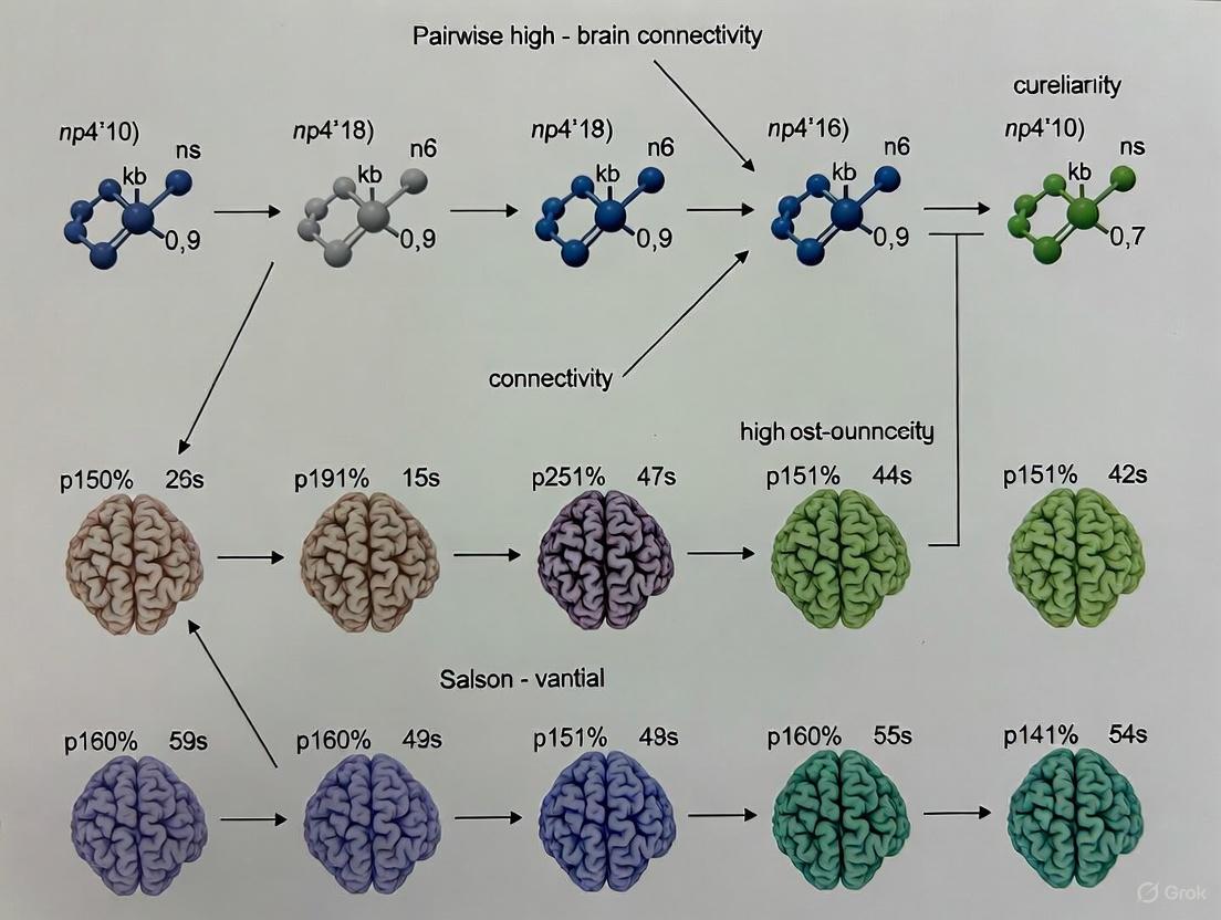
Figure 1: Workflow for pairwise mutual information analysis.
Protocol 2: Higher-Order Connectivity Analysis Using O-Information
Aim: To characterize higher-order interactions and distinguish between synergistic and redundant information in groups of three or more brain regions.
Aim: To characterize higher-order interactions and distinguish between synergistic and redundant information in groups of three or more brain regions.
Materials and Reagents:
- Preprocessed neuroimaging time series (as in Protocol 1)
- Specialized software for higher-order analysis (e.g.,
hoilibrary in Python, custom scripts for topological analysis [1])
Procedure:
- Signal Standardization: Z-score all regional time series to ensure comparability [1].
- Define Variable Sets: Select a target brain region (Xi) and a set of other regions (\mathbf{X} = {X{j}, X_{k}, ...}) with which its potential HOIs will be analyzed.
- Compute Multi-Information: Calculate the total correlation or multi-information for the set ({Xi, \mathbf{X}}), which captures the total shared information among all variables: (I({Xi, \mathbf{X}}) = \sum p({Xi, \mathbf{X}}) \log \frac{p({Xi, \mathbf{X}})}{\prod p(x)}).
- Calculate O-Information: The O-information (\Omega({X_i, \mathbf{X}})) is computed as the difference between the total correlation of the entire set and the sum of the total correlations of all subsets of size (n-1). A positive (\Omega) indicates a system dominated by redundancy, while a negative (\Omega) indicates a system dominated by synergy [1].
- Hypergraph Construction: Represent the results as a hypergraph where hyper-edges connect groups of regions (e.g., triplets, quadruplets) that exhibit significant synergistic or redundant interactions.
- Topological Analysis: Use computational topology tools to analyze the weighted simplicial complex and extract local higher-order indicators, such as "violating triangles" that represent coherent co-fluctuations not explainable by pairwise connections [1].
Figure 2: Workflow for O-information analysis.
The Scientist's Toolkit: Essential Research Reagents
Table 3: Key Computational Tools and Resources
| Tool/Resource | Type | Primary Function | Application Context |
|---|---|---|---|
| PySPI Package [2] | Software Library | Implements 239 pairwise interaction statistics, including mutual information | Large-scale benchmarking of FC methods; standardized calculation of information-theoretic measures |
| HOI Library | Software Library | Specialized for estimating O-information and other higher-order measures | Analysis of synergistic and redundant information in multivariate neural data |
| Human Connectome Project Data [2] [1] | Reference Dataset | Provides high-quality, multimodal neuroimaging data from healthy adults | Method validation; normative baselines; individual differences research |
| Schaefer Parcellation [2] | Brain Atlas | Defines functionally coherent cortical regions of interest | Standardized node definition for reproducible network construction |
| Allen Human Brain Atlas [2] | Reference Data | Provides correlated gene expression maps | Validation of FC findings against transcriptomic data |
| Tensor Decomposition Algorithms [9] | Computational Method | Identifies multilinear patterns and change points in high-order datasets | Dynamic connectivity analysis; capturing transient higher-order states |
Integrated Analysis Workflow
The following diagram integrates pairwise and higher-order approaches into a comprehensive analysis pipeline for validating brain connectivity.
Figure 3: Integrated validation pipeline for multi-scale connectivity.
The Critical Need for Single-Subject Analysis in Personalized Medicine
Personalized medicine aims to move beyond population-wide averages to deliver diagnoses and treatments tailored to the individual patient. A significant statistical challenge in this endeavor is the reliable interpretation of massive omics datasets, such as those from transcriptomics or neuroimaging, from a single subject. Traditional cohort-based statistical methods are often inapplicable or underpowered for single-subject studies (SSS), creating a critical methodological gap [10]. This document outlines the application notes and protocols for conducting robust single-subject analyses, framed within advanced research on statistical validation of pairwise and high-order brain connectivity. We provide detailed methodologies, data presentation standards, and visualization tools to empower researchers in this field.
Background and Rationale
In both transcriptomics and functional brain connectivity, the standard approach for identifying significant signals (e.g., Differentially Expressed Genes (DEGs) or functional connections) relies on having multiple replicates per condition to estimate variance and compute statistical significance. However, in a clinical setting, obtaining multiple replicates from a single patient is often prohibitively expensive, ethically challenging, or simply impractical [10] [11].
The core challenge is that statistical artefacts and biases can be easily confounded with authentic biological signals when analyzing a dataset from one individual [10]. Furthermore, in neuroimaging, traditional models that represent brain function as a network of pairwise interactions are limited in their ability to capture the complex, synergistic dynamics of the human brain [3] [1]. High-order interactions (HOIs), which involve three or more brain regions simultaneously, are increasingly recognized as crucial for a complete understanding of brain function and its relation to behavior and disease [1] [12]. The move towards personalized neuroscience therefore requires methods that can derive meaningful insights from individual brain recordings by analyzing descriptive indexes of physio-pathological states through statistics that prioritize subject-specific differences [3].
The following tables summarize key performance metrics for single-subject analysis methods as validated in benchmark studies.
Table 1: Performance of Single-Subject DEG Methods in Transcriptomics [11]
| Method Name | Median ROC-AUC (Yeast) | Median ROC-AUC (MCF7) | Key Characteristics |
|---|---|---|---|
| ss-NOIseq | > 90% | > 75% | Designed for single-subject analysis without replicates. |
| ss-DEGseq | > 90% | > 75% | Adapts a cohort method for single-subject use. |
| ss-Ensemble | > 90% | > 75% | Combines multiple methods; most robust across conditions. |
| ss-edgeR | Variable | Variable | Performance highly dependent on the proportion of true DEGs. |
| ss-DESeq | Variable | Variable | Performance highly dependent on the proportion of true DEGs. |
Table 2: Comparative Performance of Connectivity Measures in Neuroimaging [1] [2]
| Connectivity Measure Type | Task Decoding Capacity | Individual Identification | Association with Behavior |
|---|---|---|---|
| Pairwise (e.g., Pearson Correlation) | Baseline | Baseline | Baseline |
| High-Order (e.g., Violating Triangles) | Greatly Improved | Improved | Significantly Strengthened |
| Precision/Inverse Covariance | N/A | High | High [2] |
| Spectral Measures | N/A | Moderate | Moderate [2] |
Experimental Protocols
Protocol 1: Single-Subject Transcriptomic Analysis via an "All-against-One" Framework
This protocol is designed for identifying differentially expressed genes (DEGs) from two RNA-Seq samples (e.g., diseased vs. healthy) from a single patient without replicates [10] [11].
1. Prerequisite Data: Two condition-specific transcriptomes from a single subject (e.g., Subject_X_Condition_A.txt, Subject_X_Condition_B.txt).
2. Software and Environment Setup:
- Computing Environment: R programming language (v3.4.0 or later).
- Required R Packages:
referenceNof1(for constructing unbiased reference standards), and packages for ss-DEG methods (e.g.,NOISeq,DESeq2,edgeR) [10] [11].
3. Reference Standard (RS) Construction: To avoid analytical bias, do not use the same method for RS creation and discovery.
- Obtain an isogenic dataset (biological replicates from a matched background) relevant to your study context [11].
- Apply multiple, distinct DEG analytical methods (e.g., NOISeq, edgeR, DESeq) to the isogenic replicate dataset to identify a consensus set of DEGs. This consensus becomes your RS [10] [11].
- Optimize the RS by applying fold-change (FC) thresholds and expression-level cutoffs to increase concordance between methods [10].
4. Single-Subject DEG Prediction:
- Apply one or more ss-DEG methods (e.g., ss-NOIseq, ss-Ensemble) to the two samples from your single subject.
- The output is a list of predicted DEGs for that subject.
5. Validation against Reference Standard:
- Compare the predicted DEGs from Step 4 against the RS from Step 3.
- Calculate performance metrics such as Receiver-Operator Characteristic (ROC) curves and Precision-Recall plots to evaluate the accuracy of the ss-DEG method [11].
6. Recommendation: For the most robust results, use an ensemble learner approach that integrates predictions from multiple ss-DEG methods to resolve conflicting signals and improve stability [10] [11].
Protocol 2: Single-Subject High-Order Brain Connectivity Analysis
This protocol details the statistical validation of high-order functional connectivity in an individual's brain using fMRI data, enabling subject-specific investigation and treatment planning [3].
1. Data Acquisition and Preprocessing:
- Data: Resting-state or task-based fMRI time series from a single subject.
- Preprocessing: Standardize the fMRI signals from N brain regions using z-scoring [1].
2. Constructing Functional Connectivity Networks:
- Calculate all possible pairwise interactions between brain regions. Common measures include Mutual Information (MI) or Pearson's correlation coefficient [3] [2].
- Calculate High-Order Interactions (HOIs). One method involves computing k-order time series as the element-wise products of (k+1) z-scored time series, which are then re-standardized. This represents the instantaneous co-fluctuation magnitude of (k+1)-node interactions (e.g., triangles) [1].
3. Statistical Validation of Connectivity Measures:
- For Pairwise Connections (MI): Use surrogate data analysis.
- Generate surrogate time series that mimic the individual properties (e.g., power spectrum) of the original signals but are otherwise uncoupled.
- Compute the MI for the original paired signals and for a large number of surrogate pairs.
- The MI value is considered statistically significant if it exceeds the 95th percentile of the surrogate distribution [3].
- For High-Order Interactions (O-Information): Use bootstrap analysis.
- Generate multiple bootstrap-resampled versions of the original multivariate time series.
- Compute the O-information (or other HOI metric) for each resampled dataset.
- Construct confidence intervals from the bootstrap distribution.
- An HOI is significant if its confidence interval does not cross zero. Differences in HOI between experimental conditions can also be assessed this way [3].
4. Feature Extraction and Interpretation:
- Extract significant pairwise and high-order features that have passed the above statistical tests.
- These subject-specific fingerprints can be used for:
- Task Decoding: Identifying which cognitive task a subject is performing based on brain activity patterns [1].
- Individual Identification: Uniquely identifying an individual based on their functional connectome [1] [2].
- Clinical Correlation: Associating connectivity patterns with individual behavioral measures or treatment outcomes [3] [1].
Visualization of Workflows
Single-Subject Transcriptomics Analysis
High-Order Brain Connectivity Analysis
The Scientist's Toolkit: Research Reagent Solutions
Table 3: Essential Tools for Single-Subject Analysis in Personalized Medicine
| Item / Resource | Function / Description | Application Context |
|---|---|---|
referenceNof1 R Package |
Provides a robust framework for constructing method-agnostic reference standards to evaluate single-subject analyses, minimizing statistical artefact biases. | Transcriptomics [10] |
| Isogenic Biological Replicates | Publicly available datasets (e.g., Yeast BY4741 strain, MCF7 cell line) used as a ground truth for developing and validating single-subject reference standards. | Transcriptomics [11] |
| Surrogate Data Algorithms | Algorithms (e.g., Iterative Amplitude-Adjusted Fourier Transform) that generate null model time series to test the significance of pairwise functional connections. | Neuroimaging [3] |
| Bootstrap Resampling Methods | Statistical technique to estimate the sampling distribution of a statistic (e.g., O-Information) by resampling the data with replacement, used to derive confidence intervals for HOIs. | Neuroimaging [3] |
| Topological Data Analysis (TDA) | A set of computational tools (e.g., Persistent Homology) that can infer and analyze the higher-order interaction structures from neuroimaging time series data. | Neuroimaging [1] [12] |
| SPI (Statistical Pairwise Interactions) Library | A comprehensive library (e.g., pyspi) containing 239+ pairwise statistics for benchmarking and optimizing functional connectivity mapping. |
Neuroimaging [2] |
Linking Network Topology to Brain Function and Criticality
Understanding how brain functions arise from neuronal architecture is a central question in neuroscience. The Critical Brain Hypothesis (CBH) proposes that brain networks operate near a phase transition, a state supporting optimal computational performance, efficient memory usage, and high dynamic range for health and function [13]. Mounting evidence suggests that the brain's particular hierarchical modular topology—characterized by groups of nodes segregated into modules, which are in turn nested within larger modules—plays a crucial role in achieving and sustaining this critical state [13] [1]. Furthermore, traditional models based on pairwise interactions are increasingly seen as limited, with higher-order interactions (HOIs) providing a more complete picture of brain dynamics [1]. This Application Note details the quantitative evidence and provides standardized protocols for investigating the link between network topology, criticality, and brain function within the context of statistical validation of pairwise and high-order brain connectivity research.
Quantitative Data on Topology, Criticality, and Higher-Order Interactions
The following tables summarize key quantitative findings from computational and empirical studies, highlighting how specific topological features influence brain dynamics and the added value of higher-order analysis.
Table 1: Influence of Intramodular Topology on Critical Dynamics in Hierarchical Modular Networks [13]
| Network Topology Type | Robustness in Sustaining Criticality | Typical Dynamical Regime | Key Characteristic |
|---|---|---|---|
| Sparse Erdős-Rényi (ER) | High | Critical/Quasicritical | Random pairwise connection probability (ε = 0.01) |
| Sparse Regular (K-Neighbor, KN) | High | Critical/Quasicritical | Fixed degree (K = 40) for all neurons |
| Fully Connected (FC) | Low | Tends toward Supercritical | All-to-all intramodular connectivity |
Table 2: Performance Comparison of Connectivity Methods in fMRI Analysis [1]
| Analysis Method | Task Decoding (Element-Centric Similarity) | Individual Identification | Association with Behavior |
|---|---|---|---|
| Traditional Pairwise (Edge) Signals | Baseline | Baseline | Baseline |
| Higher-Order (Triangle) Signals | Greatly Improved | Improved | Significantly Stronger |
| Homological Scaffold Signals | Improved | Improved | Stronger |
Table 3: Key Statistical Measures for Comparing Quantitative Data Across Conditions [14] [15]
| Statistical Measure | Category | Description | Interpretation in Connectivity Research |
|---|---|---|---|
| Mean / Median | Measure of Center | Average / Central value in a sorted dataset | Compares the typical level of connectivity strength or activity between groups. |
| Standard Deviation | Measure of Variability | Average deviation of data points from the mean | Indicates the variability or consistency of connectivity values within a single group or condition. |
| Interquartile Range (IQR) | Measure of Variability | Range between the 25th and 75th percentiles (Q3 - Q1) | Describes the spread of the central portion of the data, reducing the influence of outliers. |
Experimental Protocols
Protocol 1: Simulating Criticality in Hierarchical Modular Networks
This protocol outlines the steps for constructing and simulating hierarchical modular neuronal networks to study their critical dynamics [13].
1. Network Construction:
- Objective: Generate a network with a nested modular structure.
- Procedure for ER and KN Networks:
- Initialization: Start with a single module of N neurons at hierarchical level H=0.
- Recursive Division: For each level from H=1 to Hmax:
- Randomly split every existing module into two new modules of equal size.
- Rewire Intermodular Connections: For each connection linking neurons in different modules, replace it with a new connection within the same module as the presynaptic neuron with a high probability (e.g., R=0.9). This reinforces intramodular density.
- Procedure for FC Networks:
- Initialization: Start by partitioning N neurons into multiple, smaller, fully connected modules.
- Recursive Clustering: At each higher hierarchical level, cluster existing modules into pairs of larger modules.
- Establish Intermodular Links: For each pair of modules being connected, create links between their neurons with a level-dependent probability (e.g., using parameters α=1 and p=1/4) to maintain a constant number of cross-level connections.
2. Neuron Model and Dynamics:
- Model: Use a network of discrete-time stochastic Leaky Integrate-and-Fire (LIF) neurons.
- Membrane Potential Update: The subthreshold membrane potential ( Vi[t] ) of neuron *i* at time *t* evolves as: ( Vi[t+1] = μVi[t] + Ii[t] + \frac{1}{N}\sum{j=1}^{N}W{ij}[t]Sj[t] ) where *μ* is the leakage constant, ( Ii ) is external input, ( W{ij} ) is the adaptive synaptic weight, and ( Sj ) is the spike state of neuron j (1 for spike, 0 otherwise) [13].
- Spiking: A spike is emitted when ( Vi[t] ) exceeds a fixed threshold ( V{th} ). The potential is then reset to ( V_{reset} ).
3. Homeostatic Adaptation:
- Objective: Implement a biological mechanism to self-organize towards criticality.
- Mechanism: Use dynamic neuronal gains or dynamic synapses (e.g., LHG dynamics). These parameters increase when network activity is low and decrease when activity is high, creating a negative feedback loop that stabilizes activity near a critical point [13].
4. Data Collection and Analysis:
- Avalanche Definition: Record spike trains and define avalanches as sequences of continuous activity bounded by silent periods.
- Criticality Signatures: Calculate the distributions of avalanche sizes and durations. A power-law distribution is a key signature of criticality. Further, analyze the scaling relationship between average avalanche size and duration [13].
Protocol 2: Assessing Brain Network Resistance with TMS-EEG
This protocol uses Transcranial Magnetic Stimulation combined with Electroencephalography (TMS-EEG) to empirically probe network dynamics and its resistance to change in humans [16].
1. Experimental Design:
- Objective: Measure changes in brain network reactivity during offline processing following a behavioral task.
- Participants: Recruit subjects according to study design (e.g., healthy controls vs. patient populations).
- Conditions: Include at least two sessions: a baseline resting-state measurement and a post-task measurement.
2. Setup Preparation:
- TMS: Use a TMS apparatus with a MRI-guided neuromavigation system to ensure precise and consistent targeting of a specific brain region (e.g., primary motor cortex).
- EEG: Apply a high-density EEG cap (e.g., 64 channels). Impedances should be kept below 5 kΩ to ensure high-quality signal acquisition.
- Artifact Control: Employ a TMS-compatible system and use techniques to minimize the large electromagnetic artifact induced by the TMS pulse on the EEG recording.
3. Data Collection:
- Stimulation Protocol: At each session, apply a series of TMS pulses (e.g., 100-200 pulses) to the target region during resting state. The inter-stimulus interval should be jittered to prevent anticipatory effects.
- EEG Recording: Continuously record EEG data before, during, and after each TMS pulse. Sampling rate should be sufficiently high (e.g., ≥ 5 kHz) to capture the initial TMS-evoked potential (TEP) and the subsequent spread of activity.
4. Data Analysis:
- Preprocessing: Filter the data (e.g., 1-100 Hz bandpass, 50/60 Hz notch). Automatically or manually reject trials with large artifacts (e.g., eye blinks, muscle activity).
- TMS-Evoked Potential (TEP): Average the EEG signals time-locked to the TMS pulses to extract the characteristic TEP components, which reflect the local and network-level response to the perturbation.
- Network Metrics: Calculate the global mean field power (GMFP) or similar indices from the TEP as a measure of the overall network activation in response to the stimulus. A change in the amplitude or duration of this response (e.g., post-task) indicates a change in the network's resistance or excitability [16].
Protocol 3: Inferring Higher-Order Interactions from fMRI Data
This protocol details a topological method to uncover HOIs from fMRI time series, moving beyond pairwise correlation [1].
1. Data Preprocessing:
- Starting Data: Use preprocessed fMRI BOLD time series from N brain regions.
- Standardization: Z-score each regional time series to have zero mean and unit variance.
2. Constructing k-Order Time Series:
- Calculation: For each timepoint, compute all possible k-order time series as the element-wise product of (k+1) z-scored time series. For example, a 2-order (triangle) time series for regions {i, j, k} is: ( TS{ijk}[t] = zi[t] \cdot zj[t] \cdot zk[t] ).
- Standardization and Signing: Z-score these new k-order time series. Then, assign a sign at each timepoint: positive if all (k+1) original time series were concordant (all positive or all negative), and negative otherwise. This highlights fully coherent group interactions.
3. Building Simplicial Complexes:
- Encoding: At each timepoint t, encode the brain's activity into a mathematical object called a weighted simplicial complex.
- Weights: Nodes (0-simplices) represent regions. Edges (1-simplices) represent pairwise interactions, weighted by the traditional co-fluctuation value. Triangles (2-simplices) represent triple interactions, weighted by the signed 2-order time series value calculated in the previous step.
4. Extracting Higher-Order Indicators:
- Global Indicator (Hyper-coherence): Identify "violating triangles"—triangles whose weight (strength of triple interaction) is greater than the weights of its three constituent edges. The fraction of such triangles quantifies global hyper-coherence.
- Local Indicators:
- Violating Triangles List: Record the identity and weight of each violating triangle. These are triplets of regions whose coordinated activity cannot be explained by pairwise relationships alone.
- Homological Scaffold: Construct a weighted graph that highlights the edges most critical to forming mesoscopic topological structures (like cycles) within the simplicial complex, assessing their importance in the overall higher-order landscape [1].
Visualization of Concepts and Workflows
Hierarchical Modular Network Construction and Criticality
HM Network Criticality: This diagram illustrates the recursive algorithm for building hierarchical modular networks and the homeostatic mechanism that regulates neuronal activity to maintain a critical state.
Higher-Order fMRI Analysis Pipeline
HOI Analysis Pipeline: This workflow outlines the key steps for inferring higher-order interactions from fMRI data, from raw signals to higher-order topological indicators.
The Scientist's Toolkit: Research Reagent Solutions
Table 4: Essential Materials and Tools for Connectivity and Criticality Research
| Item / Reagent | Function / Application | Key Characteristics / Examples |
|---|---|---|
| Stochastic LIF Neuron Model | Computational unit for simulating spiking network dynamics. | Includes membrane potential, leakage, spiking threshold, and reset; can be extended with adaptation [13]. |
| Homeostatic Plasticity Rules | A biologically-plausible mechanism to self-organize and maintain network criticality. | Dynamic neuronal gains (N parameters) or dynamic synapses like LHG (O(N²) parameters) [13]. |
| Hierarchical Network Generators | Algorithms to create computational models with nested modular architecture. | Erdős-Rényi (ER), K-Neighbor (KN), and Fully Connected (FC) generative algorithms [13]. |
| TMS-EEG System | Non-invasive tool for causal perturbation and measurement of whole-brain network dynamics. | Combines a TMS apparatus with a high-density, TMS-compatible EEG system for evoking and recording brain activity [16]. |
| Simplicial Complex Analysis | Mathematical framework for representing and analyzing higher-order interactions. | Encodes nodes, edges, triangles, etc., into a single object for topological interrogation [1]. |
| Topological Data Analysis (TDA) Libraries | Software for extracting features from inferred higher-order structures. | Used to compute indicators like hyper-coherence and homological scaffolds from simplicial complexes [1]. |
A Practical Framework for Validating Connectivity on a Single-Subject Basis
In brain connectivity research, distinguishing genuine neural interactions from spurious correlations caused by noise, finite data samples, or signal properties is a fundamental challenge. Statistical validation is therefore not merely a supplementary step but a cornerstone for ensuring the biological validity and reproducibility of findings. Within the context of pairwise and high-order brain connectivity research, two computer-intensive statistical methods have become indispensable: surrogate data analysis and bootstrap analysis [17] [3].
Surrogate data analysis is primarily used for hypothesis testing, creating synthetic data that preserve specific linear properties of the original data (e.g., power spectrum, autocorrelation) but destroy the nonlinear or dependency structures under investigation. By comparing connectivity metrics from original and surrogate data, researchers can test the null hypothesis that their results are explainable by a linear process [18] [19]. Conversely, bootstrap analysis is a resampling technique for estimating the sampling distribution of a statistic, such as a connectivity metric. It allows researchers to construct confidence intervals and assess the stability and reliability of their estimates without making strict distributional assumptions [3] [20].
This article provides detailed application notes and protocols for implementing these core validation techniques, framed within the rigorous demands of modern pairwise and high-order brain connectivity research.
Surrogate Data Analysis
Conceptual Foundation and Applications
Surrogate data methods test the null hypothesis that an observed time series is generated by a specific linear process. The core principle involves generating multiple surrogate datasets that mimic the original data's linear characteristics but are otherwise random. If a connectivity metric (e.g., synchronization likelihood, mutual information) computed from the original data is significantly different from the distribution of that metric computed from the surrogates, the null hypothesis can be rejected, providing evidence for genuine, non-random connectivity [18] [21].
This technique is crucial in brain connectivity for:
- Testing for Non-Random Connectivity: Determining whether observed functional connections exceed chance levels [21].
- Validating Nonlinear Dynamics: Assessing whether brain signals exhibit significant nonlinearity, justifying the use of nonlinear analysis methods [18].
- Establishing Statistical Thresholds: Defining significance thresholds for connectivity matrices in network analysis, thereby controlling for false positives [18] [21].
Quantitative Comparison of Surrogate Generation Algorithms
Table 1: Key Algorithms for Generating Surrogate Data
| Algorithm Name | Core Principle | Properties Preserved | Properties Randomized | Primary Use Case in Connectivity |
|---|---|---|---|---|
| Phase Randomization | Applies a Fourier transform, randomizes the phase spectrum, and performs an inverse transform [18]. | Power spectrum (and thus autocorrelation) [18] [19]. | All temporal phase relationships, destroying nonlinear dependencies [18]. | General-purpose test for nonlinearity and non-random connectivity in stationary signals [18]. |
| Autoregressive Randomization (ARR) | Fits a linear autoregressive (AR) model to the data and generates new data by driving the AR model with random noise [19]. | Autocorrelation function and the covariance structure of multivariate data [19]. | The precise temporal sequence and any higher-order moments not captured by the AR model. | Testing for nonlinearity in multivariate signals while preserving linear temporal dependencies [19]. |
| Static Null (Gaussian) | Generates random data from a multivariate Gaussian distribution with a covariance matrix equal to that of the original data [19]. | Covariance structure between signals [19]. | All temporal dynamics and non-Gaussian properties. | Testing whether observed connectivity is explainable by static, linear correlations only [19]. |
Experimental Protocol: Testing Connectivity Significance with Phase Randomization
This protocol details the steps for validating pairwise or high-order connectivity metrics using phase-randomized surrogates.
Objective: To determine if the observed functional connectivity value between two or more neural signals is statistically significant against the null hypothesis of a linear correlation structure.
Materials and Reagents:
- Preprocessed neural time-series data (e.g., EEG, MEG, fMRI BOLD).
- Computing environment with programming capabilities (e.g., MATLAB, Python, R).
- Software for connectivity metric calculation (e.g., Mutual Information, Synchronization Likelihood, O-Information for high-order interactions [3]).
Procedure:
- Compute Original Metric: Calculate the functional connectivity metric of interest (e.g., mutual information for pairwise, O-information for high-order) on the original multivariate neural dataset [3].
- Generate Surrogates: For each original time series, generate a large number (N) of phase-randomized surrogate time series [18] [21].
- Technical Note: For signals with a strong dominant frequency (e.g., alpha rhythm in EEG), ensure the segment length comprises an integer number of periods of this frequency to avoid artificial non-linearity detection [18].
- Compute Null Distribution: Calculate the same connectivity metric for each of the N surrogate datasets, creating a null distribution of connectivity values under the linear hypothesis.
- Statistical Testing: Compare the original connectivity value to the null distribution.
- For a one-tailed test (e.g., testing for connectivity greater than chance), the p-value is estimated as the proportion of surrogate-derived metrics that are greater than or equal to the original metric.
- A standard significance threshold is p < 0.05. Correct for multiple comparisons if testing many connections (e.g., using False Discovery Rate) [21].
Figure 1: Workflow for surrogate data analysis to test connectivity significance. The process tests whether the original connectivity metric is significantly different from what is expected by a linear process.
Bootstrap Analysis
Conceptual Foundation and Applications
Bootstrap analysis is a resampling method used to assess the reliability and precision of estimated parameters. By drawing multiple random samples (with replacement) from the original data, it approximates the sampling distribution of a statistic. This is particularly valuable in brain connectivity research, where the underlying distribution of many connectivity metrics is unknown or non-normal [20].
In the context of pairwise and high-order connectivity, bootstrap methods are instrumental for:
- Constructing Confidence Intervals (CIs): Providing a range of plausible values for a connectivity metric, such as the strength of a functional connection [3] [20].
- Assessing Stability in Single-Subject Analyses: Enabling robust statistical inferences at the individual level, which is crucial for personalized medicine and clinical applications [3].
- Comparing Conditions: Testing whether connectivity changes between two experimental conditions (e.g., rest vs. task) are statistically significant [22].
Quantitative Comparison of Bootstrap Methods
Table 2: Key Bootstrap Methods for Connectivity Research
| Bootstrap Method | Core Principle | Key Advantage | Considerations for Connectivity Analysis |
|---|---|---|---|
| Percentile Bootstrap | The confidence interval is directly taken from the percentiles (e.g., 2.5th and 97.5th) of the bootstrap distribution [20]. | Simple and intuitive to implement. | Can be biased if the bootstrap distribution is not centered on the original statistic [20]. |
| Bias-Corrected and Accelerated (BCa) | Adjusts the percentiles used for the CI to account for both bias and skewness in the bootstrap distribution [22] [20]. | More accurate confidence intervals for skewed statistics and small sample sizes; highly recommended for practice [22]. | Computationally more intensive than the percentile method. |
| Case Resampling | Entire experimental units (e.g., all time series from a single subject's scan) are resampled with replacement. | Preserves the inherent dependency structure within a subject's data. | Ideal for group-level analysis where each subject is an independent unit. |
| Paired Bootstrap | For paired data (e.g., baseline vs. variant under identical seeds), the deltas (differences) are resampled [22]. | Reduces variance by exploiting the positive correlation between paired measurements, increasing sensitivity to detect small changes [22]. | Essential for comparing connectivity across conditions within the same subject or under identical computational seeds. |
Experimental Protocol: Paired Bootstrap for Comparing Connectivity Across Conditions
This protocol uses a paired, BCa bootstrap to evaluate if a change in brain connectivity between two conditions is statistically significant at the single-subject or group level.
Objective: To test whether the difference in a functional connectivity metric between Condition A and Condition B is statistically significant, using a paired design to control for variability.
Materials and Reagents:
- Neural data from the same subject(s) under two different conditions (e.g., rest and task).
- Computational resources for multiple connectivity estimations.
Procedure:
- Calculate Per-Unit Deltas: For each independent experimental unit (e.g., a subject or a random seed), calculate the delta (Δ) as the connectivity metric in Condition B minus the connectivity metric in Condition A. This creates a vector of observed deltas [22].
- Generate Bootstrap Samples: Generate a large number (e.g., 1000-2000) of bootstrap samples by resampling the vector of deltas with replacement. Each sample must be the same size as the original vector.
- Compute Bootstrap Distribution: For each bootstrap sample, calculate the mean delta. This creates the bootstrap distribution of the mean difference.
- Construct BCa Confidence Interval: Calculate the BCa confidence interval (e.g., 95%) from this bootstrap distribution [22] [20].
- Hypothesis Testing: If the 95% BCa confidence interval for the mean delta does not include zero, you can reject the null hypothesis and conclude a significant difference in connectivity between the two conditions [22].
Figure 2: Workflow for a paired bootstrap analysis to compare connectivity across two conditions. This method is more powerful for detecting small changes by leveraging paired measurements.
The Scientist's Toolkit: Research Reagent Solutions
Table 3: Essential Computational and Data "Reagents" for Connectivity Validation
| Item Name | Function / Definition | Application Note |
|---|---|---|
| Preprocessed fMRI/EEG Time Series | The cleaned, artifact-free neural signal from regions of interest (ROIs). The fundamental input data. | Preprocessing (filtering, artifact rejection) is critical, as poor data quality severely biases connectivity estimates and their validation [17]. |
| Connectivity Metric Algorithms | Software implementations for calculating metrics like Mutual Information (pairwise) or O-Information (high-order) [3]. | The choice of metric (pairwise vs. high-order) dictates the complexity of interactions that can be detected. High-order metrics can reveal synergistic structures missed by pairwise approaches [3] [1]. |
| Phase Randomization Script | Code to perform Fourier transform, phase randomization, and inverse transform. | A core "reagent" for generating surrogate data. Must be carefully implemented to handle signal borders and dominant rhythms [18]. |
| BCa Bootstrap Routine | A computational function that performs the Bias-Corrected and Accelerated bootstrap procedure. | A more robust alternative to simple percentile methods for constructing confidence intervals, especially with skewed statistics [22] [20]. |
| High-Performance Computing (HPC) Cluster | Parallel computing environment. | Both surrogate and bootstrap analyses are computationally intensive, requiring 1000s of iterations. HPC drastically reduces computation time. |
| Statistical Parcellation Atlas | A predefined map of brain regions (e.g., with 100-500 regions) [23]. | Provides the nodes for the connectivity network. Higher-order parcellations allow for the investigation of finer-grained, specialized subnetworks [23]. |
Step-by-Step Guide to Assessing Pairwise Connectivity Significance
In contemporary neuroscience, particularly within the framework of the BRAIN Initiative's vision to generate a dynamic picture of brain function, the statistical validation of brain connectivity metrics is essential [24]. This is especially true for personalized neuroscience, where the goal is to derive meaningful insights from individual brain signal recordings to inform subject-specific interventions and treatment planning [3]. Analyzing the descriptive indexes of physio-pathological states requires statistical methods that prioritize individual differences across varying experimental conditions.
Functional connectivity networks, which model the brain as a complex system by investigating inter-relationships between pairs of brain regions, have long been a valuable tool [3]. However, the usefulness of standard pairwise connectivity measures is limited because they can miss high-order dependencies and are susceptible to spurious connections from finite data size, acquisition errors, or structural misunderstandings [3]. Therefore, moving from simple observation of a connectivity value to establishing its statistical significance and accuracy is a critical step for a reliable assessment of an individual's underlying condition, helping to prevent biased clinical decisions. This guide provides a detailed protocol for assessing the significance of pairwise connectivity, forming a foundational element of a broader thesis on the statistical validation of pairwise and high-order brain connectivity.
Core Statistical Methodologies
The rationale for this approach involves using surrogate data to test the significance of putative connections and bootstrap resampling to quantify the accuracy of the connectivity estimates and compare them across conditions [3].
Surrogate Data Analysis for Significance Testing
Purpose: To test the null hypothesis that two observed brain signals are independent. This procedure generates simulated data sets that preserve key individual properties of the original signals (e.g., linear autocorrelation) but are otherwise uncoupled [3] [25].
Theoretical Basis: The method creates a null distribution for the mutual information (MI) value under the assumption of independence. Suppose you compute the MI, denoted as ( I_{orig} ), for the original pair of signals. The surrogate testing procedure is as follows:
- Generate Multiple Surrogate Pairs: Create ( N_s ) (e.g., 1,000) independent pairs of surrogate time series from the original pair of signals.
- Compute Null Distribution: Calculate the MI, ( I{surr}^{(i)} ), for each of the ( Ns ) surrogate pairs.
- Determine Significance: Compare the original MI value, ( I{orig} ), to the distribution of ( I{surr} ).
A one-tailed test is typically used to identify a connectivity value significantly greater than expected by chance. The ( p )-value can be approximated as: [ p = \frac{\text{number of times } I{surr}^{(i)} \geq I{orig}}{N_s} ] A connection is deemed statistically significant if the ( p )-value is below a predefined threshold (e.g., ( p < 0.05 ), corrected for multiple comparisons).
Bootstrap Analysis for Estimating Confidence Intervals
Purpose: To assess the accuracy and stability of the estimated pairwise connectivity measure (e.g., MI) and to enable comparisons of connectivity strength across different experimental conditions (e.g., pre- vs. post-treatment) on a single-subject level [3].
Theoretical Basis: The bootstrap technique involves drawing multiple random samples (with replacement) from the original data to create a sampling distribution for the statistic of interest [3] [25].
- Generate Bootstrap Samples: Create ( N_b ) (e.g., 1,000) new data sets by randomly resampling, with replacement, the time points from the original multivariate dataset.
- Compute Bootstrap Distribution: For each bootstrap sample, re-calculate the pairwise MI, ( I_{boot}^{(i)} ), for the connection in question.
- Construct Confidence Intervals: The distribution of ( I_{boot} ) values forms an empirical sampling distribution. A ( 100(1-\alpha)\% ) confidence interval (e.g., 95% CI) can be derived using the percentile method: the interval is defined by the ( \alpha/2 ) and ( 1-\alpha/2 ) quantiles of the bootstrap distribution.
The resulting confidence intervals allow researchers to determine the reliability of individual estimates and to assess whether a change in connectivity between two states is statistically significant (e.g., if the confidence intervals do not overlap).
Experimental Protocols
Protocol 1: Surrogate Data Testing for Significant Pairwise Links
This protocol details the steps to identify which pairwise connections in a single subject's functional connectivity network are statistically significant.
- Step 1: Data Preprocessing. Begin with preprocessed resting-state fMRI (or other neuroimaging) time series data for ( N ) brain regions. Let ( X ) and ( Y ) be the ( T )-length time series (where ( T ) is the number of time points) for two specific regions of interest.
- Step 2: Calculate Observed Mutual Information. Compute the mutual information ( I(X; Y) ) for the original data, denoted as ( I_{orig} ).
- Step 3: Generate Surrogate Data. Create ( Ns = 1000 ) surrogate pairs ( (X^*i, Y^*_i) ). A common and robust method is the Iterative Amplitude Adjusted Fourier Transform (IAAFT) algorithm, which preserves the power spectrum and amplitude distribution of the original signals while breaking any nonlinear coupling.
- Step 4: Compute Surrogate Mutual Information. For each surrogate pair ( (X^_i, Y^i) ), calculate the mutual information, ( I{surr}^{(i)} ).
- Step 5: Formulate Statistical Test. Construct the null distribution from the ( Ns ) values of ( I{surr} ).
- Step 6: Correct for Multiple Comparisons. Since you are testing connections between many possible pairs of regions, apply a multiple comparisons correction (e.g., False Discovery Rate, FDR) to the resulting ( p )-values across all tested pairs.
- Step 7: Identify Significant Network. Retain only those pairwise connections with an FDR-corrected ( p )-value < 0.05. This constitutes the statistically significant pairwise functional network for the individual.
Protocol 2: Bootstrap Confidence Intervals for Cross-Condition Comparison
This protocol assesses the reliability of a connectivity estimate and tests for significant changes in connectivity between two conditions (e.g., rest vs. task, pre- vs. post-treatment) within a single subject.
- Step 1: Data Preparation. Organize the preprocessed time series data for the two conditions (Condition A and Condition B) separately. For each condition, you will have a ( T \times N ) matrix of time series.
- Step 2: Select Connection of Interest. Identify the specific pairwise connection (e.g., between Region P and Region Q) to be analyzed.
- Step 3: Bootstrap Sampling for Each Condition.
- For Condition A, generate ( Nb = 1000 ) bootstrap samples by randomly selecting ( T ) time points from the Condition A data, with replacement.
- For each bootstrap sample ( j ), compute the MI between Region P and Q, ( I{A}^{(j)} ).
- Repeat this process for Condition B to obtain ( Nb ) values of ( I{B}^{(j)} ).
- Step 4: Construct Confidence Intervals. For the distributions of ( I{A} ) and ( I{B} ), calculate the 95% confidence intervals using the 2.5th and 97.5th percentiles.
- Step 5: Compare Conditions. A conservative approach to determine a significant change is to check for non-overlapping 95% confidence intervals between Condition A and Condition B. For a more direct test, one can compute the bootstrap distribution for the difference ( I{A} - I{B} ) and check if its 95% confidence interval excludes zero.
The Scientist's Toolkit: Research Reagent Solutions
Table 1: Essential Materials and Analytical Tools for Connectivity Validation
| Item Name | Function/Benefit | Example/Notes |
|---|---|---|
| Preprocessed rsfMRI Data | Foundation for all analyses; cleaned and standardized BOLD time series. | Data should be preprocessed (motion correction, normalization, etc.) from a reliable source or pipeline [23]. |
| Mutual Information Algorithm | Core metric for quantifying pairwise, nonlinear functional connectivity. | Can be estimated using linear (parametric) or nonlinear (nonparametric) methods; parametric models are often preferred for neuroimaging data [3]. |
| IAAFT Surrogate Algorithm | Generates phase-randomized surrogate data that preserve linear autocorrelations. | Crucial for creating a valid null distribution; available in toolboxes like 'TISEAN' or as custom scripts in Python/R [3] [25]. |
| Bootstrap Resampling Routine | Method for estimating confidence intervals and stability of connectivity metrics. | Can be implemented with custom code in analysis environments; fundamental for single-subject inference [3]. |
| Multiple Comparison Correction | Controls false positives when testing many connections across the brain. | False Discovery Rate (FDR) is commonly used for network-wide hypothesis testing [25]. |
Data Presentation and Quantitative Benchmarks
Table 2: Key Quantitative Benchmarks for Method Application
| Parameter | Recommended Setting | Rationale & Impact |
|---|---|---|
| Number of Surrogates ((N_s)) | (\geq 1000) | Balances computational cost with the precision of the empirical null distribution; allows for accurate estimation of (p)-values as low as 0.001. |
| Number of Bootstraps ((N_b)) | (\geq 1000) | Ensures stable estimation of confidence intervals and reliable assessment of effect sizes across conditions. |
| Significance Threshold (per test) | (p < 0.05) | Standard initial threshold for identifying putatively significant connections, which must then be corrected for multiple comparisons. |
| Multiple Comparisons Correction | FDR (q < 0.05) | Controls the expected proportion of false discoveries among all significant findings, making it suitable for large-scale network testing. |
| Confidence Interval Level | 95% | Standard confidence level for making inferences about parameter reliability and cross-condition differences. |
Visual Workflows and Diagram Specifications
Diagram 1: Surrogate testing workflow for significant network identification.
Diagram 2: Bootstrap analysis workflow for cross-condition comparison.
Implementing High-Order Interaction Analysis with O-Information
Traditional models of human brain function have predominantly represented neural activity as a network of pairwise interactions between brain regions, known as functional connectivity (FC). However, this approach is fundamentally limited as it fails to capture the simultaneous co-fluctuation of three or more brain regions. These complex relationships, termed higher-order interactions (HOIs), are increasingly recognized as crucial for fully characterizing the brain's complex spatiotemporal dynamics [1].
Going beyond pairwise correlation, recent methodological advances allow researchers to infer these HOIs from temporal brain signals. This represents a fundamental shift in analytical approach, moving from conventional methods like Pearson's correlation to frameworks capable of capturing the information shared among multiple variables simultaneously. O-information, an extension of multivariate information theory, serves as a powerful tool for this purpose, quantifying the synergistic and redundant information structure within a set of brain regions [1].
This document provides detailed application notes and experimental protocols for implementing high-order interaction analysis with O-information, framed within the rigorous statistical validation required for robust brain connectivity research.
Computational Foundations of O-Information
Theoretical Background
O-information (short for "information about organizational structure") is an information-theoretic measure that characterizes the statistical dependencies among multiple random variables. It extends the concept of dual total correlation to provide a more nuanced view of multivariate interactions, describing the balance between synergy (information that is only available from the joint state of all variables) and redundancy (information shared across multiple variables) within a system.
For a set of n random variables, ( X = {X1, X2, ..., Xn} ), the O-information, ( \Omega(X) ), is defined as:
( \Omega(X) = (n-2)H(X) + \sum{i=1}^n [H(Xi) - H(X{-i})] )
where ( H(\cdot) ) denotes the Shannon entropy, and ( X{-i} ) represents the set of all variables except ( Xi ).
A positive Ω indicates a predominance of redundant information, where the same information is shared across multiple elements. A negative Ω signifies a synergistic system, where the whole provides more information than the sum of its parts.
Advantages Over Traditional Pairwise Methods
O-information provides several critical advantages for analyzing brain connectivity [1]:
- Specificity for High-Order Effects: Directly quantifies interactions involving three or more regions, unlike pairwise correlation which can only approximate these relationships.
- Directional Characterization: Distinguishes whether a network is primarily redundancy-dominated (information diffusion) or synergy-dominated (integrated processing).
- Beyond Functional Connectivity: Captures statistical dependencies that remain hidden when analyzing only pairwise marginals, potentially revealing novel biomarkers of brain function and dysfunction.
Experimental Protocols for fMRI Data Analysis
Data Acquisition and Preprocessing
Source Data: The protocols below are optimized for functional Magnetic Resonance Imaging (fMRI) time series, particularly from publicly available datasets like the Human Connectome Project (HCP). The analysis of 100 unrelated subjects from the HCP, encompassing both resting-state and task-based fMRI, provides a robust foundation for methodology development [1].
Preprocessing Pipeline:
- Standard Preprocessing: Perform standard fMRI preprocessing steps including slice-time correction, motion realignment, normalization to standard space (e.g., MNI), and spatial smoothing.
- Parcellation: Extract mean time series from a predefined brain atlas. Studies commonly use the Schaefer 100x7 parcelation or similar cortical parcellations combined with subcortical regions (e.g., total of N=119 regions) [1].
- Noise Reduction: Apply appropriate denoising strategies for physiological noise, motion artifacts, and other confounds (e.g., CompCor, global signal regression).
- Standardization: Z-score each regional time series to ensure comparability across regions and subjects [1].
O-Information Estimation Protocol
Objective: To compute the O-information for all possible combinations of k brain regions within a defined network.
Step-by-Step Workflow:
Time Series Preparation:
- Input: Preprocessed, z-scored fMRI time series for N brain regions across T time points [1].
- Format: Data matrix of dimensions [N regions × T time points].
Combination Generation:
- For a chosen interaction order k (starting with k=3 for triplets), generate all possible combinations of k regions from the total N regions.
- The number of combinations is given by the binomial coefficient ( C(N, k) ).
Probability Distribution Estimation:
- For each k-tuple of regions, estimate the joint probability distribution of their neural activity.
- Given the continuous nature of fMRI data, this typically requires discretization (binning) or density estimation techniques.
Entropy Calculation:
- Compute the required entropy terms from the probability distributions:
- Individual entropies: ( H(Xi) ) for each region in the k-tuple
- Marginal entropies: ( H(X{-i}) ) for each subset excluding one region
- Joint entropy: ( H(X) ) for the full k-tuple
- Compute the required entropy terms from the probability distributions:
O-Information Computation:
- Apply the O-information formula to the calculated entropy values for each k-tuple.
- Repeat for all combinations at the chosen order k.
Statistical Assessment:
- Generate null distributions for O-information values using appropriate surrogate data techniques.
- Determine the statistical significance of observed O-information values compared to the null model.
Implementation Considerations:
- Computational Complexity: The number of combinations grows combinatorially with N and k. For large-scale analysis, consider high-performance computing resources.
- Density Estimation: Kernel density estimation or Gaussian copula approaches often provide more accurate entropy estimates than simple binning.
- Multiple Comparisons: Apply false discovery rate (FDR) correction when testing significance across multiple combinations.
Validation and Statistical Testing Protocol
Objective: To ensure that observed high-order interactions are statistically robust and biologically meaningful.
Validation Framework:
Surrogate Data Generation:
- Create phase-randomized surrogate time series that preserve pairwise correlations but destroy higher-order dependencies.
- Alternatively, generate Gaussian linear stochastic processes with matched power spectra.
Null Distribution Construction:
- Apply the O-information estimation protocol to the surrogate datasets.
- Repeat for multiple surrogate realizations (typically 1000+) to build a null distribution for each k-tuple.
Significance Testing:
- Compare observed O-information values against the null distribution.
- Flag k-tuples with O-information values significantly different from the null (p < 0.05, FDR-corrected).
Robustness Verification:
- Test stability of results across different parcellation schemes.
- Verify consistency using multiple entropy estimation techniques.
- Assess test-retest reliability when multiple scan sessions are available.
Biological Validation:
- Correlate O-information patterns with behavioral measures or clinical variables.
- Compare with alternative high-order measures (e.g., topological methods) for convergent validity [1].
- Map significant high-order interactions onto known brain networks and systems.
Experimental Workflow Visualization
Figure 1: Comprehensive workflow for O-Information analysis of fMRI data, showing the pipeline from raw data acquisition to final validated results.
Comparative Analysis of Brain Connectivity Methods
Table 1: Quantitative comparison of different functional connectivity analysis methods, highlighting the advantages of O-information for capturing high-order interactions.
| Method | Interaction Type | Key Metric | Advantages | Limitations |
|---|---|---|---|---|
| Pairwise Correlation | Pairwise (2 regions) | Pearson's r | Simple, interpretable, computationally efficient | Misses higher-order interactions, limited to linear associations [1] |
| Partial Correlation | Conditional pairwise | Partial correlation coefficient | Accounts for common network influences, emphasizes direct relationships | Still limited to pairwise interactions, sensitive to network size [2] |
| Edge-Centric Approaches | Dynamic pairwise | Edge time series | Captures overlapping communities, finer temporal resolution | Remains limited to pairwise co-fluctuations [1] |
| O-Information | High-order (k≥3 regions) | Ω (O-information) | Quantifies synergy vs. redundancy, captures true multivariate dependencies | Computationally intensive, requires careful statistical validation [1] |
| Topological Methods | High-order (k≥3 regions) | Violating triangles, homological scaffolds | Encodes meaningful brain biomarkers, improves task decoding and identification | Different mathematical framework than information theory [1] |
Table 2: Performance benchmarking of connectivity methods across key neuroscience applications based on recent literature [1] [2].
| Method | Task Decoding Accuracy | Individual Identification | Structure-Function Coupling | Brain-Behavior Prediction |
|---|---|---|---|---|
| Pairwise Correlation | Baseline | Moderate | Moderate (R²: ~0.15-0.25) | Baseline |
| Partial Correlation | Moderate improvement | High | High (R²: ~0.25) | Moderate improvement |
| Precision-Based Methods | High | High | High (R²: ~0.25) | High |
| O-Information | Superior (theoretical) | Superior (theoretical) | To be investigated | Superior (theoretical) |
| Topological HOI Approaches | Significantly improved over pairwise | Significantly improved over pairwise | Similar to pairwise | Significantly strengthened associations [1] |
Table 3: Essential computational tools and resources for implementing O-information analysis in neuroimaging research.
| Resource Category | Specific Tools/Platforms | Function/Purpose |
|---|---|---|
| Neuroimaging Data | Human Connectome Project (HCP) [1], UK Biobank, ADNI | Provides high-quality, publicly available fMRI datasets for method development and validation |
| Parcellation Atlases | Schaefer 100x7 [2], Glasser MMP, AAL, Brainnetome | Standardized brain region definitions for time series extraction and network construction |
| Programming Languages | Python, R, MATLAB | Core computational environments with specialized toolboxes for information theory and neuroimaging |
| Information Theory Toolboxes | IDTxl (Information Dynamics Toolkit), JIDT (Java Information Dynamics Toolkit) | Provides optimized algorithms for entropy estimation and multivariate information measures |
| High-Performance Computing | SLURM, Kubernetes, Cloud Computing Platforms | Manages computational complexity of combinatorial analysis across large datasets |
| Statistical Validation Tools | Surrogate Data Algorithms, Phase Randomization, ARIMA Modeling | Generates appropriate null models for statistical testing of high-order interactions |
| Visualization Software | BrainNet Viewer, Connectome Workbench, Graphviz [this protocol] | Enables visualization of high-order interaction patterns in brain space and as abstract networks |
Advanced Analytical Framework for High-Order Interactions
Figure 2: Analytical framework showing parallel processing of interactions at different orders, culminating in a comprehensive O-information matrix that differentiates synergistic and redundant networks.
Application to Clinical and Cognitive Neuroscience
The O-information framework provides powerful applications across multiple domains of neuroscience research:
Clinical Applications:
- Biomarker Discovery: Identify altered high-order interaction patterns in neurological and psychiatric disorders. Previous studies using alternative HOI measures have successfully differentiated patients in different states of consciousness and detected effects associated with age and disease [1].
- Drug Development: Assess how pharmacological interventions modify high-order brain dynamics, potentially revealing novel mechanisms of action.
- Treatment Monitoring: Track changes in synergistic processing as a function of treatment response or disease progression.
Cognitive Neuroscience:
- Task Decoding: Decode cognitive states and tasks from the configuration of high-order interactions. Local higher-order indicators have been shown to outperform traditional node and edge-based methods in task decoding [1].
- Individual Differences: Characterize individual-specific patterns of high-order brain organization that contribute to behavioral variability. HOI approaches significantly improve individual identification of unimodal and transmodal functional subsystems [1].
- Brain-Behavior Mapping: Establish robust associations between high-order neural dynamics and behavioral measures, with HOI methods demonstrating significantly strengthened brain-behavior associations compared to traditional approaches [1].
Implementation Considerations for Clinical Studies:
- Ensure appropriate sample sizes for sufficient statistical power when comparing patient and control groups.
- Account for potential confounding factors such as medication effects, comorbidities, and motion artifacts.
- Validate findings across multiple independent cohorts when possible.
- Consider longitudinal designs to track changes in high-order interactions over time and in response to interventions.
The study of brain network connectivity has emerged as a critical frontier in understanding neurological disorders. This application note details the implementation of advanced pairwise high-order brain connectivity analysis within clinical case studies, bridging the pathophysiological gap between hepatic encephalopathy (HE) and classical neurodegenerative diseases. The statistical validation framework presented herein addresses the pressing need for optimized functional connectivity (FC) metrics that move beyond conventional Pearson's correlation to capture the complex, dynamic interactions underlying neuroinflammatory and neurodegenerative processes [2]. With HE affecting 30-80% of cirrhosis patients and representing a significant economic burden on healthcare systems, the development of sensitive connectivity biomarkers offers substantial clinical utility for early detection and therapeutic monitoring [26] [27]. The methodologies outlined provide a validated toolkit for researchers and drug development professionals seeking to quantify network-level disturbances across neurological conditions.
Pathophysiological Context and Connectivity Implications
Hepatic Encephalopathy: A Neuroinflammatory Model of Network Dysfunction
HE represents a unique clinical model of potentially reversible brain dysfunction mediated by systemic metabolic disturbances. The condition spans a spectrum from minimal HE (mHE) with subtle cognitive deficits to overt HE (OHE) featuring disorientation, asterixis, and coma [28] [26]. Pathophysiologically, HE involves complex neuroinflammatory mechanisms triggered by hyperammonemia and systemic inflammation, including microgliosis, astrogliosis, and proinflammatory cytokine production [29]. These processes lead to altered neurotransmission, particularly enhanced GABAergic signaling in cerebellar Purkinje neurons, which correlates with observed motor deficits [29]. Mounting evidence suggests these neuroinflammatory changes produce distinctive signatures in brain network architecture that can be quantified through advanced FC analysis.
Shared Mechanisms with Neurodegenerative Diseases
Neurodegenerative diseases, including Alzheimer's and Parkinson's disease, share hallmark neuroinflammatory features with HE, such as microglial activation, oxidative stress, and cytokine-mediated neuronal injury [30] [31]. The "prion-like" propagation of protein aggregates along connected neural networks represents a canonical example of trans-neuronal spread that can be mapped using connectomics [30]. The convergence of cell-autonomous (intrinsic neuronal vulnerability) and non-cell-autonomous (network-mediated spread) mechanisms in both HE and neurodegeneration provides a strong rationale for applying similar connectivity analysis frameworks across these conditions [30].
Table 1: Comparative Pathophysiology and Connectivity Implications
| Feature | Hepatic Encephalopathy | Neurodegenerative Diseases |
|---|---|---|
| Primary Insult | Hyperammonemia, systemic inflammation | Protein misfolding (Aβ, tau, α-synuclein), genetic mutations |
| Neuroinflammation | Microgliosis, astrogliosis, cytokine elevation (IL-6, IL-18, TNF-α) [29] [26] | Microglial activation, cytokine release, astrocyte dysfunction [30] |
| Network Targets | Cortical-cerebellar circuits, frontoparietal networks [29] [32] | Disease-specific vulnerable networks (e.g., default mode in AD) [30] |
| Connectivity Manifestations | Disrupted functional hubs, altered long-range connectivity [32] | Progressive network disintegration, hub vulnerability [30] |
| Potential for Recovery | Potentially reversible with treatment [28] | Largely progressive with limited reversal |
Statistical Framework for Pairwise High-Order Connectivity Analysis
Benchmarking Pairwise Interaction Statistics
Groundbreaking research has systematically evaluated 239 pairwise interaction statistics for FC mapping, revealing substantial quantitative and qualitative variation across methods [2]. While Pearson's correlation remains the default choice in many studies, multiple alternative statistics demonstrate superior performance for specific applications. Key findings from this benchmarking effort include:
- Covariance, precision, and distance measures display multiple desirable properties, including strong correspondence with structural connectivity [2]
- Precision-based statistics (e.g., partial correlation) effectively partial out shared network influences and emphasize direct regional relationships [2]
- Structure-function coupling varies widely across methods (R²: 0-0.25), with precision, stochastic interaction, and imaginary coherence showing strongest relationships with structural connectivity [2]
- Individual fingerprinting and brain-behavior prediction capacities are highly method-dependent [2]
Table 2: Performance Characteristics of Select Pairwise Statistics
| Statistic Family | Hub Distribution | Structure-Function Coupling (R²) | Distance Relationship | Individual Fingerprinting |
|---|---|---|---|---|
| Covariance (Pearson) | Sensory-motor, attention networks | Moderate (0.15-0.20) | Strong inverse | Moderate |
| Precision | Distributed, including transmodal | High (0.20-0.25) | Moderate | High |
| Distance Correlation | Similar to covariance | Moderate | Strong inverse | Moderate |
| Spectral Measures | Variable | Low to moderate | Weak | Variable |
| Information Theoretic | Variable | Low to moderate | Variable | High |
High-Order Hyperconnectivity Framework
Moving beyond conventional pairwise connectivity, multi-level hypernetwork analysis captures complex interactions among multiple brain regions simultaneously. This approach has demonstrated particular utility for identifying subtle network alterations in mild HE, achieving classification performance superior to conventional methods [32]. The hypernetwork framework employs hyperedges to represent higher-order relationships, with feature extraction based on node hyperdegree, hyperedge global importance, and hyperedge dispersion [32].
Dynamic Connectivity and Change Point Analysis
The temporal non-stationarity of FC represents a crucial dimension for understanding brain network reorganization in neurological disorders. Tensor-based decomposition methods enable identification of significant change points in network architecture across time, conditions, and subjects [9]. The Tucker decomposition model, when applied to multi-mode tensors representing source-space EEG connectivity, effectively captures transitions between network states in response to interventions [9].
Figure 1: Workflow for High-Order Brain Connectivity Analysis. This diagram outlines the comprehensive pipeline from multimodal data acquisition through statistical validation of connectivity measures.
Application Notes: Hepatic Encephalopathy Case Study
Experimental Protocol: Multi-level Hypernetwork Analysis for Mild HE Detection
Objective: To identify patients with minimal hepatic encephalopathy (mHE) using resting-state fMRI-based hyperconnectivity features.
Patient Population:
- 36 mHE patients and 36 cirrhotic controls without mHE [32]
- Cirrhosis confirmed by clinical, laboratory, and imaging criteria
- mHE diagnosis based on specialized psychometric tests (PHES, critical flicker frequency)
Data Acquisition:
- Resting-state fMRI: TR/TE=2000/30ms, voxel size=3×3×3mm³, 8-minute acquisition
- High-resolution T1-weighted structural images: 1mm³ isotropic resolution
- Preprocessing: slice-time correction, motion realignment, spatial normalization to MNI space, nuisance regression (WM, CSF, motion parameters), band-pass filtering (0.01-0.1Hz)
Hypernetwork Construction:
- Brain Parcellation: Divide preprocessed fMRI data into 100 cortical and subcortical regions using the Schaefer atlas [2] [32]
- Time-series Extraction: Compute regional mean BOLD signals for each parcel
- Multi-level Hyperedge Formation:
- Level 1: Conventional pairwise connectivity using precision correlation
- Level 2: High-order hyperedges capturing simultaneous co-activation patterns across multiple regions (>2 nodes)
- Level 3: Dynamic hyperedges tracking temporal synchronization patterns
- Feature Extraction:
- Node hyperdegree: Number of hyperedges incident to each node
- Hyperedge global importance: Centrality measure quantifying hyperedge influence
- Hyperedge dispersion: Variability metric assessing hyperedge stability
Statistical Analysis and Classification:
- Feature Selection: Gradient boosting decision tree for identifying discriminative hypernetwork features
- Classification: Leave-one-out cross-validation with ensemble classifier
- Validation: External validation using autism spectrum disorder dataset to assess generalizability [32]
Expected Outcomes:
- Classification accuracy exceeding 85% for distinguishing mHE from cirrhotic controls [32]
- Identification of key network hubs in frontoparietal and cerebellar networks most affected in mHE
- Correlation between hyperedge dispersion and psychometric test performance
Integration with Multimodal Biomarkers
The hypernetwork analysis should be complemented by assessment of established HE biomarkers to enhance pathophysiological interpretation:
- Blood Ammonia: Venous ammonia levels following standardized collection protocols (refrigerated transport, rapid processing) [26]
- Inflammatory Markers: IL-6, IL-18, TNF-α quantification via ELISA [26]
- Genetic Profiling: Glutaminase gene microsatellite analysis for OHE risk stratification [27]
- Metabolic Profiling: Serum metabolomics identifying alterations in glucose, lactate, trimethylamine-N-oxide [26]
Application Notes: Neurodegeneration Case Study
Experimental Protocol: Dynamic Connectivity Change Point Analysis in Therapeutic Monitoring
Objective: To identify significant transitions in brain network states during therapeutic interventions for neurodegenerative conditions.
Patient Population:
- 30 patients with early-stage neurodegenerative conditions (AD, PD, or FTD)
- Age- and education-matched healthy controls
- Comprehensive neuropsychological assessment at baseline
Data Acquisition and Preprocessing:
- High-density EEG: 256 channels, sampling rate=1000Hz, impedance<10kΩ [9]
- Experimental Conditions:
- Resting-state: 5 minutes eyes closed
- Task-based: Cognitive paradigm engaging disease-relevant domains
- Intervention: Pharmacological challenge or neuromodulation
- Source Reconstruction: Boundary element method head model, weighted minimum norm estimation
- Time-Frequency Decomposition: Morlet wavelet transform across canonical frequency bands
Dynamic Connectivity and Change Point Detection:
- Tensor Construction:
- Mode 1: Source-space connectivity matrices (all-to-all connectivity)
- Mode 2: Time points across task/intervention
- Mode 3: Frequency bands (delta, theta, alpha, beta, gamma)
- Mode 4: Subjects
Tensor Decomposition:
- Tucker decomposition with non-negativity constraints
- Core tensor representing multilinear interactions across modes
- Factor matrices capturing patterns in each mode
Change Point Identification:
- ANOVA-based detection of significant network reorganization points
- Grassmann distance quantification of subspace differences
- Statistical thresholding via permutation testing (p<0.05, FDR-corrected)
Network State Summarization:
- Characterize stable network configurations between change points
- Compute graph theory metrics for each stable epoch
- Correlate network transitions with behavioral measures
Validation Framework:
- Test-retest reliability in stable patient subgroup
- Correlation with established PET biomarkers (amyloid, tau, FDG) where available [30]
- Prediction of clinical progression over 6-12 month follow-up
The Scientist's Toolkit: Research Reagent Solutions
Table 3: Essential Materials and Analytical Tools for Connectivity Research
| Category | Item | Specification/Function | Representative Examples |
|---|---|---|---|
| Data Acquisition | High-density EEG System | 256-channel recording for source-space analysis [9] | Electrical Geodesics, Brain Products systems |
| MRI Scanner | 3T with multiband sequences for high-temporal resolution fMRI | Siemens Prisma, GE MR750, Philips Achieva | |
| MEG System | Whole-head neuromagnetometer for electrophysiological connectivity | Elekta Neuromag, CTF MEG systems | |
| Computational Tools | Connectivity Toolbox | Library of pairwise interaction statistics | PySPI (239 statistics across 6 families) [2] |
| Tensor Decomposition Library | Multiway data analysis for dynamic connectivity | TensorLy, N-way Toolbox for MATLAB [9] | |
| Hypernetwork Analysis | Higher-order connectivity mapping | Custom MATLAB/Python scripts [32] | |
| Biological Assays | Cytokine Panel | Quantification of inflammatory markers | Multiplex ELISA (IL-6, IL-18, TNF-α) [26] |
| Metabolic Profiling | Serum metabolomics for systemic biomarkers | LC-MS platforms [26] | |
| Genetic Analysis | Risk allele identification | Glutaminase gene microsatellite profiling [27] | |
| Validation Tools | PET Tracers | In vivo protein aggregation and metabolic imaging | [¹¹C]PIB (amyloid), [¹⁸F]AV1451 (tau) [30] |
| Cognitive Batteries | Behavioral correlation with connectivity measures | PHES for HE, MoCA for neurodegeneration [28] [26] |
Signaling Pathways in Hepatic Encephalopathy and Neurodegeneration
Figure 2: Neuroinflammatory Signaling Pathways in Hepatic Encephalopathy. This diagram illustrates key molecular mechanisms linking systemic triggers to network-level dysfunction and clinical manifestations.
Statistical Validation Framework
Methodological Benchmarking Protocol
To ensure robust and reproducible connectivity findings, implement comprehensive benchmarking of pairwise statistics:
Multi-method Assessment: Apply a representative subset of pairwise statistics (minimum 10-15 methods spanning covariance, precision, spectral, and information-theoretic families) to your dataset [2]
Performance Metrics Evaluation:
- Hub consistency: Similarity of hub identification across methods (Jaccard index)
- Structure-function coupling: Correlation with DTI-based structural connectivity
- Individual fingerprinting: Test-retest identifiability using differential identifiability metric
- Brain-behavior prediction: Variance explained in clinical scores across methods
Optimized Statistic Selection: Choose the pairwise statistic that maximizes performance for your specific research question while maintaining biological interpretability
Cross-Disorder Validation
Enhance methodological rigor through comparative application across disorders:
- Convergent validity: Test whether connectivity measures capture shared neuroinflammatory pathways across HE and neurodegeneration [29] [30]
- Divergent validity: Verify disorder-specific connectivity signatures correspond to distinct clinical features
- Longitudinal stability: Assess sensitivity to change in interventional contexts
The application of statistically validated high-order brain connectivity analysis provides a powerful framework for elucidating network-level disturbances across hepatic encephalopathy and neurodegenerative diseases. The protocols outlined herein enable researchers to move beyond conventional connectivity approaches to capture the complex, dynamic interactions that underlie neuroinflammatory processes. As the field advances, the integration of multi-omic data with connectivity measures [31], combined with increasingly sophisticated in vitro models [31], will further enhance our capacity to identify novel therapeutic targets and monitor treatment response. The rigorous statistical benchmarking and validation frameworks ensure that connectivity biomarkers can be deployed with confidence in both basic research and clinical drug development contexts.
Integrating Connectivity Biomarkers into Drug Development Pipelines
The integration of brain connectivity biomarkers into drug development pipelines represents a transformative approach for accelerating therapeutic discovery, particularly in neuroscience. Connectivity biomarkers, derived from functional magnetic resonance imaging (fMRI) and other neuroimaging techniques, provide quantitative measures of brain network organization and function that can serve as objective indicators of disease state, treatment target engagement, and therapeutic efficacy. The emergence of artificial intelligence (AI) and machine learning (ML) has further enhanced our ability to extract meaningful biomarkers from complex neuroimaging data, enabling a more precise and personalized approach to drug development [33]. This paradigm shift aligns with the broader movement toward precision medicine, where therapies are tailored to individual patient characteristics based on robust biological signatures [34].
Connectivity biomarkers offer distinct advantages for drug development. Unlike traditional clinical endpoints that may take years to manifest, connectivity biomarkers can provide early indicators of pharmacological effects on neural systems, potentially shortening clinical trial timelines. Furthermore, they can help identify patient subgroups most likely to respond to specific therapeutic interventions, enabling more efficient clinical trial designs and increasing the probability of success. The validation of these biomarkers requires rigorous statistical approaches, particularly when distinguishing between pairwise and higher-order interactions in brain networks, which capture different aspects of brain organization and function [1].
Quantitative Characterization of Connectivity Biomarkers
The table below summarizes key connectivity biomarker characteristics and their implications for drug development:
Table 1: Connectivity Biomarker Characteristics and Drug Development Applications
| Biomarker Category | Spatial Scale | Temporal Dynamics | Statistical Validation Approach | Drug Development Application |
|---|---|---|---|---|
| Pairwise Functional Connectivity | Regional pairs | Static or dynamic | Correlation analysis, graph theory metrics | Target engagement, safety biomarkers |
| Higher-Order Interactions [1] | Multiple regions (3+) | Instantaneous co-fluctuations | Topological data analysis, hypergraphs | Mechanism of action, patient stratification |
| Network Topology [35] | Whole-brain | Stable traits | Graph theory, modularity analysis | Disease progression, treatment response |
| Functional Network Connectivity [23] | Network-level | Task or rest states | Independent component analysis | Pharmacodynamic biomarkers |
| Spatiotemporal Patterns [36] | Multiscale | Dynamic | Graph convolutional networks | Predictive biomarkers for clinical outcomes |
Table 2: Performance Characteristics of Advanced Connectivity Biomarker Analytical Approaches
| Analytical Method | Classification Accuracy (AUC) | Sensitivity to Disease Stage | Multi-Center Robustness | Technical Requirements |
|---|---|---|---|---|
| STGC-GCAM Framework [36] | 0.95-0.98 (CN vs AD) | High | Validated across 6 sites | High computational resources |
| Higher-Order Topological Approach [1] | Superior to pairwise for task decoding | Not specified | Tested on HCP data | Specialized topological algorithms |
| BASIC Score Framework [35] | Continuous scale | Sensitive to 5 AD stages | Validated across 2 centers | Moderate computational resources |
| Very High-Order ICA (500 components) [23] | Enhanced detection of schizophrenia patterns | Fine-grained network alterations | Large-scale validation (100k+ subjects) | Extensive computational resources |
Experimental Protocols for Connectivity Biomarker Validation
Protocol 1: Higher-Order Connectivity Analysis for Target Engagement Studies
Purpose: To identify and validate higher-order functional connectivity patterns as biomarkers of target engagement in early-phase clinical trials.
Materials and Equipment:
- Resting-state fMRI data (minimum 10-minute acquisition)
- High-resolution T1-weighted structural images
- Computational resources for large-scale data processing
- Quality control metrics (framewise displacement < 0.25mm) [23]
Procedure:
- Data Preprocessing: Perform standard preprocessing steps including slice timing correction, realignment, normalization to standard space (e.g., MNI), and spatial smoothing.
- Time Series Extraction: Extract BOLD time series from a parcellation scheme with 100-500 regions depending on desired spatial specificity.
- Higher-Order Signal Construction: Compute k-order time series as element-wise products of k+1 z-scored time series, followed by re-z-scoring for cross-order comparability [1].
- Simplicial Complex Formation: Encode instantaneous k-order time series into weighted simplicial complexes at each time point.
- Topological Feature Extraction: Apply computational topology tools to extract local and global indicators, including hyper-coherence and violating triangles.
- Statistical Validation: Compare higher-order features between treatment and control groups using appropriate multiple comparison correction.
Expected Outcomes: Identification of treatment-sensitive higher-order interactions that may not be detectable through traditional pairwise connectivity analysis.
Protocol 2: Multi-Center Biomarker Validation for Phase III Trials
Purpose: To establish reliability and generalizability of connectivity biomarkers across multiple clinical sites and scanner platforms.
Materials and Equipment:
- Resting-state fMRI data from multiple imaging centers
- Harmonized imaging protocols when possible
- Traveling subject data for calibration (recommended) [37]
- Computational framework for handling site effects
Procedure:
- Data Collection: Acquire resting-state fMRI data using standardized acquisition parameters across sites. Include traveling subjects when feasible.
- Quality Control Implementation: Apply consistent exclusion criteria (e.g., head motion > 3mm translation, 1.0° rotation) [36].
- Site Effect Modeling: Apply linear fixed effects models to quantify and account for participant, protocol, and scanner factors in connectivity variation [37].
- Biomarker Feature Selection: Use sparse machine learning algorithms to select connectivity features robust to site-specific variations.
- Ensemble Classifier Training: Develop ensemble classifiers that maintain performance across sites through weighted summation of selected features and ensemble averaging.
- Cross-Validation: Implement leave-one-site-out cross-validation to estimate real-world performance.
Expected Outcomes: Connectivity biomarkers with demonstrated reliability across imaging platforms and clinical sites, suitable for regulatory submission.
Visualization Frameworks
Biomarker Integration Pipeline
Analytical Validation Framework
Research Reagent Solutions
Table 3: Essential Research Tools for Connectivity Biomarker Development
| Tool/Category | Specific Examples | Function in Pipeline | Technical Specifications |
|---|---|---|---|
| Data Processing Platforms | DPARSF, fMRIPREP, CONN | Preprocessing pipeline implementation | Motion correction, normalization, denoising |
| Connectivity Analysis Software | FSL, AFNI, SPM, BrainConnectivityToolbox | Pairwise and network analysis | Graph theory metrics, statistical testing |
| Higher-Order Analysis Tools | Topological Data Analysis libraries | Higher-order interaction quantification | Simplicial complex construction, persistence homology |
| Machine Learning Frameworks | STGC-GCAM, Sparse Ensemble Classifiers | Feature selection and classification | Graph convolutional networks, cross-validation |
| Multi-Center Harmonization | COINSTAC, Traveling Subject Protocols | Cross-site data integration | Covariate adjustment, batch effect correction |
| Validation Frameworks | BASIC Score, LOOCV, Bootstrapping | Biomarker performance assessment | Kendall's rank correlation, hazard ratios |
Discussion and Future Directions
The integration of connectivity biomarkers into drug development pipelines represents a paradigm shift in neuroscience therapeutics. The statistical validation of both pairwise and higher-order connectivity measures provides a robust foundation for quantifying therapeutic effects on neural systems. Higher-order approaches have demonstrated particular promise, as they "greatly enhance our ability to decode dynamically between various tasks, to improve the individual identification of unimodal and transmodal functional subsystems, and to strengthen significantly the associations between brain activity and behavior" [1].
Future developments in this field will likely focus on several key areas. First, the standardization of acquisition and processing protocols across multiple centers will be essential for regulatory acceptance of connectivity biomarkers. Second, the integration of connectivity biomarkers with other data modalities, including genomics, proteomics, and digital health metrics, will enable more comprehensive biomarkers of disease progression and treatment response. Finally, the application of AI and machine learning approaches will continue to enhance our ability to extract meaningful signals from complex neuroimaging data, with frameworks like STGC-GCAM already demonstrating exceptional classification performance (AUC values of 0.95-0.98 for Alzheimer's disease detection) [36].
As these biomarkers mature, they have the potential to transform drug development by providing objective, quantitative measures of target engagement and treatment response early in the development process, ultimately accelerating the delivery of effective therapies to patients with neurological and psychiatric disorders.
Navigating Pitfalls and Enhancing Robustness in Connectivity Analysis
In brain connectivity research, distinguishing genuine neural interactions from spurious correlations is a fundamental challenge. Spurious connectivity refers to statistical dependencies that are incorrectly identified as true neural connections, arising from methodological artifacts rather than underlying biology. Within the broader context of statistical validation for pairwise and high-order brain connectivity research, understanding and mitigating these artifacts is paramount for generating reliable, reproducible, and biologically plausible findings. This document details the common sources of spurious connectivity, with a focused analysis on the effects of finite data size and noise, and provides application notes and experimental protocols for their identification and mitigation.
The table below summarizes the primary sources of spurious connectivity, their effects on connectivity estimates, and recommended mitigation strategies.
Table 1: Common Sources of Spurious Connectivity in Neuroimaging Data
| Source Category | Specific Source | Impact on Connectivity Estimates | Recommended Mitigation Strategies |
|---|---|---|---|
| Data Properties | Finite Data Size | Increased estimation variance; spurious correlations due to overfitting [3]. | Use surrogate data analysis; bootstrap confidence intervals [3]. |
| Low Signal-to-Noise Ratio | Inflation or suppression of connectivity values; reduced detectability of true interactions [38]. | Source space projection with beamforming [39]; optimal preprocessing. | |
| Signal Acquisition & Mixing | Volume Conduction & Field Spread | Instantaneous, false-positive connections between sensors due to signal mixing [17] [38]. | Use of connectivity measures robust to zero-lag correlations (e.g., PLI); source localization [17] [38]. |
| Common Sources of Noise | Artificially high connectivity among channels affected by common noise (e.g., cardiac, motion) [39]. | Source space projection with adaptive spatial filters (e.g., beamformer) [39]. | |
| Data Processing | Improper Preprocessing | Introduction of structured, spurious network patterns, especially in high-frequency bands [40]. | Frequency-specific nuisance regression; validation of preprocessing pipelines. |
The Scientist's Toolkit: Essential Reagents and Analytical Solutions
Table 2: Key Research Reagents and Analytical Solutions for Connectivity Validation
| Item Name | Type/Class | Primary Function in Connectivity Research |
|---|---|---|
| Surrogate Data | Analytical Method | Generates null-hypothesis data with preserved properties (e.g., linear, autocorrelation) to test significance of connectivity [3]. |
| Bootstrap Resampling | Analytical Method | Estimates confidence intervals and accuracy of connectivity metrics on a single-subject basis [3]. |
| Beamformer (e.g., LCMV) | Spatial Filter Algorithm | Reconstructs source-space time series while suppressing biological and environmental noise, reducing spurious connectivity [39]. |
| Phase Lag Index (PLI) | Connectivity Metric | Measures phase synchronization while being robust to false positives from volume conduction [38]. |
| Multivariate Autoregressive (MVAR) Model | Modeling Framework | Enables estimation of multivariate, directed connectivity (e.g., DTF, PDC), mitigating common drive effects [38]. |
| Simplicial Complex / Hypergraph | Mathematical Model | Encodes higher-order interactions beyond pairwise connections for a more complete network analysis [1] [41]. |
Experimental Protocols for Assessing and Mitigating Spurious Connectivity
Protocol 1: Surrogate Data Analysis for Significance Testing
Objective: To statistically validate whether an estimated connectivity value represents a true interaction rather than a random correlation.
Workflow Diagram: Surrogate Data Analysis
Detailed Methodology:
- Compute Original Metric: Calculate the connectivity matrix of interest (e.g., mutual information, coherence) from the original preprocessed neural time series
X_original[3]. - Generate Surrogate Data: Create a large number (e.g., N=1000) of surrogate datasets. A common method is the Fourier transform (FT) surrogate:
- Perform a Fourier Transform (FT) on
X_original. - Randomly rotate the phase of each Fourier component while preserving the amplitude spectrum.
- Perform an inverse FT to create a new time series
X_surrogate. This new dataset preserves the linear properties and power spectrum of the original but destroys any non-linear coupling between signals [3].
- Perform a Fourier Transform (FT) on
- Compute Null Distribution: For each surrogate dataset, compute the same connectivity metric, resulting in a distribution of connectivity values under the null hypothesis of no true coupling.
- Statistical Testing: For each potential connection, compare the original connectivity value against the surrogate null distribution. A connection is deemed statistically significant if the original value exceeds the (1-α) percentile (e.g., 95th for α=0.05) of the surrogate distribution [3].
Protocol 2: Bootstrap Analysis for Confidence Interval Estimation
Objective: To assess the reliability and precision of a connectivity estimate from a single subject, accounting for finite data size variability.
Workflow Diagram: Bootstrap Confidence Intervals
Detailed Methodology:
- Resampling: From the original single-subject time series of length
T, drawTdata points randomly with replacement to form a new bootstrap sample of the same length. - Metric Computation: Calculate the connectivity matrix for this bootstrap sample.
- Iteration: Repeat steps 1 and 2 a large number of times (e.g., M=1000).
- Confidence Interval Estimation: For each connection in the network, the M bootstrap estimates form an empirical distribution. The confidence interval (e.g., 95%) is derived from the percentiles of this distribution (e.g., 2.5th and 97.5th percentiles) [3]. A wide confidence interval indicates low reliability, often linked to finite data effects or high noise.
Protocol 3: Source-Space Projection to Mitigate Volume Conduction and Noise
Objective: To reduce spurious connectivity caused by field spread (EEG/MEG) and common noise sources by reconstructing and analyzing signals in brain source space.
Workflow Diagram: Source-Space Connectivity
Detailed Methodology:
- Forward Model: Construct a head model from individual or template anatomical MRI scans. Compute the lead field matrix, which defines how electrical currents at each possible source location in the brain project to the sensors [39].
- Source Reconstruction: Use an inverse method to estimate the source time series. Beamforming (e.g., Linearly Constrained Minimum Variance, LCMV) is highly effective:
- The beamformer algorithm constructs a spatial filter for each voxel in the brain that passes activity from that location while suppressing activity from all other locations, including noise sources [39].
- The weighting parameters for the spatial filter are derived from the data covariance matrix, allowing it to adaptively suppress correlated noise [39].
- Connectivity Estimation: Calculate connectivity metrics between the reconstructed source time series. Even after source localization, it is prudent to use metrics like Phase Lag Index (PLI) or multivariate Granger causality to further guard against residual spurious correlations [38].
Finite data size and noise are pervasive sources of spurious connectivity that can severely compromise the interpretation of brain network findings. The experimental protocols outlined herein—surrogate data analysis, bootstrap resampling, and source-space projection—provide a robust methodological framework for statistically validating both pairwise and high-order connectivity measures. Integrating these validation steps as standard practice is essential for advancing personalized neuroscience and developing reliable biomarkers for drug development and clinical applications.
Addressing the Signal-to-Noise Ratio and Volume Conduction Problems
In the field of brain connectivity research, two fundamental methodological challenges persistently limit the accuracy and interpretability of findings: the low signal-to-noise ratio (SNR) inherent in neurophysiological data and the confounding effects of volume conduction (VC), where electrical signals from a single neural source are detected by multiple sensors. These issues are particularly critical in pairwise and high-order connectivity analyses, as they can lead to the identification of spurious connections and the misrepresentation of neural network dynamics [3] [42]. Overcoming these challenges is a prerequisite for generating statistically robust and physiologically valid models of brain network function, which in turn is essential for advancing biomarker discovery in neuropsychiatric drug development [43] [44].
This document provides application notes and detailed experimental protocols designed to address these issues within a framework of rigorous statistical validation. The methodologies outlined herein are foundational for research aimed at characterizing pairwise and high-order brain connectivity in both healthy and pathological states.
The table below summarizes core methodological approaches for addressing SNR and volume conduction, along with key benchmarking results that inform their selection.
Table 1: Key Metrics and Methodological Performance for Addressing SNR and Volume Conduction
| Method Category | Specific Technique | Primary Function | Key Performance Metric/Outcome | Reference / Benchmark Context |
|---|---|---|---|---|
| Pairwise Connectivity | Precision/Inverse Covariance | Reduces VC by modeling direct relationships, partialling out network influence. | High structure–function coupling (R² up to 0.25). | [2] |
| Imaginary Coherence | Mitigates VC by ignoring zero-lag, volume-conducted signals. | High structure–function coupling. | [2] | |
| Phase Lag Index (PLI) | Similar to Imaginary Coherence, robust to zero-lag correlations from VC. | N/A | [42] | |
| High-Order Interaction (HOI) Analysis | O-Information (OI) | Quantifies system-level dominance of redundancy vs. synergy. | Reveals "shadow structures" of synergy missed by pairwise methods. | [3] |
| Topological Data Analysis (TDA) / Q-analysis | Extracts multi-scale topological features without thresholding; models HOIs via simplicial complexes. | Identifies disruptions in high-dimensional functional organization in MDD. | [42] [45] | |
| Statistical Validation | Surrogate Data Analysis | Tests significance of connections against null hypothesis of uncoupled signals. | Essential for establishing significance of individual connections. | [3] |
| Bootstrap Analysis | Generates confidence intervals for connectivity metrics; enables condition comparison. | Crucial for assessing accuracy of individual estimates and cross-condition differences. | [3] | |
| Machine Learning Frameworks | Global Constraints oriented Multi-resolution (GCM) | Learns optimal brain network structures from data under global priors, mitigating noise. | 30.6% accuracy improvement, 96.3% compute time reduction vs. baselines. | [41] |
| Brain Connectivity Network Structure Learning (BCNSL) | Adaptively derives optimal, individualized brain network structures. | Outperforms state-of-the-art in cross-dataset brain disorder diagnosis. | [46] |
Experimental Protocols for Robust Connectivity Analysis
Protocol: Statistically Validated Single-Subject Pairwise and High-Order Connectivity Analysis
This protocol is designed for the analysis of resting-state fMRI (rs-fMRI) or local field potential (LFP) data on a single-subject level, incorporating surrogate and bootstrap tests for statistical rigor [3].
I. Materials and Equipment
- Neuroimaging Data: Preprocessed rs-fMRI BOLD time series or multivariate LFP recordings.
- Computing Environment: Software for information-theoretic analysis (e.g., MATLAB, Python with PySPI package [2]) and custom scripts for surrogate/bootstrapping.
II. Procedure
- Data Preprocessing:
- For fMRI: Perform standard steps including slice-timing correction, motion realignment, normalization to standard space, and band-pass filtering.
- For LFP: Apply appropriate band-pass filtering based on the frequency bands of interest (e.g., theta, gamma).
Define Network Nodes:
- Parcellate the brain into regions of interest (ROIs) using a standardized atlas. Each ROI's average time series will serve as a network node.
Calculate Connectivity Matrices:
- Pairwise Connectivity: Compute the Mutual Information (MI) between all pairs of ROIs to create a pairwise functional connectivity (FC) matrix [3].
- High-Order Connectivity: Compute the O-Information (OI) for triplets (or larger sets) of ROIs to assess the dominance of redundant or synergistic interactions [3].
Statistical Validation with Surrogates:
- Generate Surrogate Data: Create multiple (e.g., 1000) phase-randomized surrogate time series for each original ROI time series. These surrogates preserve the linear properties of the original data but destroy any nonlinear coupling between signals [3].
- Compute Null Distribution: Calculate the MI and OI for the same pairs/groups of ROIs using the surrogate datasets.
- Test for Significance: For each real connection (pairwise or high-order), compare its strength against the distribution of null values from the surrogates. Connections with a strength exceeding the 95th percentile of the null distribution are deemed statistically significant (α = 0.05).
Bootstrap for Confidence Intervals:
- Generate Bootstrap Samples: Create multiple (e.g., 1000) bootstrap samples by randomly resampling the original time series data with replacement.
- Compute Bootstrap Distribution: Recalculate the MI and OI for each bootstrap sample.
- Estimate Confidence Intervals: For each connection, determine the 95% confidence interval from the bootstrap distribution. This allows for the assessment of the accuracy of the estimate and the comparison of connectivity strength across different experimental conditions (e.g., pre- vs. post-treatment) within the same subject [3].
Protocol: Mitigating Volume Conduction in EEG/MEG Functional Connectivity
This protocol is tailored for sensor-level or source-reconstructed EEG/MEG data where volume conduction is a primary concern.
I. Materials and Equipment
- Preprocessed and artifact-cleaned EEG/MEG time-series data.
- Source reconstruction software (if moving to source space).
- Toolboxes supporting connectivity metrics robust to volume conduction (e.g., FieldTrip, HERMES).
II. Procedure
- Data Preprocessing:
- Standard filtering, artifact rejection (e.g., for eye blinks, muscle activity), and epoching.
Source Reconstruction (Recommended):
- To fundamentally address VC, project sensor-level data to source space using methods like weighted Minimum Norm Estimate (wMNE) or beamforming. This localizes neural activity, reducing the VC artifact present at the sensor level.
Apply VC-Robust Connectivity Metrics:
- If analysis must be performed in sensor space, or as an additional safeguard in source space, use metrics insensitive to zero-lag correlations.
- Imaginary Part of Coherency (Imaginary Coherence): This measure discards the real part of coherency, which is most affected by VC, thus capturing only lagged interactions [2].
- Phase-Lag Index (PLI): Quantifies the asymmetry of the distribution of phase differences between two signals. A phase difference of 0 or π radians (indicative of VC) will result in a PLI of zero [42].
- Phase Slip Index (PSI) or Weighted PLI (wPLI): These are derivatives of PLI that offer improved robustness and reduced sensitivity to noise.
Statistical Validation:
- Apply the same surrogate data testing procedure described in Protocol 3.1 to establish the significance of connections identified with the above metrics.
Visual Workflows for Connectivity Analysis Pipelines
High-Order Connectivity Analysis Workflow
The following diagram illustrates the integrated pipeline for statistically-validated high-order brain connectivity analysis, from raw data to clinically relevant insights.
Volume Conduction Mitigation Pathway
This pathway details the specific steps for addressing the problem of volume conduction in electrophysiological data.
The Scientist's Toolkit: Research Reagent Solutions
Table 2: Essential Computational Tools and Analytical Reagents
| Tool/Reagent | Category/Type | Primary Function in Connectivity Research |
|---|---|---|
| PySPI Package [2] | Software Library | A comprehensive Python library for calculating 239 pairwise interaction statistics, enabling benchmarking and selection of optimal connectivity measures. |
| Surrogate Data Algorithms [3] | Computational Method | Algorithms (e.g., Iterative Amplitude-Adjusted Fourier Transform - IAAFT) to generate phase-randomized surrogate data for statistical hypothesis testing of connections. |
| Bootstrap Resampling [3] | Statistical Method | A resampling technique used to estimate the confidence intervals and accuracy of computed connectivity metrics at the individual subject level. |
| O-Information (OI) [3] | Information-Theoretic Metric | A multivariate metric that quantifies the balance between redundant and synergistic information sharing within a group of brain regions, capturing high-order dependencies. |
| Q-analysis Package [45] | Topological Analysis Tool | A Python package for analyzing higher-order interactions in networks using simplicial complexes and algebraic topology, providing metrics like structure vectors and topological entropy. |
| VC-Robust Metrics (PLI, Imaginary Coh) [2] [42] | Signal Processing Metric | A family of connectivity metrics designed to be insensitive to the spurious, zero-lag correlations caused by volume conduction in EEG/MEG data. |
| Global Constraints Model (GCM) [41] | Machine Learning Framework | An end-to-end framework that learns optimal functional brain network structures directly from data under global constraints (e.g., signal sync, subject identity), mitigating noise. |
Optimizing Thresholding Strategies for Sparse Connectome Generation
The generation of a sparse connectome from dense connectivity data is a critical step in brain network research. Traditional models of human brain activity often represent it as a network of pairwise interactions; however, going beyond this limitation requires methods that can infer higher-order interactions (HOIs) involving three or more brain regions [1]. The process of thresholding—converting weighted connectivity matrices into binary graphs—fundamentally shapes the topological properties of the resulting network and subsequent biological interpretations. This protocol outlines optimized thresholding strategies for generating sparse connectomes that preserve biologically relevant architecture while eliminating spurious connections, with particular emphasis on integrating these approaches within statistical validation frameworks for pairwise and high-order brain connectivity research.
The challenge in connectome thresholding stems from the inherent trade-offs between removing false positives, retaining true connections, and maintaining network connectedness. Arbitrary threshold selection can artificially alter network properties, leading to biased conclusions about brain organization [21]. This protocol compares multiple thresholding methodologies, provides experimental workflows for their implementation, and establishes guidelines for method selection based on research objectives, with special consideration for advancing high-order interaction analysis in neurodegenerative disease and drug development contexts.
Theoretical Foundations of Connectome Sparsification
The Thresholding Imperative in Connectomics
Brain connectivity matrices derived from neuroimaging data typically represent continuous statistical relationships between brain regions. These include correlation coefficients from fMRI, coherence values from EEG, or tractography streamlines from diffusion MRI. Converting these continuous values into binary edges (connected/not connected) is necessary for graph-theoretical analyses that reveal brain network organization [21]. The thresholding process directly controls the trade-off between network density and specificity, with implications for both pairwise and high-order connectivity analyses.
Sparse connectomes offer several advantages over dense networks: they are more biologically plausible given the brain's economical wiring constraints, computationally more efficient to analyze, statistically more robust to false connections, and better suited for identifying salient network architecture. For high-order interactions, appropriate thresholding is particularly crucial as it affects the identification of network motifs and simplex structures that form the building blocks of complex brain dynamics [1] [47].
Comparative Analysis of Thresholding Methodologies
Table 1: Thresholding Methods for Sparse Connectome Generation
| Method Category | Specific Approach | Key Parameters | Advantages | Limitations |
|---|---|---|---|---|
| Fixed Threshold | Percentage-based | Top 5-30% of connections | Simple implementation, preserves strongest connections | Arbitrary, ignores individual network properties |
| Absolute value | Correlation > 0.5, PDC > threshold | Intuitive, consistent across networks | Sensitive to data scaling, may fragment network | |
| Fixed Network Topology | Fixed edge density | k = 5-30% of possible edges | Enables direct comparison between groups | May include spurious or exclude true connections |
| Fixed average degree | k = 5-15 | Controls for node degree distribution | Same as fixed density | |
| Statistical Validation | Surrogate data testing | p < 0.05 with FDR correction | Controls false discovery rate, dataset-specific | Computationally intensive, requires null model |
| Sparse Inverse Covariance Estimation (SICE) | Regularization parameter (λ) | Model-based, handles small sample sizes | Assumes multivariate normality | |
| Multi-scale | Proportional thresholding | Density range: 5-30% in 1% increments | Enables robustness checking, captures consistency | Does not provide single network, more complex analysis |
Statistical validation approaches generally outperform fixed threshold methods by providing dataset-specific, principled criteria for edge inclusion [21]. The surrogate data approach, which creates null distributions by disrupting temporal relationships in the original data, is particularly effective for controlling false positives in functional connectivity studies. For structural connectivity, SICE provides a robust framework for small sample sizes common in clinical studies [48].
Experimental Protocols for Thresholding Implementation
Protocol 1: Statistical Validation via Surrogate Data
Purpose: To generate sparse connectomes using statistical significance testing against appropriate null models.
Materials and Reagents:
- Preprocessed neuroimaging data (fMRI, EEG, MEG, or DTI)
- Computing environment with statistical software (Python, R, MATLAB)
- Phase randomization algorithms for surrogate generation
Procedure:
- Compute Original Connectivity Matrix: Calculate pairwise connectivity values (correlation, coherence, PDC, etc.) for all region pairs [21].
- Generate Surrogate Datasets: Create multiple surrogate datasets (typically 1,000-10,000) using phase randomization techniques that preserve individual signal properties while disrupting inter-regional temporal relationships.
- Compute Null Distribution: For each connection, calculate connectivity values from all surrogate datasets to build a null distribution representing the case of no true connection.
- Determine Statistical Threshold: For each connection, compare the original connectivity value against the null distribution. Apply false discovery rate (FDR) correction for multiple comparisons (typically q < 0.05).
- Construct Binary Adjacency Matrix: Retain connections that survive statistical testing and set all others to zero.
- Verify Network Connectivity: Ensure the thresholded network maintains full connectivity (no isolated nodes); if not, iteratively add back the strongest connections until connectedness is achieved.
Validation: Apply the same procedure to negative control data (e.g., phantom measurements or randomized data) to verify the method correctly identifies no significant connections.
Protocol 2: Sparse Inverse Covariance Estimation (SICE)
Purpose: To estimate sparse connectivity networks from cross-sectional data (e.g., PET, resting-state fMRI) using regularized inverse covariance estimation.
Materials and Reagents:
- Multivariate brain data (regional glucose metabolism from PET, BOLD signals from fMRI)
- Software with SICE/Graphical Lasso implementation (Python scikit-learn, R glasso)
Procedure:
- Data Preparation: Organize data into n × p matrix (n subjects, p brain regions). Check multivariate normality assumption.
- Parameter Tuning: Use cross-validation to select the optimal regularization parameter (λ) that balances model fit and sparsity.
- Model Estimation: Solve the SICE optimization problem: Θ̂ = argmaxΘ>0 log(det(Θ)) - tr(SΘ) - λ||Θ||₁, where Θ is the inverse covariance matrix, S is the sample covariance matrix [48].
- Extract Binary Network: Identify non-zero entries in the estimated Θ as significant connections in the sparse connectome.
- Strength Quantification: For non-zero connections, use the magnitude of entries in Θ as measures of connection strength.
Validation: Apply stability selection or bootstrapping to assess robustness of identified connections to variations in the sample.
Protocol 3: Multi-scale Density-Based Thresholding
Purpose: To assess connectome properties across a range of sparsity levels for robust feature identification.
Materials and Reagents:
- Weighted connectivity matrix
- Graph analysis toolkit (Brain Connectivity Toolbox, NetworkX)
Procedure:
- Define Sparsity Range: Select a biologically plausible range of network densities (typically 5% to 30% in 1% increments).
- Threshold Proportionally: At each density level, retain the strongest connections up to the target density.
- Compute Graph Metrics: Calculate network properties (modularity, efficiency, small-worldness) at each threshold level.
- Identify Consistent Features: Determine network features that persist across multiple density levels.
- Integrate Results: Use consistent features for downstream analysis or select a single representative density based on prior literature or convergence of network properties.
Validation: Compare results across multiple thresholding methods to identify robust findings insensitive to specific methodological choices.
Workflow Integration and Decision Pathways
Higher-Order Connectome Analysis
From Pairwise to Higher-Order Interactions
Traditional connectomes represent brain connectivity as pairwise interactions between regions. However, higher-order interactions (HOIs) involving three or more regions simultaneously provide a more complete characterization of brain dynamics [1] [3]. HOIs can be represented mathematically as simplicial complexes or hypergraphs, where k-simplices represent (k+1)-node interactions [1].
Protocol for Higher-Order Connectome Generation:
- Compute Edge-Level Signals: Calculate time series of pairwise co-fluctuations between brain regions [1].
- Construct Higher-Order Time Series: Compute k-order time series as element-wise products of k+1 z-scored time series, representing instantaneous co-fluctuation magnitude of (k+1)-node interactions.
- Encode Simplicial Complexes: For each timepoint, encode all instantaneous k-order time series into a weighted simplicial complex.
- Extract Topological Indicators: Apply computational topology tools to extract local and global higher-order indicators.
- Statistical Validation: Use bootstrap and surrogate data analyses to establish significance of higher-order features on a single-subject basis [3].
Table 2: Higher-Order Interaction Metrics and Their Applications
| Metric Category | Specific Metric | Description | Research Application |
|---|---|---|---|
| Local HOI Indicators | Violating Triangles (Δv) | Triangles whose weight exceeds expected value from pairwise edges | Identifies irreducible higher-order interactions [1] |
| Homological Scaffold | Weighted graph highlighting edges' importance to mesoscopic topological structures | Reveals backbone of higher-order architecture [1] | |
| Global HOI Indicators | Hyper-coherence | Fraction of higher-order triplets co-fluctuating more than expected from pairwise | Quantifies global higher-order dependency [1] |
| O-Information (OI) | Measures balance between redundancy and synergy in multivariate systems | Characterizes informational properties of HOIs [3] | |
| Temporal HOI Features | Instantaneous Hypergraphs | Time-varying higher-order structures | Captures dynamic reorganization of HOIs [1] |
Integration with Thresholding Strategies
Higher-order connectome analysis requires careful thresholding at multiple stages:
- Pairwise Network Sparsification: Apply statistical validation thresholding to the underlying pairwise network before HOI detection.
- HOI Significance Testing: Use bootstrap confidence intervals to establish significant higher-order interactions beyond chance [3].
- Multi-order Integration: Maintain consistent sparsity levels across different orders (pairwise, triplets, quadruplets) for fair comparison.
The Researcher's Toolkit
Table 3: Essential Resources for Sparse Connectome Generation
| Resource Category | Specific Tool/Resource | Purpose | Implementation Notes |
|---|---|---|---|
| Software Libraries | Connectome Viewer Toolkit | Management, analysis, and visualization of connectomes [49] | Python-based, supports multi-modal data integration |
| Brain Connectivity Toolbox | Graph theory metrics for brain networks | MATLAB/Python, comprehensive metric collection | |
| SICE/Graphical Lasso | Sparse inverse covariance estimation | Available in scikit-learn, R glasso package | |
| Statistical Packages | Surrogate Data Toolboxes | Phase randomization and statistical testing | EEGLAB, FieldTrip, or custom Python/R code |
| FDR Correction Tools | Multiple comparison correction | Standard in most statistical environments | |
| Data Standards | Connectome File Format (CFF) | Standardized container for multi-modal connectome data [49] | XML-based, enables data sharing and reproducibility |
| NIFTI, GIFTI | Standard neuroimaging data formats | Foundation for CFF | |
| Visualization Tools | Connectome Visualization Toolkit | Interactive exploration of brain networks [50] | Supports both anatomical and abstract representations |
| Graphviz | Diagram generation for workflows and networks | Used in this protocol |
Applications in Neurodegenerative Disease and Drug Development
The optimization of thresholding strategies for sparse connectome generation has particular relevance for neurodegenerative disease research and therapeutic development. In Alzheimer's disease (AD), SICE has revealed decreased functional connectivity within the temporal lobe (especially hippocampus-related pathways) and increased connectivity within the frontal lobe, suggesting compensatory mechanisms [48]. Genuine high-order interaction analysis in AD and frontotemporal dementia demonstrates distinctive hyper- and hypo-connectivity patterns across spatiotemporal scales that outperform standard pairwise approaches for classification accuracy [47].
For drug development applications, sparse connectomes provide sensitive biomarkers for tracking treatment response. The single-subject statistical validation approaches enable personalized treatment planning and monitoring [3]. Higher-order interaction metrics may capture network-level effects of pharmacological interventions that would be missed by conventional pairwise connectivity analysis.
Protocol for Clinical Connectome Biomarker Development:
- Population-Specific Threshold Calibration: Establish thresholding parameters on a healthy control cohort matched for age and acquisition protocol.
- Multi-modal Integration: Combine structural (SICE-based) and functional (surrogate-validated) sparse connectomes for comprehensive network characterization.
- Longitudinal Consistency: Maintain consistent thresholding across timepoints in intervention studies.
- HOI Feature Selection: Identify higher-order motifs that differentiate patient groups or correlate with clinical outcomes.
- Validation Against External Standards: Correlate connectome findings with established biomarkers (amyloid PET, tau PET, cognitive scores).
Optimizing thresholding strategies is essential for generating biologically meaningful sparse connectomes that accurately represent both pairwise and higher-order brain network architecture. Statistical validation approaches outperform fixed threshold methods by providing principled, data-driven criteria for edge selection while controlling false positives. The integration of these thresholding strategies with emerging higher-order interaction analysis methods represents a promising frontier for understanding complex brain network organization in health and disease.
Future methodological developments should focus on dynamic thresholding approaches that adapt to individual network properties, unified frameworks for consistent multi-order thresholding, and standardized validation protocols for clinical applications. As connectome mapping technologies advance toward single-neuron resolution [51] [52], optimized sparse representation strategies will become increasingly crucial for managing complexity while preserving biologically significant network features.
Improving Directionality Inference in Effective Connectivity
Effective connectivity (EC) describes the causal influence one neural system exerts over another, providing directionality to brain network models that is absent from traditional functional connectivity analyses [53]. Inferring this directionality is crucial for understanding information flow in the brain, yet remains methodologically challenging due to the complex interplay between asymmetric structural connections and intrinsic regional heterogeneity [53]. This Application Note frames these challenges within the broader context of statistical validation for pairwise and high-order brain connectivity research, providing practical solutions for researchers investigating neural circuits in both basic and clinical neuroscience.
The limitation of assuming homogeneous brain regions in traditional EC estimation methods has become increasingly apparent. Regional heterogeneity—variation in intrinsic features such as neurotransmitter receptor profiles, neuron density, and myelin content—significantly shapes neural dynamics and can confound directionality estimation if not properly accounted for [53]. Simultaneously, there is growing recognition that pairwise interactions alone cannot fully capture the complex, multi-region coordination of brain function [54] [1]. High-order interactions (HOIs) involving three or more brain regions appear fundamental to functional integration and complexity in brain networks [54].
This Note presents integrated frameworks that simultaneously address regional heterogeneity and directional connectivity while incorporating statistical validation techniques for both pairwise and high-order connectivity measures on a single-subject basis, which is particularly valuable for clinical applications and personalized treatment planning [54].
Quantitative Benchmarking of Connectivity Methods
Performance Comparison of Pairwise Interaction Statistics
Table 1: Benchmarking of selected pairwise connectivity measures across key neuroscientific applications [2].
| Pairwise Statistic Family | Structure-Function Coupling (R²) | Individual Fingerprinting Accuracy | Brain-Behavior Prediction | Key Applications |
|---|---|---|---|---|
| Precision/Inverse Covariance | 0.25 | 92% | 0.38 | Network isolation, direct influence mapping |
| Covariance (Pearson) | 0.18 | 85% | 0.29 | General functional connectivity mapping |
| Stochastic Interaction | 0.24 | 89% | 0.35 | Information-theoretic applications |
| Distance Correlation | 0.16 | 82% | 0.27 | Nonlinear dependency detection |
| Imaginary Coherence | 0.22 | 87% | 0.32 | Oscillatory coupling, phase-based interactions |
Higher-Order Connectivity Performance Metrics
Table 2: Performance advantage of higher-order connectivity methods over traditional pairwise approaches in fMRI analysis [1].
| Analysis Type | Pairwise Method Performance | Higher-Order Method Performance | Performance Advantage | Key Higher-Order Measures |
|---|---|---|---|---|
| Task Decoding | 0.68 (ECS) | 0.83 (ECS) | +22% | Violating triangles, Homological scaffolds |
| Individual Identification | 75% accuracy | 89% accuracy | +14% | Hyper-coherence, Simplicial complexes |
| Behavior Prediction | r = 0.31 | r = 0.45 | +45% | Local topological indicators |
| Resting-State Dynamics | Moderate temporal resolution | Fine-timescale dynamics | Significantly improved | Edge-time series, k-order interactions |
Integrated Theoretical Framework
Unified Model of Regional Heterogeneity and Asymmetric Connectivity
The relationship between regional heterogeneity and asymmetric connections can be formalized through the Jacobian matrix of linearized neural dynamics [53]:
Where Jij represents the effective connectivity from region j to region i, hi quantifies the effective heterogeneity of region i, and Cij represents the asymmetric structural connection from region j to region i [53]. This formulation explicitly disentangles the contributions of intrinsic nodal properties from directional anatomical influences.
High-Order Interactions and Information Dynamics
High-order interactions in neural systems manifest through two primary modes of information sharing [54]:
- Redundancy: Information replicated across multiple system elements, detectable through subgroup communications
- Synergy: Information emerging only from the joint state of three or more variables, undetectable in pairwise analyses
The O-information (OI) metric provides a framework to quantify whether a neural system is redundancy- or synergy-dominated, with synergistic interactions being particularly relevant for understanding how the brain generates novel information through coordinated multi-region activity [54].
Experimental Protocols
Protocol 1: Simultaneous Estimation of Heterogeneity and Directionality
Purpose: To concurrently estimate regional heterogeneity and asymmetric connectivity from neural activity and symmetric structural connectivity data [53].
Workflow:
- Input Data Preparation: Acquire resting-state fMRI time series and diffusion MRI-based symmetric structural connectivity matrices
- Network Model Specification: Implement the large-scale circuit model with synaptic gating variables:
dSi/dt = -Si/τs + γ(1-Si)H(xi) + σνi(t)whereH(xi)represents the population firing rate function [53] - Model Linearization: Compute the Jacobian matrix around system fixed points to obtain the linearized dynamics
- Parameter Estimation: Solve the inverse problem to estimate effective heterogeneity
hiand asymmetric connectionsCij - Validation: Compare estimates with ground truth data (e.g., macaque cortical connectivity) and perform sensitivity analysis
Key Parameters:
- Temporal resolution: Adjust for sampling interval effects
- Node heterogeneity parameters: Local recurrent strength (wi) range: 0.0652-0.1581 nA
- External input current (Ii) range: 0.30-0.35 nA
- Kinetic parameters: τs = 0.1s, γ = 0.641, σ = 0.01 [53]
Protocol 2: Single-Subject Statistical Validation of Pairwise and High-Order Connectivity
Purpose: To assess the significance of pairwise and high-order functional connectivity patterns on a single-subject basis using surrogate and bootstrap methods [54].
Workflow:
- Data Acquisition: Collect single-subject resting-state fMRI time series (minimum 10-15 minutes)
- Pairwise Connectivity Estimation: Compute mutual information (MI) between all region pairs
- High-Order Connectivity Estimation: Calculate O-information (OI) to quantify redundancy/synergy in triplets or larger regions sets
- Surrogate Data Testing: Generate phase-randomized surrogate time series to test significance of pairwise connections
- Bootstrap Analysis: Create bootstrap samples to establish confidence intervals for high-order interaction measures
- Multiple Comparison Correction: Apply false discovery rate (FDR) correction across all connections
- Cross-Condition Comparison: Statistically compare connectivity estimates across different experimental conditions or time points
Validation Metrics:
- Significance thresholds: p < 0.05 (FDR-corrected)
- Confidence intervals: 95% bootstrap CIs
- Effect sizes: Cohen's d for cross-condition comparisons
Protocol 3: Topological Inference of High-Order Interactions
Purpose: To reconstruct and quantify high-order interactions from fMRI time series using topological data analysis [1].
Workflow:
- Signal Standardization: Z-score all original fMRI time series
- k-Order Time Series Computation: Calculate element-wise products of k+1 z-scored time series for all combinations
- Sign Assignment: Assign positive signs to fully concordant group interactions, negative to discordant interactions
- Simplicial Complex Construction: Encode k-order time series into weighted simplicial complexes at each timepoint
- Topological Indicator Extraction: Compute hyper-coherence, violating triangles, and homological scaffolds
- Recurrence Analysis: Construct recurrence plots for task decoding and individual identification
Key Analysis Parameters:
- Cortical parcellation: 100 cortical + 19 subcortical regions
- Temporal filtering: Standard fMRI preprocessing pipelines
- Thresholding: 95th percentile for recurrence plot binarization
- Community detection: Louvain algorithm for task block identification
Visualization Frameworks
Unified Estimation Workflow
High-Order Connectivity Inference Pipeline
Statistical Validation Framework
The Scientist's Toolkit
Table 3: Essential research reagents and computational tools for directionality inference in effective connectivity studies.
| Category | Resource | Specification/Parameters | Application |
|---|---|---|---|
| Neuroimaging Data | HCP S1200 Release | 100 unrelated subjects, resting-state & tasks | Method benchmarking [1] [2] |
| Reference Connectomes | Macaque Cortical Connectivity | Directed SC + regional heterogeneity | Ground truth validation [53] |
| Computational Tools | PySPI Package | 239 pairwise statistics, 49 measures | Comprehensive FC estimation [2] |
| Topological Analysis | TDA Pipeline | Simplicial complexes, persistence homology | HOI quantification [1] |
| Statistical Validation | Surrogate & Bootstrap | Phase randomization, resampling | Significance testing [54] |
| Network Modeling | Large-Scale Circuit Model | Synaptic gating variables, firing rate models | Neural dynamics simulation [53] |
Implementation Guidelines
Data Quality Considerations
The accuracy of directionality inference depends critically on data quality and preprocessing. Key considerations include:
- Temporal Resolution: Address sampling interval effects by matching analysis temporal resolution to underlying neural dynamics [53]
- Parcellation Scheme: Use consistent anatomical atlases (e.g., Schaefer 100x7) to enable cross-study comparisons [2]
- Signal Quality: Implement rigorous motion correction, physiological noise removal, and quality control metrics
Computational Requirements
The methods described herein have significant computational demands:
- Memory: High-order interaction analysis requires substantial RAM for storing k-order time series and simplicial complexes
- Processing Power: Bootstrap and surrogate analyses benefit from parallel computing architectures
- Storage: Large-scale connectivity matrices and time series demand significant storage capacity
Clinical Translation
For clinical applications, particularly single-subject analysis for treatment planning [54]:
- Establish subject-specific baselines through repeated measurements
- Implement standardized processing pipelines to minimize technical variability
- Develop normative databases for comparison in pathological populations
- Focus on reproducible connectivity signatures with established test-retest reliability
The investigation of effective connectivity in brain networks is a crucial tool for understanding brain function in neuroimaging studies employing functional magnetic resonance imaging (fMRI). Granger causality (GC) analysis has gained prominence as a key approach, based on the principle of temporal precedence where knowledge of the past temporal evolution of a signal from one brain region increases the predictability of another region's future signal evolution [55]. However, fMRI does not measure neuronal activity directly but rather signals resulting from the smoothing of neuronal activity by the hemodynamic response function (HRF) and subsequent down-sampling due to MR acquisition speed [55].
A significant challenge in fMRI-based connectivity research stems from the regional variability of the hemodynamic response, which varies across brain regions and individuals due to multiple factors including vasculature differences, baseline cerebral blood flow, hematocrit, caffeine ingestion, partial volume imaging of veins, and physiological differences [55]. This HRF variability has the potential to confound inferences of neuronal causality from fMRI data, necessitating robust methodological approaches that demonstrate resilience to these confounding effects.
Simulation studies have quantitatively characterized that in the absence of HRF confounds, even tens of milliseconds of neuronal delays can be inferred from fMRI. However, in the presence of HRF delays opposing neuronal delays, the minimum detectable neuronal delay increases to hundreds of milliseconds [55]. This underscores the critical importance of developing and applying methodologies that are resilient to these confounding factors, particularly for research applications in drug development where accurate characterization of brain network connectivity can inform target engagement and treatment efficacy.
Impact of Experimental Parameters on GC Sensitivity
Table 1: Effect of Experimental Parameters on Granger Causality Analysis Sensitivity [55]
| Parameter | Condition | Minimum Detectable Neuronal Delay | Detection Accuracy |
|---|---|---|---|
| HRF Confounds | Absent | Tens of milliseconds | High |
| HRF Confounds | Present (opposing neuronal delays) | Hundreds of milliseconds | Reduced |
| Sampling Period (TR) | Faster sampling | Improved detection | Up to 90% accuracy |
| Sampling Period (TR) | Slower sampling | Reduced detection | Below 90% accuracy |
| Signal-to-Noise Ratio (SNR) | Low measurement noise | Improved sensitivity | High |
| Signal-to-Noise Ratio (SNR) | High measurement noise | Reduced sensitivity | Lower |
Performance Comparison of Connectivity Approaches
Table 2: Comparison of Connectivity Methodologies for fMRI Data Analysis [3] [1]
| Methodology | Interaction Type | Key Advantages | Limitations |
|---|---|---|---|
| Pairwise Functional Connectivity | Statistical dependencies between pairs of regions | Computational efficiency; Straightforward interpretation | Cannot detect high-order dependencies beyond pairwise correlations |
| Granger Causality | Temporal precedence between regions | No assumption about connections; Works with large ROIs | Confounded by HRF variability; Requires specific experimental contexts |
| High-Order Interactions (HOIs) | Simultaneous interactions among ≥3 regions | Captures emergent properties; Reveals synergistic subsystems | Computational complexity; Combinatorial challenges |
| Topological Data Analysis | Temporal higher-order patterns | Superior task decoding; Enhanced brain fingerprinting | Complex implementation; Computationally intensive |
Alzheimer's Disease Drug Development Pipeline (2025)
Table 3: Current Landscape of Alzheimer's Disease Therapeutic Development [56]
| Therapy Category | Number of Drugs | Percentage of Pipeline | Key Characteristics |
|---|---|---|---|
| Biological Disease-Targeted Therapies | ~41 | 30% | Monoclonal antibodies, vaccines, ASOs |
| Small Molecule Disease-Targeted Therapies | ~59 | 43% | Typically oral administration; <500 Daltons |
| Cognitive Enhancement Agents | ~19 | 14% | Symptomatic relief |
| Neuropsychiatric Symptom Management | ~15 | 11% | Address agitation, psychosis, apathy |
| Repurposed Agents | ~46 | 33% | Approved for other indications |
Experimental Protocols
Protocol 1: Assessing Resilience to HRF Variability in GC Analysis
Purpose: To evaluate the sensitivity of Granger causality analysis to neuronal causal influences under conditions of hemodynamic response variability and differential noise.
Materials and Methods:
Neuronal Signal Acquisition: Utilize local field potentials (LFPs) recorded at a sampling frequency of 1 kHz from appropriate model systems as ground truth for neuronal causal relationships [55].
Signal Generation:
- Implement bivariate models using single-channel LFP data x(n) with delayed replication: y(n) = x(n-dₙ), where dₙ represents neuronal delay
- Implement multivariate models incorporating intrinsic dynamics: y(n) = [C × x(n-dₙ)] + [(1-C) × yᵢₙₜᵣᵢₙₛᵢc(n)], where C controls causal influence strength (varied from 0.5 to 0.9) [55]
fMRI Data Simulation:
- Convolve neuronal signals with canonical hemodynamic response function
- Introduce systematic variations in hemodynamic delays between HRFs
- Manipulate signal-to-noise ratio (SNR) through additive noise
- Apply down-sampling to replicate typical fMRI repetition times (TRs) [55]
Experimental Manipulations:
- Neuronal delay (dₙ): Varied systematically
- Hemodynamic delay: Set at empirical upper limit of normal physiological range, opposing neuronal delay direction
- TR: Multiple sampling periods evaluated
- SNR: Multiple noise levels tested [55]
Granger Causality Analysis:
- Apply both bivariate and multivariate GC implementations
- Use vector autoregressive (VAR) models
- Assess detection accuracy of known neuronal delays [55]
Validation: Compare inferred connectivity patterns with ground truth neuronal causal relationships established through electrophysiological measurements.
Protocol 2: Statistical Validation of Pairwise and High-Order Connectivity
Purpose: To provide robust statistical validation of both pairwise and high-order brain connectivity patterns on a single-subject basis, addressing the need for personalized neuroscience applications.
Materials and Methods:
Data Acquisition: Acquire resting-state fMRI (rest-fMRI) data using appropriate imaging parameters, focusing on multivariate fMRI signals from multiple brain regions [3].
Connectivity Assessment:
- Pairwise Connectivity: Calculate mutual information (MI) between all pairs of brain regions: I(Sᵢ;Sⱼ) = H(Sᵢ) - H(Sᵢ|Sⱼ), where H denotes entropy
- High-Order Interactions: Compute O-information (OI) to quantify redundancy and synergy in groups of three or more regions [3]
Statistical Validation through Surrogate Data:
- Generate surrogate time series that preserve individual properties of original signals but are otherwise uncoupled
- Create empirical null distributions for connectivity measures
- Assess significance of putative connections against surrogate distributions [3]
Bootstrap Analysis:
- Apply bootstrap techniques to generate confidence intervals for connectivity estimates
- Enable comparison of individual estimates across different experimental conditions
- Establish accuracy bounds for individual subject assessments [3]
Single-Subject Inference:
- Focus on subject-specific differences between conditions
- Establish reliable assessment of individual's underlying (patho)physiological state
- Enable optimization of individual treatment plans in clinical applications [3]
Applications: Particularly valuable for longitudinal assessment of treatment effects in clinical trials and personalized therapeutic planning.
Protocol 3: Topological Assessment of Higher-Order Functional Interactions
Purpose: To characterize higher-order interactions in fMRI data using topological methods that surpass traditional pairwise approaches in task decoding and individual identification.
Materials and Methods:
Data Preprocessing:
- Standardize N original fMRI signals through z-scoring
- Use appropriate cortical parcellation (e.g., 100 cortical and 19 subcortical regions) [1]
Higher-Order Time Series Computation:
- Compute k-order time series as element-wise products of k+1 z-scored time series
- Apply additional z-scoring for cross-k-order comparability
- Assign sign based on parity rule: positive for fully concordant group interactions, negative for discordant interactions [1]
Simplicial Complex Construction:
- Encode instantaneous k-order time series into weighted simplicial complexes at each timepoint t
- Define weight of each simplex as value of associated k-order time series at timepoint t [1]
Topological Indicator Extraction:
- Global Indicators: Calculate hyper-coherence (quantifying fraction of higher-order triplets co-fluctuating beyond pairwise expectations) and coherent/decoherent contributions
- Local Indicators: Extract violating triangles (identifying higher-order coherent co-fluctuations indescribable by pairwise connections) and homological scaffolds (assessing edge relevance to mesoscopic topological structures) [1]
Performance Assessment:
- Construct recurrence plots from local indicators (BOLD, edges, triangles, scaffold signals)
- Create time-time correlation matrices and binarize at appropriate percentiles
- Apply community detection algorithms (e.g., Louvain method)
- Evaluate task decoding performance using element-centric similarity measure [1]
Validation Metrics: Compare higher-order approaches with traditional node and edge-based methods for task decoding accuracy, individual identification capability, and brain-behavior association strength.
Visualization Diagrams
Workflow for Resilience Assessment in Connectivity Research
Topological Pipeline for Higher-Order fMRI Analysis
Statistical Validation Framework for Single-Subject Analysis
Research Reagent Solutions
Table 4: Essential Research Tools for Connectivity Resilience Studies
| Research Tool | Function/Purpose | Application Context |
|---|---|---|
| Local Field Potentials (LFPs) | Ground truth neuronal activity measurement | Validation of fMRI-based connectivity inferences |
| Canonical Hemodynamic Response Function | Simulation of BOLD signal generation | Testing resilience to hemodynamic variability |
| Vector Autoregressive (VAR) Models | Implementation of Granger causality analysis | Assessment of effective connectivity |
| Surrogate Data Algorithms | Generation of null distributions for statistical testing | Significance assessment of connectivity patterns |
| Bootstrap Resampling Methods | Estimation of confidence intervals for connectivity measures | Single-subject inference and longitudinal comparison |
| Topological Data Analysis Tools | Extraction of higher-order interaction patterns | Going beyond pairwise connectivity limitations |
| O-Information Calculator | Quantification of redundancy vs. synergy in multivariate systems | Characterization of high-order information sharing |
| Simplicial Complex Construction | Mathematical representation of higher-order relationships | Topological analysis of brain network organization |
Benchmarking Biomarkers: From Technical Validation to Clinical Qualification
Establishing Reproducibility and Modifiability for Pharmacological fMRI
Pharmacological functional magnetic resonance imaging (pharmaco-fMRI) serves as a critical tool in central nervous system (CNS) drug development, enabling non-invasive assessment of a compound's effects on brain circuit function [57] [58]. Its application spans from confirming central pharmacology in early-phase trials to demonstrating disease-related signal normalization in later phases [57]. However, the interpretability and reproducibility of pharmaco-fMRI studies hinge upon establishing two fundamental properties of the fMRI readouts: reproducibility (the reliability and stability of measurements across time and sites) and modifiability (the capacity to detect biologically plausible changes induced by pharmacological intervention) [57]. Within the advancing framework of brain connectivity research, these properties must be demonstrated not only for traditional pairwise functional connectivity but also for high-order interactions (HOIs) that capture complex, synergistic dependencies among multiple brain regions [3] [1]. This protocol details the methodological and statistical procedures to robustly establish these properties, ensuring that pharmaco-fMRI can fulfill its potential as a validated biomarker in drug development.
Core Principles and Definitions
Key Properties of a Pharmaco-fMRI Biomarker
- Reproducibility: The fMRI readout must demonstrate high test-retest reliability under stable conditions. This requires standardized and broadly accepted protocols to facilitate comparison between studies conducted at different sites, by different sponsors, and with different molecules [57].
- Modifiability: The fMRI readout must be sensitive to dose-dependent and exposure-dependent changes following pharmacological intervention. Demonstrating a dose-response relationship is of particular value for informing dose selection in later-phase clinical trials [57].
- Biological Interpretability: Observed signal changes must be linked to a compound's mechanism of action, either through direct target engagement or downstream effects on relevant brain circuits [58].
Connectivity Frameworks in fMRI
- Pairwise Functional Connectivity: Models the brain as a network of pairwise statistical dependencies (e.g., correlations) between regional time series. While established and interpretable, it is inherently limited to dyadic interactions [3] [1].
- High-Order Interactions (HOIs): Captures synergistic information shared simultaneously among three or more brain regions. HOIs may provide a more nuanced view of brain network complexity and have been shown to improve the characterization of brain states and their association with behavior [3] [1].
Experimental Protocols for Establishing Reproducibility
Good Imaging Practice (GIP) for Data Acquisition
A procedural framework for GIP is essential for reproducible pharmaco-fMRI data collection, especially in multi-center trials [59].
3.1.1 Site Qualification and Technical Setup
- Scanner Procurement and Calibration: Ensure scanner stability through regular quality assurance (QA) phantoms. Document all hardware and software specifications [59].
- Pulse Sequence Harmonization: Standardize the functional sequence (e.g., BOLD or ASL) across all sites. Key parameters (e.g., TR, TE, flip angle, voxel size, FOV) must be identical. A pre-trial phantom and human volunteer calibration study is recommended to confirm cross-site signal harmony [59].
- Subject Preparation and Positioning: Implement standardized subject instructions, head fixation procedures (e.g., foam padding), and consistent coil setup to minimize inter-session and inter-subject variability [59].
3.1.2 The fMRI Scan-Day Process A controlled, stepwise procedure on the day of scanning is critical.
Figure 1: Standardized workflow for an fMRI scan day to ensure procedural reproducibility.
Statistical Validation of Connectivity Measures
For a connectivity metric to be considered reproducible, its significance must be statistically validated on a single-subject basis, which is crucial for personalized medicine and clinical trial enrichment [3].
3.2.1 Surrogate Data Analysis for Pairwise Connectivity
- Objective: To test the null hypothesis that the observed pairwise connectivity (e.g., Mutual Information) between two brain regions is not significantly different from what would be expected by chance, given the individual properties of each signal [3].
- Procedure:
- For a pair of recorded time series, generate multiple surrogate datasets (e.g., via Fourier Transform or Iterative Amplitude Adjusted Fourier Transform (IAAFT) methods) that preserve the linear autocorrelations and amplitude distribution of the original signals but destroy any non-linear interdependence [3].
- Compute the connectivity metric (e.g., MI) for the original pair and for all surrogate pairs.
- Establish a significance threshold (e.g., 95th percentile) from the surrogate distribution.
- The original connectivity is deemed significant if it exceeds this threshold [3].
3.2.2 Bootstrap Analysis for High-Order Interactions
- Objective: To generate confidence intervals for high-order connectivity metrics (e.g., O-information), allowing for the assessment of their stability and for comparisons across conditions within a single subject [3].
- Procedure:
- From the original multivariate time series, generate a large number (e.g., 1000) of bootstrap samples by resampling data points with replacement.
- For each bootstrap sample, compute the high-order interaction metric.
- From the resulting distribution of the metric, calculate bootstrap confidence intervals (e.g., 95% CI via the percentile method).
- A significant change in HOIs between two conditions (e.g., pre- vs. post-drug) is inferred if the confidence intervals do not overlap [3].
Experimental Protocols for Demonstrating Modifiability
Pharmaco-fMRI Study Design
Demonstrating that an fMRI readout is modifiable by a drug requires a carefully controlled experimental design.
4.1.1 Basic Crossover Design A within-subject, placebo-controlled, crossover design is often the most powerful early-phase approach.
- Components:
- Subjects: Healthy volunteers or patient population, carefully screened.
- Interventions: Placebo, one or more active doses of the investigational drug, and potentially an active control (e.g., a drug with known mechanism).
- fMRI Sessions: Each subject undergoes fMRI under each intervention condition, in a randomized and counterbalanced order, with adequate washout periods [57] [58].
- fMRI Paradigm: The choice of paradigm (task-based, resting-state, or phMRI) should be linked to the drug's putative mechanism and target brain circuits [57].
4.1.2 Data Analysis for Modifiability
- Preprocessing: Utilize standardized pipelines (e.g., AFNI, FSL, SPM) that include motion correction, normalization, and filtering. Control for non-neural confounds via regression of physiological noise (e.g., white matter, CSF signals) [60] [61].
- Connectivity Analysis:
- Statistical Modeling: Employ mass-univariate (e.g., SPM) or multivariate models (e.g., MANOVA, machine learning) to identify connectivity features that show a significant main effect of drug vs. placebo and/or a significant dose-response relationship [57] [58].
Benchmarking Modifiability with Reference Compounds
To validate a paradigm's modifiability, it is instructive to test it with compounds whose neurophysiological effects are reasonably well-understood [58]. The table below summarizes exemplary findings from the literature.
Table 1: Exemplary pharmaco-fMRI effects of reference compounds on functional networks.
| Drug Class | Example Compound | Target Network/Circuit | Observed fMRI Effect | Key Brain Regions |
|---|---|---|---|---|
| SSRI/NaRI | Citalopram, Reboxetine | Emotional Processing Circuits | Attenuation of limbic activation (e.g., amygdala); Enhanced prefrontal activation [58] | Amygdala, Prefrontal Cortex, Anterior Cingulate |
| Benzodiazepine | Lorazepam | Emotional Processing Circuits | Attenuation of amygdala activation during negative emotional stimuli [58] | Amygdala, Insula |
| Stimulant | Methylphenidate | Attention Networks | Normalization of hypoactivation in fronto-parietal and cingulo-opercular networks in ADHD [58] | Dorsal Anterior Cingulate, Lateral Prefrontal Cortex |
| Antiepileptic | Carbamazepine | Cognitive Networks | Altered 'hubness' in limbic circuit and default mode network; Negative correlation with serum levels [58] | Medial Temporal Lobe, Cingulate, Precuneus |
The Scientist's Toolkit: Essential Reagents & Materials
Table 2: Key research reagents and solutions for pharmaco-fMRI studies.
| Item | Specification / Example | Primary Function in Protocol |
|---|---|---|
| MR Scanner | 3T or higher field strength; Standardized head coil | Hardware platform for acquiring BOLD and structural images. |
| QA Phantom | Spherical phantom with specific relaxometry properties | Regular monitoring of scanner stability and performance for reproducibility. |
| Analysis Software | AFNI, FSL, SPM, CONN, custom MATLAB/Python scripts | Data preprocessing, statistical analysis, and visualization of results. |
| Paradigm Stimuli | Validated cue databases (e.g., for drug cue reactivity) [61] | Standardized presentation of cognitive/emotional tasks during fMRI. |
| Connectivity Toolboxes | BRAPH, BrainConnectivity Toolbox, specialized HOI code [1] | Computation of pairwise and high-order functional connectivity metrics. |
| Pharmacological Agent | Reference compound (active control) or Novel Investigational Compound | The intervention whose effect on brain connectivity is being tested. |
| Placebo | Matched in appearance to the active drug | Control for non-specific effects (e.g., expectancy, scanning environment). |
Integrated Data Analysis and Interpretation Workflow
The final step involves an integrated analysis to confirm that reproducible connectivity signatures are meaningfully modified by the pharmacological intervention. The following workflow synthesizes the protocols for reproducibility and modifiability.
Figure 2: Integrated data analysis workflow for establishing reproducible and modifiable connectivity signatures. MoA: Mechanism of Action.
This structured approach, combining rigorous acquisition standards, advanced statistical validation for both pairwise and high-order connectivity, and controlled pharmacological challenges, provides a solid foundation for establishing pharmaco-fMRI as a reproducible and modifiable biomarker. This, in turn, enhances its utility in de-risking and accelerating CNS drug development [57] [3] [58].
The analysis of dynamic functional connectivity in the human brain has evolved significantly with the advent of sophisticated statistical methods. This application note provides a comprehensive comparative analysis between traditional pairwise approaches and emerging high-order interaction (HOI) methods for detecting change-points in brain connectivity. Within the broader context of statistical validation in brain connectivity research, we examine how these methodologies perform across various experimental conditions, their computational requirements, and their applicability to clinical and pharmaceutical development settings. We present structured quantitative comparisons, detailed experimental protocols, and analytical frameworks to guide researchers in selecting appropriate methods for specific research objectives, particularly focusing on their ability to capture the complex, multi-dimensional nature of neural interactions that underlie cognitive functions and pathological states.
Functional connectivity (FC) analysis has become a cornerstone of modern neuroscience, providing insights into the coordinated activity patterns between spatially separated brain regions. Traditional approaches have predominantly relied on pairwise measures such as Pearson correlation, mutual information, and coherence to quantify these relationships [62] [63]. These methods model the brain as a graph where nodes represent regions and edges represent statistical dependencies between pairs of regions. While computationally efficient and straightforward to interpret, pairwise approaches are inherently limited by their constructional requirement that every interaction must be between two elements [3]. This limitation has prompted the development of high-order interaction (HOI) methods that can capture simultaneous statistical dependencies among three or more brain regions [3] [1].
The detection of change-points in functional connectivity is particularly important for understanding how brain networks reconfigure in response to stimuli, tasks, or pathological states. Change-points represent temporal boundaries where the statistical properties of connectivity undergo significant shifts, potentially reflecting transitions between cognitive states or disease progression markers [64] [65]. Within the framework of statistical validation for brain connectivity research, it is crucial to determine whether observed dynamic changes represent genuine neural phenomena or merely random fluctuations inherent to stationary processes [62].
This application note systematically compares pairwise and high-order methods for change-point detection in functional connectivity, with particular emphasis on their statistical validation, implementation requirements, and applicability to pharmaceutical research where the identification of robust biomarkers is essential for drug development.
Theoretical Foundations and Comparative Framework
Pairwise Connectivity Methods
Pairwise functional connectivity approaches examine statistical dependencies between two brain regions' signals. The most established methods include:
- Pearson Correlation: Measures linear dependence between two time series, widely used due to computational simplicity and straightforward interpretation [62] [63].
- Mutual Information (MI): Quantifies both linear and non-linear statistical dependencies based on information theory, defined as I(Si;Sj) = H(Si) - H(Si|Sj), where H denotes entropy [3].
- Partial Correlation: Measures the association between two regions while controlling for the influence of other regions, potentially reducing spurious connections [62].
These methods represent FC as a graph where nodes correspond to brain regions and edges represent the strength of pairwise statistical relationships. For change-point detection, these measures are typically computed within sliding windows across the time series, with statistical tests applied to identify significant changes in connectivity patterns [64] [65].
High-Order Interaction Methods
High-order interaction methods capture complex, simultaneous dependencies among multiple brain regions that cannot be reduced to pairwise interactions:
- O-Information (OI): An information-theoretic measure that quantifies whether a system is dominated by redundancy (information replicated across elements) or synergy (information emerging only from joint interactions) [3] [66].
- Hyper-Coherence: Quantifies the fraction of higher-order triplets that co-fluctuate beyond what is expected from corresponding pairwise co-fluctuations [1].
- Topological Data Analysis: Uses computational topology tools to analyze weighted simplicial complexes representing simultaneous interactions among multiple regions [1] [41].
These approaches recognize that certain neural computations may involve irreducible interactions among multiple regions simultaneously, similar to exclusive-OR (XOR) operations where the relationship between three areas cannot be determined from any pair alone [67] [1].
Key Conceptual Differences
The fundamental distinction between pairwise and high-order approaches lies in their representation of brain interactions. Pairwise methods assume that all network properties can be derived from dyadic relationships, implicitly treating the brain as a system dominated by redundant information. In contrast, high-order methods explicitly model synergistic information that emerges only when multiple regions interact simultaneously [3] [66]. This theoretical distinction has practical implications for change-point detection sensitivity, as certain state transitions may be detectable only through changes in synergistic interactions that remain invisible to pairwise measures [1] [66].
Quantitative Comparative Analysis
Table 1: Performance Comparison of Pairwise and High-Order Methods
| Metric | Pairwise Methods | High-Order Methods | Comparative Advantage |
|---|---|---|---|
| Task Decoding Accuracy | Moderate (Baseline) | 30.6% relative improvement [1] | High-Order |
| Computational Time | Baseline | 96.3% reduction with GCM framework [41] | High-Order (Modern implementations) |
| Individual Identification | Moderate discrimination of functional "fingerprints" [63] | Enhanced discrimination of unimodal/transmodal subsystems [1] | High-Order |
| Brain-Behavior Association | Moderate correlations | Significantly strengthened associations [1] | High-Order |
| Clinical Discrimination | Able to distinguish conditions like eMCI [63] | Enhanced differentiation of states of consciousness [66] | High-Order |
| HOI Strength During Tasks | Dominant mode [67] | Very weak at macroscopic level [67] | Pairwise (for certain tasks) |
| Sensitivity to Consciousness States | Limited differentiation | Significant changes in synergy/redundancy patterns [66] | High-Order |
Table 2: Statistical Power Across Mental Disorders (Resting-State fMRI)
| Method Category | ADHD vs. HC | Bipolar Disorder vs. HC | Schizophrenia vs. HC | Consciousness States (Meditation/Hypnosis) |
|---|---|---|---|---|
| Pairwise (Dynamic FC) | Moderate separation [62] | Moderate separation [62] | Moderate separation [62] | Limited differentiation [66] |
| High-Order (Synergy/Redundancy) | Not reported | Not reported | Not reported | Significant changes: synergy increases during meditation, decreases during hypnosis/AICT [66] |
Experimental Protocols and Methodological Guidelines
General Workflow for Change-Point Detection in Functional Connectivity
The following diagram illustrates the comprehensive workflow for change-point detection in functional connectivity studies, integrating both pairwise and high-order methods:
Protocol 1: Pairwise Change-Point Detection with Sliding Windows
Application: Suitable for initial exploratory analysis, large-scale studies with computational constraints, and when established pairwise biomarkers are available.
Materials and Reagents:
- Preprocessed fMRI BOLD time series (after standard preprocessing pipeline)
- Region of Interest (ROI) atlas (e.g., AAL, Harvard-Oxford, HCP-MMP)
- Computing environment with statistical software (Python/R, FSL, AFNI)
Procedure:
- Time Series Extraction: Extract mean BOLD time series for each ROI, resulting in a V × T matrix (V regions, T time points).
- Sliding Window Setup: Define window parameters:
- Connectivity Calculation: For each window:
- Change-Point Detection:
- Statistical Validation:
- Generate surrogate data with preserved autocorrelation but random phase [3]
- Establish significance threshold via permutation testing
- Correct for multiple comparisons across time points
Validation Notes: Test sensitivity to window length parameters; verify that detected change-points are not artifacts of non-stationarity in individual time series [62].
Protocol 2: High-Order Change-Point Detection with O-Information
Application: Recommended when investigating complex cognitive processes, states of consciousness, or when pairwise methods yield inconclusive results.
Materials and Reagents:
- Preprocessed fMRI BOLD time series
- ROI atlas with 50-200 regions (balance between resolution and computational feasibility)
- Computing environment with information theory toolboxes (e.g., ITK, GPU acceleration for large computations)
Procedure:
- Time Series Preparation: Same as Protocol 1
- O-Information Calculation:
- Temporal Dynamics:
- Compute O-information within sliding windows (similar to Protocol 1)
- Alternatively, use instantaneous measures based on co-fluctuation patterns [1]
- Change-Point Detection:
- Monitor significant changes in synergy/redundancy balance across time
- Apply statistical tests to identify points where HOI patterns reorganize
- Statistical Validation:
- Use bootstrap resampling to establish confidence intervals for HOI measures [3]
- Implement surrogate data tests specific to information-theoretic measures
Validation Notes: Computational demands scale combinatorially with network size; consider pre-selection of relevant regions based on prior knowledge or preliminary pairwise analysis [3] [41].
Protocol 3: Multi-Subject Change-Point Alignment
Application: Essential for group studies, clinical trials, and identifying population-level connectivity dynamics.
Materials and Reagents:
- Processed change-point statistics from multiple subjects
- Task paradigm timing information (for task-based studies)
- Temporal alignment algorithms (dynamic time warping or linear alignment)
Procedure:
- Individual Change-Point Detection: Apply either Protocol 1 or 2 to each subject individually
- Test Statistic Extraction: For each subject, extract the change-point test statistic as a function of time [64]
- Temporal Alignment:
- Align change-point statistics across subjects using dynamic time warping or landmark registration
- Use known task transitions as alignment landmarks when available
- Group-Level Inference:
- Compute mean aligned change-point function across subjects
- Identify consistently detected change-points across the population
- Validation:
- Compare detected group change-points with experimental paradigm
- Assess consistency across resampled subsets of subjects
Validation Notes: Account for inter-subject variability in hemodynamic response; verify that aligned change-points reflect neural phenomena rather than alignment artifacts [64].
Table 3: Key Research Reagents and Computational Tools
| Category | Item | Specification/Function | Example Applications |
|---|---|---|---|
| Data Acquisition | fMRI Scanner | 3T or higher, standard echo-planar imaging protocols | BOLD signal acquisition for FC analysis [62] |
| Preprocessing Tools | FSL FEAT Software | Automated preprocessing pipeline: slice timing, motion correction, filtering | Standardized preprocessing for consistent results [63] |
| ROI Atlases | AAL, Harvard-Oxford, HCP-MMP | Parcellation schemes dividing brain into 50-400 regions | Standardized region definition for cross-study comparisons [1] |
| Pairwise Analysis | Pearson Correlation Code | MATLAB/Python implementation with Fisher transformation | Basic pairwise connectivity estimation [62] [63] |
| Information Theory | O-Information Toolkit | MATLAB/Python implementation of OI and related measures | Quantifying synergy/redundancy in neural signals [3] [66] |
| Topological Analysis | TDA Libraries | Java/Python libraries for persistent homology | Simplicial complex analysis of high-order interactions [1] |
| Statistical Validation | Surrogate Data Generators | Algorithmic generation of phase-randomized surrogate data | Significance testing of connectivity measures [3] |
| High-Performance Computing | GPU Acceleration | CUDA/OpenCL implementations of combinatorial calculations | Feasible computation of high-order measures [41] |
Integration in Pharmaceutical Research and Development
The application of connectivity change-point detection in pharmaceutical contexts requires special consideration of reliability, scalability, and biomarker validation:
Clinical Trial Applications
- Target Engagement Biomarkers: High-order methods may detect subtle changes in brain network organization following pharmacological interventions, particularly for drugs targeting neurotransmitter systems with diffuse projections (e.g., serotonin, norepinephrine) [66].
- Patient Stratification: Change-point patterns in resting-state connectivity can identify patient subgroups with distinct network dynamics, potentially predicting treatment response [63].
- Longitudinal Monitoring: Dynamic connectivity measures can track disease progression or treatment effects over time, with change-points indicating transitions between disease states [63] [65].
Method Selection Guidelines for Drug Development
- Phase I Studies: Prioritize established pairwise methods for initial safety and tolerability studies where neural biomarkers are secondary endpoints.
- Phase II Studies: Incorporate high-order methods for proof-of-concept studies where detecting subtle cognitive effects is crucial.
- Phase III Studies: Implement multi-modal approaches combining both pairwise and high-order methods for comprehensive biomarker assessment.
Regulatory Considerations
- Standardization: Implement consistent preprocessing and analysis pipelines across study sites to minimize technical variability.
- Validation: Establish test-retest reliability of change-point detection methods in healthy controls before application to patient populations.
- Multi-site Harmonization: Use phantom scans and traveling subject studies to quantify and correct for scanner-specific effects on connectivity measures.
The comparative analysis of pairwise and high-order methods for detecting change-points in functional connectivity reveals a complementary rather than competitive relationship between these approaches. Pairwise methods offer computational efficiency, established statistical frameworks, and proven utility in various clinical applications [63] [65]. High-order methods provide enhanced sensitivity to complex network dynamics, particularly for states characterized by synergistic information processing [3] [1] [66].
For researchers in pharmaceutical development, the selection of methods should be guided by specific research questions, target engagement hypotheses, and practical constraints. Pairwise methods serve as robust initial approaches for large-scale studies and when established biomarkers are available. High-order methods offer promising avenues for investigating complex neuropsychiatric conditions and consciousness-altering compounds where traditional approaches may miss critical aspects of neural reorganization.
Future directions should focus on optimizing computational efficiency of high-order methods, establishing standardized analytical pipelines, and validating these approaches in large-scale clinical trials to fully realize their potential as biomarkers in drug development.
Linking Connectivity Signatures to Therapeutic Outcomes and Symptom Severity
Connectivity signatures, derived from high-dimensional biological data, are revolutionizing the understanding of disease mechanisms and therapeutic interventions. These signatures, which quantify functional relationships between genes, proteins, or brain regions, provide a systems-level view of pathological states and treatment responses. Within statistical validation frameworks for pairwise high-order brain connectivity research, connectivity signatures serve as crucial biomarkers for linking molecular perturbations to clinical outcomes. This application note details protocols for deriving, validating, and applying these signatures to predict therapeutic outcomes and symptom severity, with specific applications in oncology, neurology, and psychiatry. The integration of high-order connectivity metrics enables researchers to move beyond pairwise correlations to capture the complex, multi-node interactions that characterize biological systems, thereby enhancing predictive accuracy and therapeutic insights.
Background and Significance
Defining Connectivity Signatures
Connectivity signatures are multidimensional representations of functional relationships within biological systems. In transcriptomics, they capture the coordinated expression patterns of genes in response to perturbations [68] [69]. In neuroimaging, they represent synchronized activity between brain regions [2] [70]. The analytical power of these signatures lies in their ability to detect higher-order interactions beyond simple pairwise correlations, capturing the emergent properties of complex biological networks.
The transition from pairwise to high-order connectivity analysis represents a paradigm shift in computational biology. While pairwise statistics like Pearson correlation measure linear relationships between two variables, high-order methods capture interactions among multiple nodes simultaneously [70]. This is particularly relevant in brain network analysis, where hyper-network curvature measures local topologies of nodes in brain hyper-networks, capturing high-order interactions among multiple brain regions [70]. Similarly, in genomics, functional representation approaches capture pathway-level activities beyond individual gene identities [69].
Analytical Foundations
The statistical validation of high-order connectivity relies on advanced computational frameworks. Benchmarking studies have demonstrated substantial variation in functional connectivity networks depending on the choice of pairwise statistic, affecting hub identification, structure-function coupling, and individual fingerprinting [2]. Covariance, precision, and distance-based measures often show desirable properties including correspondence with structural connectivity and capacity to differentiate individuals [2].
In hyper-network analysis, curvature-based approaches build bridges between topology and geometry, providing powerful invariants that describe global properties through local measurements [70]. The bounded nature of hyper-network curvature and the positive definiteness of its derived kernel improve classification accuracy in brain disease diagnosis [70]. For genomic connectivity, deep learning models that represent gene signatures projected onto their biological functions, rather than their identities, overcome limitations of traditional identity-based similarity measurements [69].
Application Protocols
Drug Discovery and Repurposing Using Transcriptomic Connectivity
Protocol 1: Connectivity Map (CMap) Analysis for Therapeutic Candidate Identification
- Objective: Identify compounds with potential therapeutic efficacy by matching disease-associated gene signatures to drug-induced perturbation profiles.
Workflow Overview:
- Generate disease-specific gene expression signature from case-control transcriptomic data
- Query reference database of drug perturbation profiles
- Calculate connectivity scores to identify inverse drug-disease relationships
- Validate candidates in disease-relevant models
Step-by-Step Methodology:
Signature Generation:
- Obtain transcriptomic data (RNA-seq or microarray) from disease tissues and appropriate controls
- Identify differentially expressed genes (DEGs) using appropriate statistical methods (e.g., moderated t-tests for small sample sizes)
- Apply significance thresholds (e.g., p < 0.01 for unadjusted analyses; p < 0.005 for covariate-adjusted analyses) and fold-change cutoffs [71]
- Split DEGs into upregulated (
h↑) and downregulated (h↓) gene sets
Database Query:
- Access CMap/LINCS L1000 database (Build 2: ~7,000 profiles from 1,309 compounds) [68] [72]
- For each reference profile, genes are rank-ordered by fold-change relative to controls
- Use nonparametric, rank-based pattern-matching algorithms (e.g., Kolmogorov-Smirnov test) to compare query signature to reference profiles [68]
Connectivity Scoring:
- Calculate normalized connectivity scores ranging from -1 to +1
- Positive scores indicate similarity between disease and drug signatures
- Negative (inverse) scores suggest potential therapeutic relevance [68]
- Apply permutation testing to assess significance
Validation:
- Screen top candidate compounds in disease-relevant cell lines (e.g., 10 ovarian cancer cell lines for EOC study [71])
- Perform dose-response assays to determine IC50 values
- Assess functional endpoints (viability, apoptosis, pathway modulation)
Key Analysis Considerations:
- Signature specificity strongly influences prediction accuracy
- Cell line context affects translational relevance
- Dosage and timing parameters critical for clinical extrapolation
Clinical Outcome-Based Connectivity Mapping for Oncology
Protocol 2: Survival-Associated Transcriptomic Signature Analysis
Objective: Leverage clinical outcome data to identify gene signatures associated with disease progression and connect to therapeutic compounds.
Step-by-Step Methodology:
Cohort Selection:
- Identify patient cohorts with transcriptomic data and clinical outcomes (e.g., time to recurrence, overall survival)
- Ensure sufficient sample size (e.g., n > 300 for adequate power) [71]
- Collect relevant clinical covariates (age, stage, treatment history)
Signature Development:
- Perform gene-by-gene Cox proportional hazards regression with time-to-event endpoint
- Adjust for clinical covariates in multivariate models
- Select genes meeting statistical thresholds (e.g., p < 0.01 for unadjusted; p < 0.005 for adjusted analyses) [71]
- Code genes with hazard ratio (HR) > 1 as "positively" associated and HR < 1 as "negatively" associated
CMap Query and Analysis:
- Convert signature to Affymetrix identifiers compatible with CMap
- Execute CMap analysis as described in Protocol 1
- Prioritize compounds with negative connectivity scores to signature associated with poor outcomes
Therapeutic Validation:
- Screen candidates across panel of disease-relevant cell lines
- Include resistant and sensitive models when available
- Evaluate effects on viability, clonogenic survival, and disease-specific functional endpoints
Application Example - Ovarian Cancer:
High-Order Brain Connectivity for Symptom Severity Assessment
Protocol 3: Hyper-Network Curvature Analysis for Brain Disorders
Objective: Quantify high-order interactions in brain networks to identify biomarkers of symptom severity and treatment response.
Step-by-Step Methodology:
Data Acquisition and Preprocessing:
- Acquire resting-state fMRI data with appropriate parameters (TR/TE, resolution, coverage)
- Implement standard preprocessing pipeline: realignment, normalization, smoothing
- Perform nuisance regression (head motion, physiological signals)
- Apply band-pass filtering (typically 0.01-0.1 Hz) to focus on low-frequency fluctuations
Hyper-Network Construction:
- Parcellate brain into regions of interest (ROIs) using validated atlas (e.g., Schaefer 100 × 7)
- Calculate pairwise functional connectivity between all ROIs
- Construct hyper-edges using network-based generation: connect ROIs with similar connectivity patterns [70]
- Formally, a hyper-edge connects a reference ROI with its k-nearest neighbors based on connectivity profile similarity
Curvature Calculation:
- Apply Wasserstein distance to calculate optimal transport cost between local connectivity patterns
- Compute hyper-network curvature for each node using Ollivier-Ricci curvature principles [70]
- The curvature between nodes x and y is defined as: ( \kappa{xy} = 1 - \frac{W1(mx, my)}{d(x,y)} )
- Where ( W1 ) is the Wasserstein distance, ( mx ) and ( m_y ) are probability measures, and ( d(x,y) ) is the graph distance
Clinical Correlation:
- Extract curvature values for ROIs previously associated with disorder pathophysiology
- Correlate regional curvature with clinical symptom severity scores
- Use machine learning classifiers (SVM with curvature kernel) to discriminate patient groups [70]
Validation Approaches:
- Compare curvature measures between patients and matched controls
- Assess relationship with established clinical metrics and disease progression
- Evaluate predictive value for treatment response in longitudinal designs
Data Presentation and Analysis
Quantitative Comparison of Connectivity Mapping Approaches
Table 1: Comparative Analysis of Connectivity Signature Methodologies
| Method | Data Input | Statistical Approach | Output Metrics | Key Applications | Advantages | Limitations |
|---|---|---|---|---|---|---|
| CMap (Classic) [68] [71] | Transcriptomic profiles (microarray/RNA-seq) | Kolmogorov-Smirnov rank-based pattern matching | Connectivity scores (-1 to +1) | Drug repurposing, mechanism of action studies | Does not require prior knowledge of drug targets | Limited drug coverage; dosage-dependent effects |
| FRoGS [69] | Gene signatures | Deep learning functional representation | Similarity scores based on functional embedding | Target prediction, pathway analysis | Superior sensitivity for weak pathway signals | Computational intensity; requires specialized training |
| Hyper-Network Curvature [70] | fMRI time series | Wasserstein distance-based curvature calculation | Curvature values, kernel similarity measures | Brain disease classification, symptom correlation | Captures high-order interactions; bounded values | Complex implementation; theoretical sophistication |
| Pairwise Statistics Benchmark [2] | fMRI time series | 239 pairwise interaction statistics | Multiple network topology metrics | Individual fingerprinting, brain-behavior prediction | Comprehensive benchmarking | No single optimal method for all applications |
Performance Metrics for Connectivity-Based Predictions
Table 2: Validation Metrics for Connectivity Signature Applications
| Application Domain | Validation Approach | Key Performance Metrics | Exemplary Results |
|---|---|---|---|
| Drug Discovery [71] | In vitro cytotoxicity assays | IC50 values, viability reduction, clonogenic survival | 5/11 CMap-predicted compounds active in EOC cell lines [71] |
| Target Prediction [69] | Known compound-target pairs | Recall of known targets, precision-recall curves | FRoGS significantly outperformed identity-based methods for weak signals (λ = 5) [69] |
| Brain Disorder Classification [70] | Patient vs. control classification | Accuracy, AUC, sensitivity, specificity | Hyper-network curvature kernel improved classification accuracy vs. state-of-the-art graph methods [70] |
| Symptom Severity Assessment [73] | Correlation with clinical scales | Correlation coefficients (r), p-values, effect sizes | Digital biomarkers (activity, sleep, speech) correlated with PHQ-9, GAD-7, YMRS scores [73] |
The Scientist's Toolkit
Table 3: Key Resources for Connectivity Signature Research
| Resource | Type | Primary Application | Key Features | Access |
|---|---|---|---|---|
| CMap/LINCS L1000 [68] [72] | Database | Transcriptomic connectivity | >1.5M gene expression profiles from ~5,000 small molecules | https://www.broadinstitute.org/connectivity-map-cmap |
| CLUE Platform [72] | Computational infrastructure | CMap data analysis | Cloud-based suite of web applications and tools | Broad Institute platform |
| FRoGS [69] | Computational method | Functional signature representation | Deep learning model projecting genes to functional space | Custom implementation |
| PySPI Package [2] | Software library | Functional connectivity analysis | 239 pairwise interaction statistics for FC estimation | Python package |
| Human Connectome Project [2] | Reference dataset | Brain connectivity benchmarking | Standardized fMRI data from healthy young adults | Public data repository |
| TriVerity/Myrna [74] | Diagnostic platform | Infection severity assessment | 29 host mRNA measurements with machine learning interpretation | FDA-cleared device |
Advanced Integrative Applications
Multi-Modal Connectivity Integration
The integration of connectivity signatures across biological scales represents the cutting edge of biomarker development. Emerging approaches combine transcriptomic connectivity with neuroimaging-based connectivity to bridge molecular mechanisms with systems-level phenotypes. For example, compounds identified through CMap analysis of cancer signatures can be evaluated for their effects on functional brain networks in neurological complications of cancer, creating closed-loop validation systems.
Protocol 4: Cross-Modal Connectivity Integration
- Generate Disease-Specific Molecular Signatures: Apply Protocols 1 or 2 to identify candidate compounds and their associated pathway perturbations
- Assess Network-Level Effects: Evaluate how these pathway perturbations affect functional brain networks using resting-state fMRI and Protocol 3
- Correlate with Clinical Outcomes: Establish relationships between molecular signatures, network alterations, and symptom severity
- Validate Predictive Value: Prospectively test multimodal signatures for treatment response prediction
Digital Biomarkers and Connectivity
Digital biomarkers from wearable devices and smartphones provide ecological measures of symptom severity that can be correlated with connectivity signatures. In mood disorders, physical activity, sleep patterns, geolocation, and speech characteristics collected via digital platforms show correlation with established clinical scales [73]. These can be integrated with brain connectivity measures for comprehensive monitoring.
- Implementation Framework:
- Passive data collection: activity, sleep, communication patterns
- Active assessment: ecological momentary assessment, cognitive tests
- Multimodal integration: machine learning models combining digital biomarkers with connectivity signatures
- Clinical translation: development of actionable thresholds and monitoring platforms
Connectivity signatures provide a powerful framework for linking molecular and systems-level perturbations to clinical outcomes across diverse therapeutic areas. The protocols outlined herein for transcriptomic connectivity mapping and high-order brain network analysis offer validated approaches for identifying therapeutic candidates and quantifying symptom severity. As the field advances, the integration of multi-modal connectivity data with digital biomarkers promises to enhance personalized medicine approaches through improved disease stratification, treatment selection, and outcome prediction. Statistical validation remains paramount, particularly for high-order connectivity measures where methodological choices significantly impact results [2]. The continued refinement of these approaches will strengthen their utility in both basic research and clinical applications.
Functional magnetic resonance imaging (fMRI) biomarkers, particularly those derived from functional connectivity (FC), represent a transformative approach in neuroscience drug development. These biomarkers objectively measure brain activity and functional organization, providing critical insights into neural system dynamics in both healthy and pathological states. The Biomarker Qualification Program (BQP) established by the U.S. Food and Drug Administration (FDA) provides a formal regulatory pathway for qualifying biomarkers for specific contexts of use in drug development [75]. This program aims to advance public health by encouraging efficiencies and innovation in drug development processes. Qualified biomarkers have the potential to revolutionize clinical trials by providing objective, quantifiable measures of brain function that can serve as surrogate endpoints, potentially reducing trial duration and cost while providing mechanistic insights into therapeutic effects.
The development of reliable fMRI biomarkers faces significant challenges. As noted in recent research, "the low test–retest reliability of resting-state functional connectivity (rsFC)" presents a major obstacle to biomarker development [76]. Furthermore, multicenter studies have identified hierarchical variations in individual functional connectivity, ranging from within-subject across-run variations and individual differences to disease effects, inter-scanner discrepancies, and protocol differences [76]. Understanding and addressing these sources of variability is essential for developing fMRI biomarkers that meet regulatory standards for qualification and can be reliably used across research sites and patient populations.
Regulatory Framework for Biomarker Qualification
The Biomarker Qualification Program (BQP)
The FDA's Biomarker Qualification Program operates under the 21st Century Cures Act and provides a structured framework for the review and qualification of biomarkers for specific contexts of use (COU) in drug development [75]. The program's mission is to work with external stakeholders to develop biomarkers as drug development tools, with qualified biomarkers having the potential to advance public health by encouraging efficiencies and innovation in drug development [75].
The BQP focuses on several key goals: supporting outreach to stakeholders for identifying and developing new biomarkers, providing a framework for reviewing biomarkers for use in regulatory decision-making, and qualifying biomarkers for specific contexts of use that address specified drug development needs [75]. The qualification process involves several stages, beginning with submission of a Letter of Intent (LOI), followed by development of a Qualification Plan (QP), and culminating in final biomarker qualification [77].
Current Landscape and Challenges
Recent analyses of the BQP reveal significant challenges in the qualification pathway. As of July 2025, only eight biomarkers had been successfully qualified through the program, with 61 projects accepted into the BQP [77]. The majority of these projects represented safety biomarkers (30%), diagnostic biomarkers (21%), and pharmacodynamic/response biomarkers (20%), with projects primarily using molecular (46%) and radiologic/imaging (39%) methods [77].
Critical challenges in the biomarker qualification pathway include extended timelines and low success rates. LOI and Qualification Plan reviews frequently exceed FDA targets by three months and seven months, respectively [77]. For projects reaching the QP stage, QP development takes a median of 32 months, with surrogate endpoints requiring even longer at 47 months [77]. These extended timelines, coupled with the fact that half of all accepted projects remain at the initial Letter of Intent stage, demonstrate the significant challenges in advancing biomarkers through the regulatory qualification process.
Experimental Design and Methodologies
Core Experimental Protocols for fMRI Biomarker Development
Table 1: Key Experimental Protocols for fMRI Biomarker Development
| Protocol Component | Description | Key Parameters | Regulatory Considerations |
|---|---|---|---|
| Subject Recruitment | Well-defined inclusion/exclusion criteria targeting specific patient populations | Age-matched controls, clinical assessments, medication history | Population representativeness, ethical approvals, informed consent |
| Data Acquisition | Resting-state fMRI using standardized protocols | 10-minute eyes-open rest, consistent scanner parameters, physiological monitoring | Scanner harmonization, protocol standardization, quality control metrics |
| Preprocessing | Pipeline for data quality and normalization | Motion correction, slice timing, normalization to standard space | Reproducibility, transparency, documentation of all processing steps |
| Connectivity Estimation | Calculation of functional connectivity matrices | Multiple pairwise statistics (covariance, precision, spectral) | Methodological justification, sensitivity analyses, multiple comparison correction |
| Statistical Validation | Assessment of connectivity significance and reliability | Surrogate data testing, bootstrap confidence intervals, cross-validation | Type I/II error control, reliability assessment, multiple comparison correction |
Functional Connectivity Estimation Methods
The choice of functional connectivity estimation method significantly impacts biomarker properties and performance. Recent benchmarking studies have evaluated 239 pairwise statistics from 49 pairwise interaction measures across 6 families of statistics [2]. These methods range from conventional Pearson correlation to more sophisticated approaches such as precision (inverse covariance), distance correlation, and mutual information estimators.
Different FC estimation methods exhibit substantially different properties in terms of hub identification, weight-distance relationships, structure-function coupling, and individual fingerprinting capacity [2]. Covariance-based measures show strong correspondence with structural connectivity and effectively differentiate individuals, while precision-based statistics demonstrate enhanced detection of hubs in default and frontoparietal networks [2]. This methodological diversity underscores the importance of selecting FC estimation approaches aligned with specific research questions and neurophysiological mechanisms.
Statistical Validation Frameworks for Brain Connectivity
Single-Subject Statistical Validation
Advanced statistical validation approaches are essential for establishing robust fMRI biomarkers. Single-subject analysis methods enable statistical assessment of pairwise and high-order connectivity patterns in individual participants through surrogate and bootstrap data analyses [54]. Surrogate time series, which mimic individual properties of original signals while being otherwise uncoupled, assess whether dynamics of interacting nodes are significantly coupled [54]. The bootstrap technique generates confidence intervals that allow significance assessment of high-order interactions and comparison of individual estimates across experimental conditions [54].
This single-subject approach has demonstrated remarkable clinical relevance for subject-specific investigations and treatment planning. Research has confirmed that "the brain contains a plethora of high-order, synergistic subsystems that would go unnoticed using a pairwise graph structure" [54]. This suggests that high-order interactions may be essential for fully capturing brain complexity and recovery modalities following interventions.
Multicenter Validation and Reliability Assessment
Multicenter studies are essential for establishing generalizable fMRI biomarkers but introduce additional variability sources. Recent research has quantified hierarchical variations in individual functional connectivity, identifying multiple factors contributing to FC variability [76]:
- Within-subject across-run variations: Median magnitude = 0.138
- Individual differences: Median magnitude = 0.107
- Scanner-related variations: Median magnitude = 0.0259
- Protocol-related variations: Median magnitude = 0.016
- Unexplained residuals: Median magnitude = 0.160 [76]
Advanced machine learning approaches can mitigate these variability sources through optimal functional connectivity selection, weighted summation of selected FCs, and ensemble averaging [76]. These approaches effectively invert the natural hierarchy of variability factors, prioritizing disease effects over technical and individual variability sources.
Analytical Frameworks and Computational Tools
High-Order Connectivity Analysis
Table 2: Advanced Analytical Frameworks for Connectivity Biomarkers
| Analytical Framework | Key Measures | Application Context | Regulatory Advantages |
|---|---|---|---|
| Pairwise Connectivity | Mutual Information, Pearson Correlation, Precision | Initial screening, well-established networks | Methodological transparency, established validation approaches |
| High-Order Interactions (HOI) | O-Information (synergy/redundancy), Multipoint connectivity | Complex cognitive functions, network integration | Captures emergent properties, enhanced sensitivity to network disruptions |
| Contrast Subgraph Analysis | Maximally different subgraphs between cohorts | Disorder classification, individualized networks | Explicit hyper/hypo-connectivity patterns, mesoscale network features |
| Whole-Brain Dynamical Models | Bifurcation parameters, Hopf normal form | Brain state characterization, treatment response | Model-based parameters, mechanistic interpretation of dynamics |
| Digital Biomarker Integration | Smartphone-based active and passive monitoring | Ecological momentary assessment, real-world functioning | Continuous monitoring, high ecological validity, multimodal validation |
Novel Connectivity Detection Methods
Contrast subgraph analysis represents an advanced network comparison technique that identifies maximally different mesoscopic connectivity structures between typically developed individuals and clinical populations [78]. This approach can reconcile seemingly conflicting reports of hyper-connectivity, hypo-connectivity, and combinations of both that often characterize neurodevelopmental and psychiatric disorders [78].
In application to autism spectrum disorder, contrast subgraphs have identified significantly larger connectivity among occipital cortex regions and between the left precuneus and superior parietal gyrus in ASD subjects, alongside reduced connectivity in the superior frontal gyrus and temporal lobe regions [78]. This method enables group-level discrimination while also generating individual-level networks that can be studied in relation to cognitive and social performance measures.
Implementation Protocols for Biomarker Qualification
Experimental Workflow for Biomarker Validation
The following diagram illustrates the comprehensive experimental workflow for developing and validating fMRI connectivity biomarkers:
Regulatory Submission Framework
The pathway from biomarker development to regulatory qualification involves multiple stages with specific requirements at each step:
Essential Research Reagents and Computational Tools
Table 3: Research Reagent Solutions for fMRI Biomarker Development
| Category | Specific Tools/Resources | Function in Biomarker Development | Regulatory Considerations |
|---|---|---|---|
| Data Acquisition | Standardized fMRI protocols (HCP, BMB), Harmonized pulse sequences | Multicenter data consistency, protocol standardization | Scanner calibration, phantom testing, acquisition parameter documentation |
| Preprocessing Pipelines | fMRIPrep, HCP Minimal Preprocessing, AFNI, FSL | Data quality control, artifact removal, spatial normalization | Pipeline transparency, version control, parameter documentation |
| Connectivity Estimation | PySPI package (239 statistics), MIToolbox, Connectome Mapping Toolkit | Functional connectivity matrix calculation, multiple method comparison | Methodological justification, sensitivity analyses, benchmarking |
| Statistical Validation | Surrogate data algorithms, Bootstrap resampling, Cross-validation frameworks | Significance testing, reliability assessment, generalizability testing | Type I/II error control, multiple comparison correction, power analysis |
| Machine Learning | Ensemble sparse classifiers, SVM, Random Forest, Deep Learning architectures | Multivariate pattern classification, individual-level prediction | Overfitting prevention, hyperparameter optimization, cross-validation |
| Biomarker Evaluation | ROC analysis, Positive/Negative predictive values, Likelihood ratios | Biomarker performance quantification, clinical utility assessment | Confidence interval reporting, minimal clinically important difference |
The qualification of fMRI biomarkers represents a crucial frontier in advancing drug development for neurological and psychiatric disorders. The regulatory pathway, while challenging, provides a structured framework for establishing biomarkers with specific contexts of use that can reliably inform regulatory decision-making. Success in this endeavor requires rigorous attention to methodological standardization, comprehensive statistical validation, and transparent reporting of analytical procedures.
Future developments in fMRI biomarker qualification will likely focus on several key areas: (1) enhanced multicenter harmonization techniques to minimize scanner-related variability, (2) advanced dynamical systems approaches that model whole-brain network dynamics, (3) integration with digital biomarkers from wearable sensors and smartphone-based assessments [79], and (4) application of artificial intelligence methods for pattern recognition in high-dimensional connectivity data. As these methodological advances mature, the regulatory framework will similarly evolve to address emerging challenges and opportunities in biomarker development, ultimately accelerating the delivery of novel therapeutics for brain disorders.
In neuropharmacology, quantifying changes in the brain's functional architecture provides a powerful framework for understanding a substance's mechanism of action. Network integrity (within-network functional connectivity) and segregation (between-network functional connectivity) have emerged as key metrics for characterizing these changes [80] [81]. This case study details the application of resting-state functional magnetic resonance imaging (rs-fMRI) and graph theory to compare the effects of three neuropharmacological agents—lysergic acid diethylamide (LSD), d-amphetamine, and 3,4-methylenedioxymethamphetamine (MDMA)—against a placebo control. The protocols are framed within a broader research thesis emphasizing statistical validation for both pairwise and high-order brain connectivity analyses on a single-subject basis [54].
Theoretical Background and Key Metrics
The brain's intrinsic functional organization can be modeled as a graph where distinct resting-state networks (RSNs) serve as interconnected nodes [82]. Pharmacological agents can alter this organization by modifying the balance of integration and segregation among RSNs.
- Network Integrity: Refers to the strength of functional connectivity within a defined RSN. Reduced integrity indicates decreased internal coherence of the network [80] [81].
- Network Segregation: Refers to the functional separation between different RSNs. Reduced segregation indicates increased cross-talk or blurred boundaries between networks [80] [81].
Psychedelics like psilocybin (a pro-drug for psilocin) have been shown to reduce the integrity of the Default Mode Network (DMN), a change that correlates with both plasma psilocin levels and subjective reports of ego-dissolution [80]. This provides a template for comparing other substances.
Experimental Protocol for Agent Comparison
Study Design and Data Acquisition
This protocol is adapted from a published clinical trial (NCT03019822) comparing LSD, d-amphetamine, and MDMA [81].
Design: A double-blind, placebo-controlled, crossover study. Participants: 25 healthy adults (12 female, mean age 28.2 ± 4.35 years). Substance Administration:
- LSD: 0.1 mg
- d-amphetamine: 40 mg
- MDMA: 125 mg
- Placebo The order of administration is randomized and counterbalanced across participants.
fMRI Acquisition:
- Scanner: 3T MRI system (e.g., Siemens Magnetom Prisma)
- rs-fMRI Parameters: Acquired during the peak drug effect. Key parameters include TR/TE = 800/37 ms, multiband acceleration factor 8, 2.4 mm isotropic voxels, and 8 min of resting-state scan [81].
- Preprocessing: Utilize a standardized pipeline (e.g., Configurable Pipeline for the Analysis of Connectomes - C-PAC) including slice-timing correction, motion correction (Friston 24-parameter model), nuisance regression (aCompCor for WM/CSF signals), bandpass filtering (0.01–0.1 Hz), and normalization to MNI space [81].
Functional Connectivity and Graph Analysis Workflow
The following diagram illustrates the core analytical workflow from preprocessed data to final metrics.
Defining Networks and Estimating Connectivity
- Network Parcellation: Apply the Yeo et al. 7-network atlas (Visual-VIS, Auditory/Sensorimotor-ASM, Dorsal Attention-DAN, Salience-SAL, Frontoparietal-FPN, and Default Mode-DMN) to parcellate the brain [81]. The Limbic network is often excluded due to lower signal-to-noise ratio [81].
- Time Series Extraction: Use FSL's dual regression to obtain subject- and condition-specific time series for each RSN [81].
- Functional Connectivity Estimation: While Pearson's correlation is common, benchmarking studies suggest exploring other pairwise statistics. Precision-based statistics (inverse covariance) often show superior structure–function coupling and individual fingerprinting [2]. For high-order dependencies, consider O-information to quantify synergistic interactions [54].
Calculating Integrity and Segregation
- Network Integrity: For a given RSN, calculate the average functional connectivity (e.g., correlation coefficient) between all possible pairs of nodes within that network [81].
- Network Segregation: For a given RSN, calculate the average functional connectivity between its nodes and the nodes of all other RSNs [81].
Statistical Validation and Single-Subject Analysis
Robust statistical validation is critical, particularly for clinical translation where subject-specific inferences are required [54].
- Group-Level Analysis: Use repeated-measures ANOVA to test for main effects of the drug condition (LSD, d-amphetamine, MDMA, placebo) on integrity and segregation measures for each RSN, followed by post-hoc pairwise t-tests [81].
- Single-Subject Validation:
- Surrogate Data Analysis: Generate phase-randomized surrogate time series to create an empirical null distribution of connectivity values. An observed connectivity value is considered statistically significant if it falls outside the 2.5th and 97.5th percentiles of this null distribution [54].
- Bootstrap Confidence Intervals: Use a bootstrap procedure (e.g., 1000 resamples) to estimate confidence intervals for connectivity metrics. Significant changes across conditions for a single subject are inferred when confidence intervals do not overlap [54].
Comparative Results and Data Presentation
The application of the above protocol reveals distinct profiles for each substance. The table below summarizes hypothetical results based on published findings [81], illustrating how data can be structured for comparison.
Table 1: Comparative Effects on Network Integrity and Segregation
| Resting-State Network (RSN) | Placebo (Mean ± SD) | LSD (Mean ± SD) | d-amphetamine (Mean ± SD) | MDMA (Mean ± SD) | Statistical Outcome (p-value) |
|---|---|---|---|---|---|
| Default Mode (DMN) Integrity | 0.58 ± 0.05 | 0.42 ± 0.06 | 0.55 ± 0.05 | 0.53 ± 0.06 | p < 0.001, LSD < Placebo* |
| Frontoparietal (FPN) Integrity | 0.51 ± 0.04 | 0.48 ± 0.05 | 0.45 ± 0.05 | 0.44 ± 0.04 | p < 0.01, Amph, MDMA < Placebo* |
| Dorsal Attention (DAN) Integrity | 0.49 ± 0.04 | 0.47 ± 0.05 | 0.46 ± 0.04 | 0.48 ± 0.05 | p = 0.12 (n.s.) |
| DMN Segregation | 0.15 ± 0.03 | 0.08 ± 0.02 | 0.13 ± 0.03 | 0.11 ± 0.03 | p < 0.001, LSD < Placebo* |
| FPN Segregation | 0.12 ± 0.02 | 0.07 ± 0.02 | 0.10 ± 0.02 | 0.09 ± 0.02 | p < 0.01, LSD < Placebo* |
| Somatomotor (SMN) Integration | 0.10 ± 0.02 | 0.16 ± 0.03 | 0.11 ± 0.02 | 0.15 ± 0.03 | p < 0.01, LSD, MDMA > Placebo* |
Bold text highlights a notable change from the placebo condition. n.s. = not significant.
Key Findings from Comparative Analysis:
- LSD uniquely reduces integrity and segregation of high-order associative networks like the DMN and FPN, indicating a profound disruption of their normal functional boundaries [81].
- Amphetamines (d-amphetamine and MDMA) show a more pronounced effect on reducing integrity of the FPN, a network critical for cognitive control [81].
- LSD and MDMA both increase integration of the somatomotor network (SMN) with other systems, which may correlate with altered bodily awareness [81] [82].
The Scientist's Toolkit: Research Reagent Solutions
Table 2: Essential Materials and Analytical Tools
| Item | Function/Description | Example/Note |
|---|---|---|
| 3T MRI Scanner | Acquisition of high-resolution T2*-weighted BOLD images for rs-fMRI. | Siemens Magnetom Prisma, GE Discovery MR750. |
| Neurobiological Parcellation | Atlas to define nodes for network construction. | Yeo 7- or 17-Network Atlas [81]. |
| Preprocessing Pipeline | Software for standardized image preprocessing and denoising. | C-PAC, fMRIPrep, HCP Minimal Preprocessing [81]. |
| Pairwise Interaction Measures | Algorithms to compute functional connectivity between brain regions. | Covariance (Pearson), Precision (Partial Correlation) [2], Distance Correlation. |
| High-Order Interaction Measures | Algorithms to capture synergistic information beyond pairwise correlations. | O-Information [54], Multi-Information. |
| Statistical Validation Suites | Tools for surrogate and bootstrap testing on a single-subject level. | Custom scripts in Python/R (e.g., using pyspi [2] for pairwise statistics). |
| Graph Theory Toolbox | Computation of network topology metrics (integrity, segregation, etc.). | MATLAB Toolboxes (e.g., Brain Connectivity Toolbox), Python (NetworkX). |
Advanced Analytical Framework
The analytical approach can be expanded to capture the complex, high-order interactions that underlie the brain's functional architecture, moving beyond standard pairwise correlation. The following diagram outlines this advanced framework.
This framework integrates:
- Standard Pairwise Connectivity: Providing the foundational graph structure of brain networks.
- High-Order Interaction (HOI) Analysis: Using metrics like O-information to identify significant synergistic subsystems where information is shared collectively among three or more brain regions and cannot be reduced to pairwise interactions [54]. This reveals a "shadow structure" of brain organization missed by standard analyses.
- Rigorous Statistical Validation: Applying surrogate and bootstrap methods to both pairwise and HOI metrics ensures the significance and reliability of the identified connections on a single-subject basis, which is crucial for clinical application [54].
This application note provides a validated protocol for using network integrity and segregation to compare neuropharmacological agents. The case study demonstrates that LSD, d-amphetamine, and MDMA produce distinct neurofunctional signatures, with LSD having a particularly marked effect on the DMN. Integrating statistically robust single-subject analysis with both pairwise and high-order connectivity metrics offers a comprehensive framework for characterizing drug effects, with significant potential for informing targeted therapeutic development.
Conclusion
The rigorous statistical validation of both pairwise and high-order brain connectivity is paramount for transforming neuroimaging into a reliable tool for basic neuroscience and clinical drug development. This synthesis confirms that high-order models are indispensable for capturing the brain's true complexity, revealing synergistic interactions that remain invisible to standard pairwise approaches. The adoption of robust single-subject statistical frameworks, such as surrogate and bootstrap methods, provides the necessary foundation for personalized assessment and treatment monitoring. Future directions must focus on standardizing these analytical pipelines across multisite studies, strengthening the evidence base for regulatory qualification of connectivity biomarkers, and further integrating AI to uncover dynamic, predictive patterns of treatment response. Ultimately, these advances promise to accelerate the development of novel therapeutics for neurological and psychiatric disorders by providing sensitive, mechanistically informative, and clinically actionable biomarkers.
References
- 1. Higher-order connectomics of human brain function ... [https://www.nature.com/articles/s41467-024-54472-y]
- 2. Benchmarking methods for mapping functional connectivity ... [https://www.nature.com/articles/s41592-025-02704-4]
- 3. Statistical Approaches to Identify Pairwise and High-Order ... [https://pmc.ncbi.nlm.nih.gov/articles/PMC10608185/]
- 4. Increasing robustness of pairwise methods for effective ... [https://pmc.ncbi.nlm.nih.gov/articles/PMC6779268/]
- 5. The topology of synergy - PubMed Central - NIH [https://pmc.ncbi.nlm.nih.gov/articles/PMC12643269/]
- 6. Functional brain connectivity predictors of prospective ... [https://pmc.ncbi.nlm.nih.gov/articles/PMC11160855/]
- 7. View Submission [https://www.cmstatistics.org/RegistrationsV2/CFECMStatistics2025/viewSubmission.php?in=1495&token=5p61r6569358n419sr7r51p42q0q57or]
- 8. Muti-band Morlet mutual information functional connectivity ... [https://www.sciencedirect.com/science/article/pii/S0010482525013939]
- 9. A New Method for Dynamic Brain Connectivity Analysis ... [https://pmc.ncbi.nlm.nih.gov/articles/PMC12373379/]
- 10. Personalized beyond Precision: Designing Unbiased Gold ... [https://pmc.ncbi.nlm.nih.gov/articles/PMC7823282/]
- 11. Evaluating single-subject study methods for personal ... [https://bmcmedgenomics.biomedcentral.com/articles/10.1186/s12920-019-0513-8]
- 12. High-order brain network feature extraction and ... [https://www.frontiersin.org/journals/human-neuroscience/articles/10.3389/fnhum.2024.1452197/full]
- 13. Influence of topology on the critical behavior of hierarchical ... [https://www.nature.com/articles/s42005-025-02074-5]
- 14. 14 Comparing quantitative data between individuals [https://bookdown.org/pkaldunn/SRM-Textbook/BetweenQuantData.html]
- 15. Comparing Summary Statistics for Two or More Sets of ... [https://study.com/skill/learn/comparing-summary-statistics-for-two-or-more-stets-of-quantitative-data-explanation.html]
- 16. Protocol to assess changes in brain network resistance ... [https://www.sciencedirect.com/science/article/pii/S2666166725000280]
- 17. Connectivity Analysis in EEG Data: A Tutorial Review of the ... [https://pmc.ncbi.nlm.nih.gov/articles/PMC10044923/]
- 18. Surrogate Data Method Requires End-Matched Segmentation ... [https://pmc.ncbi.nlm.nih.gov/articles/PMC6170787/]
- 19. Surrogate data analyses of the energy landscape analysis ... [https://www.frontiersin.org/journals/neural-circuits/articles/10.3389/fncir.2025.1500227/full]
- 20. The use of bootstrap methods for analysing health-related ... [https://pmc.ncbi.nlm.nih.gov/articles/PMC543443/]
- 21. How the Statistical Validation of Functional Connectivity ... [https://pmc.ncbi.nlm.nih.gov/articles/PMC3420234/]
- 22. When +1% Is Not Enough: A Paired Bootstrap Protocol for ... [https://arxiv.org/html/2511.19794v1]
- 23. Ultrafine brain intrinsic connectivity networks template via ... [https://www.frontiersin.org/journals/neuroscience/articles/10.3389/fnins.2025.1672129/full]
- 24. BRAIN 2025: A Scientific Vision - BRAIN Initiative - NIH [http://braininitiative.nih.gov/vision/nih-brain-initiative-reports/brain-2025-scientific-vision]
- 25. An approach to evaluate the topological significance of motifs and... [https://bmcsystbiol.biomedcentral.com/articles/10.1186/1752-0509-3-53]
- 26. Ammonia and beyond – biomarkers of hepatic encephalopathy [https://pmc.ncbi.nlm.nih.gov/articles/PMC11735499/]
- 27. Ammonia and beyond – biomarkers of hepatic ... [https://link.springer.com/article/10.1007/s11011-024-01512-7]
- 28. Hepatic Encephalopathy - StatPearls - NCBI Bookshelf - NIH [https://www.ncbi.nlm.nih.gov/books/NBK430869/]
- 29. The Role of Neuroinflammation in the Pathogenesis of ... [https://pmc.ncbi.nlm.nih.gov/articles/PMC12553031/]
- 30. Connectomics and Molecular Imaging in Neurodegeneration [https://pmc.ncbi.nlm.nih.gov/articles/PMC7613672/]
- 31. Tackling neurodegeneration in vitro with omics: a path ... [https://www.frontiersin.org/journals/molecular-neuroscience/articles/10.3389/fnmol.2024.1414886/full]
- 32. Identification of Mild Hepatic Encephalopathy Based on ... [https://pubmed.ncbi.nlm.nih.gov/40833449/]
- 33. Integrating artificial intelligence in drug discovery and early ... [https://biomarkerres.biomedcentral.com/articles/10.1186/s40364-025-00758-2]
- 34. Integrating artificial intelligence into small molecule ... [https://www.nature.com/articles/s44386-025-00029-y]
- 35. Unified framework for brain connectivity-based biomarkers in ... [https://pmc.ncbi.nlm.nih.gov/articles/PMC9530143/]
- 36. A novel spatiotemporal graph convolutional network framework for... [https://alzres.biomedcentral.com/articles/10.1186/s13195-024-01425-8]
- 37. Computational mechanisms of neuroimaging biomarkers uncovered... [https://www.nature.com/articles/s41380-025-03134-6?error=cookies_not_supported&code=fceb50fb-20ff-4445-b0ba-cdbb9c515b3d]
- 38. From Coherence to Multivariate Causal Estimators of EEG ... [https://www.frontiersin.org/journals/physiology/articles/10.3389/fphys.2022.868294/full]
- 39. Introduction to MEG Connectivity Measurements [https://radiologykey.com/introduction-to-meg-connectivity-measurements/]
- 40. Nuisance Regression of High-Frequency Functional ... [https://pmc.ncbi.nlm.nih.gov/articles/PMC5312601/]
- 41. Extracting High-Order Functional Brain Network Structures ... [https://arxiv.org/html/2510.09175v1]
- 42. Methods for Brain Connectivity Analysis with Applications ... [https://www.mdpi.com/1099-4300/27/4/328]
- 43. Advanced neuroimaging techniques to decipher brain ... [https://www.sciencedirect.com/science/article/pii/S2213158225001342]
- 44. 5 expert predictions cover synthetic data, hybrid trials and more [https://www.drugdiscoverytrends.com/drug-development-in-2025-5-expert-predictions-cover-synthetic-data-hybrid-trials-and-more/]
- 45. Frontiers | A Q-analysis package for higher - order interactions analysis... [https://www.frontiersin.org/journals/network-physiology/articles/10.3389/fnetp.2025.1691159/full]
- 46. Brain Connectivity Network Structure Learning For ... [https://arxiv.org/html/2509.16735v1]
- 47. Genuine high-order interactions in brain networks and ... [https://www.sciencedirect.com/science/article/pii/S0969996122003102]
- 48. Learning brain connectivity of Alzheimer's disease by sparse ... [https://pmc.ncbi.nlm.nih.gov/articles/PMC3068623/]
- 49. The Connectome Viewer Toolkit: An Open Source ... [https://www.frontiersin.org/journals/neuroinformatics/articles/10.3389/fninf.2011.00003/full]
- 50. Visualizing the human connectome [https://www.sciencedirect.com/science/article/pii/S1053811913004709]
- 51. Reconstruction of a connectome of single neurons in ... [https://www.nature.com/articles/s41592-025-02784-2]
- 52. Connectome-driven neural inventory of a complete visual ... [https://www.nature.com/articles/s41586-025-08746-0]
- 53. Simultaneously determining regional heterogeneity and ... [https://pmc.ncbi.nlm.nih.gov/articles/PMC12574905/]
- 54. Statistical Approaches to Identify Pairwise and High-Order ... [https://www.mdpi.com/2075-1729/13/10/2075]
- 55. of Effect on Granger Causality Analysis of... Hemodynamic Variability [https://pmc.ncbi.nlm.nih.gov/articles/PMC3098126/]
- 56. Alzheimer's disease drug pipeline: development - PMC 2025 [https://pmc.ncbi.nlm.nih.gov/articles/PMC12131090/]
- 57. The role of fMRI in drug development - PMC - PubMed Central [https://pmc.ncbi.nlm.nih.gov/articles/PMC5931333/]
- 58. Pharmaco fMRI: Determining the functional anatomy of the ... [https://pmc.ncbi.nlm.nih.gov/articles/PMC5067101/]
- 59. A procedural framework for good imaging practice in ... [https://www.sciencedirect.com/science/article/abs/pii/S1359644611001656]
- 60. The colors of our brain: an integrated approach for ... [https://pmc.ncbi.nlm.nih.gov/articles/PMC9107439/]
- 61. A methodological checklist for fMRI drug cue reactivity ... [https://www.nature.com/articles/s41596-021-00649-4]
- 62. A Statistical Characterization of Dynamic Brain Functional ... [https://pmc.ncbi.nlm.nih.gov/articles/PMC11786241/]
- 63. A pairwise functional connectivity similarity measure ... [https://www.frontiersin.org/journals/neuroscience/articles/10.3389/fnins.2022.1081788/full]
- 64. Discovering common change-point patterns in functional ... [https://www.sciencedirect.com/science/article/abs/pii/S1361841519300659]
- 65. Dynamic Functional Connectivity Change-Point Detection ... [https://www.frontiersin.org/journals/neuroscience/articles/10.3389/fnins.2021.565029/full]
- 66. Changes in high-order interaction measures of synergy ... [https://www.sciencedirect.com/science/article/pii/S1053811924001186]
- 67. Pairwise Interactions among Brain Regions Organize ... [https://www.sciencedirect.com/science/article/abs/pii/S0306452219303276]
- 68. A review of connectivity map and computational approaches ... [https://pmc.ncbi.nlm.nih.gov/articles/PMC5952941/]
- 69. Drug target prediction through deep learning functional ... [https://www.nature.com/articles/s41467-024-46089-y]
- 70. Hyper-network Curvature: A New Representation Method ... [https://www.sciencedirect.com/science/article/abs/pii/S0031320325010581]
- 71. Drug discovery using clinical outcome-based Connectivity ... [https://bmcgenomics.biomedcentral.com/articles/10.1186/s12864-016-3149-5]
- 72. Connectivity Map (CMAP) [https://www.broadinstitute.org/connectivity-map-cmap]
- 73. The implementation of digital biomarkers in the diagnosis, ... [https://www.frontiersin.org/journals/digital-health/articles/10.3389/fdgth.2025.1595243/full]
- 74. Clinical validation of an AI-based blood testing device for ... [https://www.nature.com/articles/s41591-025-03933-y]
- 75. Biomarker Qualification Program [https://www.fda.gov/drugs/drug-development-tool-ddt-qualification-programs/biomarker-qualification-program]
- 76. Computational mechanisms of neuroimaging biomarkers ... [https://www.nature.com/articles/s41380-025-03134-6]
- 77. Timelines and Takeaways from the Biomarker Qualification ... [https://pubmed.ncbi.nlm.nih.gov/41139768/]
- 78. Contrast subgraphs catch patterns of altered functional ... [https://pmc.ncbi.nlm.nih.gov/articles/PMC12234876/]
- 79. Digital Biomarkers and AI for Remote Monitoring of Fatigue ... [https://pmc.ncbi.nlm.nih.gov/articles/PMC12110069/]
- 80. Psilocybin-induced changes in brain network integrity and ... [https://www.sciencedirect.com/science/article/pii/S0924977X21002509]
- 81. Large-scale brain connectivity changes following the ... [https://www.nature.com/articles/s41380-024-02734-y]
- 82. Delineating network integration and segregation in the ... [https://pmc.ncbi.nlm.nih.gov/articles/PMC12107243/]