A Technical Guide to Simultaneous VR-fMRI Recording: Protocols, Applications, and Best Practices for Neuroscience Research
Simultaneous Virtual Reality and functional Magnetic Resonance Imaging (VR-fMRI) is an emerging paradigm that combines the ecological validity of immersive environments with the powerful neuroimaging capabilities of fMRI.
A Technical Guide to Simultaneous VR-fMRI Recording: Protocols, Applications, and Best Practices for Neuroscience Research
Abstract
Simultaneous Virtual Reality and functional Magnetic Resonance Imaging (VR-fMRI) is an emerging paradigm that combines the ecological validity of immersive environments with the powerful neuroimaging capabilities of fMRI. This article provides a comprehensive technical guide for researchers and drug development professionals, detailing the foundational principles, methodological protocols, and optimization strategies for successful simultaneous recording. It covers the integration of MR-compatible VR hardware, advanced artifact removal techniques for clean data acquisition, and the application of this technology in clinical and cognitive neuroscience, including the study of Alzheimer's disease and mild cognitive impairment. The article also addresses common troubleshooting challenges and outlines validation frameworks to ensure data quality and interpretability, offering a roadmap for leveraging this cutting-edge tool in biomedical research.
The Convergence of Immersion and Imaging: Core Principles of VR-fMRI
Technical Support Center: Troubleshooting Guides and FAQs
Frequently Asked Questions (FAQs)
Q1: What is the primary scientific value of combining VR with fMRI? Combining VR with fMRI provides a unique opportunity to enhance the ecological validity of brain research. While fMRI is a powerful tool for understanding neural underpinnings, traditional experiments can lack real-world context. VR creates immersive, navigable environments where memories can be formed and retrieved, allowing researchers to study brain activity in more naturalistic settings while maintaining experimental control. This is particularly valuable for researching context-dependent processes like memory and navigation [1].
Q2: My VR headset is not being detected by the system in the scanner control room. What should I check? This is a common setup issue. First, verify that the link box (an interface unit) is powered ON. Then, unplug all connections from the link box and securely reconnect them. Finally, reset the headset using the SteamVR software interface [2].
Q3: Participants report a blurry image in the VR headset. How can this be resolved? A blurry image is typically caused by a poor physical fit. Instruct the participant to move the headset up and down on their face until they find the position of clearest vision. Then, tighten the headset's dial and adjust the side straps to secure it in this position [2].
Q4: We are experiencing lagging images or tracking issues in our VR simulation. How can we diagnose this? Check the simulation's frame rate by pressing the 'F' key on the keyboard. For a smooth experience, the frame rate should be at least 90 fps. If the frame rate is low, restart the computer. If the issue persists, check the base station setup for obstructions or perform a room setup in SteamVR [2].
Q5: Can I use a standard VR headset and data glove inside an fMRI scanner? No, standard commercial equipment is not safe or functional inside the high magnetic field of an MRI scanner. You must use MR-conditional (also known as MRI-compatible) equipment specifically designed to operate without risks like becoming dangerous projectiles or degrading image quality. Examples include the NordicNeuroLab VisualSystem HD for visual presentation and the 5DT Data Glove 16 MRI, which is metal-free and uses fiberoptic sensors [3] [4].
Troubleshooting Guide
The table below summarizes common technical problems and their solutions.
Table 1: VR-fMRI Technical Troubleshooting Guide
| Problem Area | Specific Issue | Troubleshooting Steps |
|---|---|---|
| VR Headset | Image not centered [2] | In the module, have the participant look straight ahead and press the 'C' button on the keyboard to re-center the view. |
| VR Headset | Menu appears unexpectedly [2] | A button on the side of the headset was likely pressed. Have the participant look away from the menu, then press the button again to close it. |
| Base Stations | Base station not detected [2] | 1. Check power connection (green light should be on).2. Ensure it has a clear line of sight to the tracking area.3. Run the automatic channel configuration in SteamVR. |
| Controllers & Trackers | Hand controller/tracker not detected [2] | 1. Ensure the device is turned on and fully charged.2. Re-pair the device through the SteamVR interface. |
| Force Plates | Inaccurate weight display [2] | Tare the force plates. Ensure no one is standing on them, then open a relevant VR module and press the 'tare' button. |
| Software | SteamVR errors [2] | 1. Check all cable connections to the link box and restart SteamVR.2. Restart the VR headset via the link box button.3. Restart the PC.4. Check for and install Windows updates. |
Experimental Protocols & Methodologies
Core Protocol: fMRI-compatible VR System for Motor Imitation
This protocol, adapted from a foundational study, details how to set up a system for studying action observation and imitation using real-time virtual avatar feedback inside the scanner [3].
1. System Setup and Hardware
- VR Simulation Software: Use a development platform like Virtools with a VR plugin (e.g., VRPack) that communicates via the open-source VRPN (Virtual Reality Peripheral Network).
- Motion Capture: Use an MRI-compatible data glove, such as the 5DT Data Glove 16 MRI, to measure hand and finger joint angles. The glove uses fiberoptic sensors and long cables to relay data from the scanner room to the control room.
- Visual Presentation: Subjects view the virtual environment via MR-compatible video goggles.
2. Experimental Task Design
- Paradigm: A blocked design is used, alternating between conditions.
- Conditions:
- Observation with Intent to Imitate (OTI): The subject observes a virtual hand avatar (1st-person perspective) performing a finger sequence, animated by pre-recorded data.
- Imitation: The subject imitates the previously observed sequence while viewing a virtual hand avatar animated in real-time by their own movements from the data glove.
- Rest/Control: Subjects view static virtual hands or moving non-anthropomorphic objects.
- Synchronization: The experiment script in the control room receives TTL pulse triggers from the fMRI scanner via a serial port to synchronize task timing with image acquisition.
3. Data Acquisition and Analysis
- fMRI Parameters: Data is acquired using a 3T MRI scanner. A standard T1-weighted anatomical scan (e.g., MPRAGE) is collected alongside T2*-weighted functional scans (BOLD contrast).
- Analysis Focus: Analyze brain activity within a distributed frontoparietal network (the "action observation-execution network") and regions associated with the sense of agency, such as the angular gyrus and insular cortex [3].
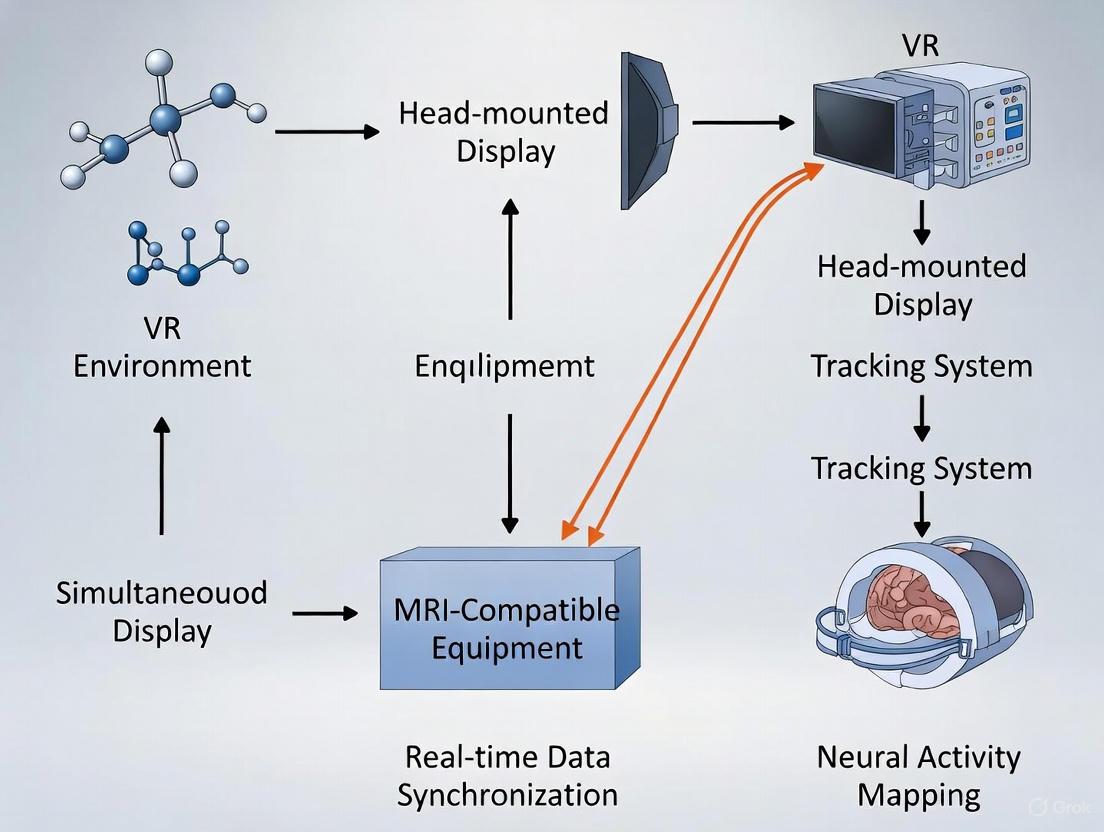
Workflow Diagram: VR-fMRI Integration for Motor Imitation Study
The diagram below illustrates the flow of information and tasks in a typical VR-fMRI experiment involving real-time hand tracking.
The Scientist's Toolkit: Essential Research Reagents & Materials
For researchers embarking on a VR-fMRI project, having the right "research reagents"—the core hardware and software components—is essential for success. The following table lists key items and their functions.
Table 2: Essential Materials for VR-fMRI Research
| Item Name | Type | Critical Function & Notes |
|---|---|---|
| MR-Compatible VR Goggles (e.g., NordicNeuroLab VisualSystem HD) [4] | Hardware | Provides stereoscopic visual stimuli. Must be MR-conditional to function safely at high field strengths (e.g., 3T) without degrading image quality. |
| MR-Compatible Data Glove (e.g., 5DT Data Glove 16 MRI) [3] | Hardware | Tracks complex hand and finger kinematics. Uses safe, fiberoptic sensors and long cables to connect to the control room. |
| Motion Tracking System (e.g., Flock of Birds by Ascension Tech) [3] | Hardware | Tracks 6-degrees-of-freedom limb or body position. Cameras and sensors must be placed outside the scanner room; system must be MR-safe. |
| VR Development Software (e.g., C++/OpenGL, Virtools) [3] | Software | Platform for creating and rendering custom virtual environments and controlling experimental logic. |
| Virtual Reality Peripheral Network (VRPN) [3] | Software | An open-source library that provides a unified interface for various VR hardware devices, simplifying integration. |
| fMRI Analysis Software Suite (e.g., FSL, SPM, AFNI, FreeSurfer) [5] [6] | Software | Used for preprocessing and statistical analysis of the acquired fMRI data. Proficiency in one or more of these is required. |
| High-Performance Computing (HPC) Resources [6] | Infrastructure | Essential for data storage and processing. Large datasets (e.g., from 100+ subjects) require cluster or cloud computing (AWS, Google Cloud). |
Troubleshooting Guides
FAQ 1: What are the primary types of interference in simultaneous VR-fMRI, and how can they be mitigated?
Issue: Simultaneous VR-fMRI experiments are plagued by electromagnetic interference that corrupts both EEG (if used) and fMRI data quality. This multi-modal interference presents a complex technical challenge.
Solutions:
- For EEG Data Corruption: Implement a multi-pronged approach involving both hardware and software solutions.
- Hardware Mitigation: Use compact EEG setups with short, shielded transmission leads to reduce the induction of artifacts. Incorporate dedicated reference sensors to actively monitor artifacts for subsequent correction [7].
- Software/Post-Processing Mitigation: Apply advanced denoising algorithms like DeepCor, a deep generative model that has been shown to outperform other methods (e.g., outperforming CompCor by 215% in enhancing BOLD signal responses to stimuli) [8].
- For fMRI Data Corruption: The presence of EEG equipment can disrupt the MRI's radiofrequency (RF) pulses.
- Hardware Mitigation: Use EEG caps with adapted, slim cables compatible with dense head RF arrays. Employ resistive materials for EEG leads or add resistors to segment their length, thereby mitigating RF interactions [7].
- Quality Assessment: Always conduct a control experiment to quantify the fMRI data degradation. One study reported EEG-induced fMRI temporal signal-to-noise ratio (tSNR) losses of 6–11% [7].
Table 1: Summary of Interference Types and Mitigation Strategies
| Interference Type | Primary Effect | Recommended Mitigation Strategy | Reported Efficacy |
|---|---|---|---|
| Gradient Artifact (EEG) | EEG signal swamped by time-varying MRI gradients [7] | Compact EEG setup with short leads; Model-based post-processing [7] | Allows detection of hallmarks like resting-state alpha [7] |
| Pulse Artifact (EEG) | EEG signal corrupted by ballistocardiogram (cardiac) [7] | Reference sensors; Data-based approaches (e.g., ICA) [7] | Corrects artifacts to a degree comparable with outside recordings [7] |
| RF Disruption (fMRI) | Perturbation of B1 field by EEG leads, reducing SNR [7] | Adapted EEG leads (resistive materials); Compatibility with dense RF coils [7] | Limits tSNR loss to 6-11% [7] |
| fMRI Thermal Noise | General noise affecting BOLD signal [8] | DeepCor denoising algorithm [8] | Outperforms CompCor by 215% [8] |
FAQ 2: What safety protocols are essential for VR-fMRI research?
Issue: Immersive VR experiments in an MRI environment introduce novel physical and psychological risks that traditional Institutional Review Board (IRB) protocols may not adequately address [9].
Solutions:
- Physical Safety: Ensure all VR equipment is MR-compatible. This includes using non-magnetic materials and verifying that devices will not heat up or become projectiles inside the magnetic field. A comprehensive safety evaluation is mandatory before any human subject testing [7].
- Psychological Safety: VR experiences are embodied and can feel like real memories, posing a risk of psychological distress (e.g., panic attacks in high-stress simulations) [9].
- Informed Consent: Implement the "3 C's of Ethical Consent in XR": Context (ensure participants understand the immersive experience), Control (maintain participant agency, including a clear and easy way to exit the simulation), and Choice (allow fully informed decisions about data sharing and future use) [9].
- Safeguards: Have a clear and immediate exit procedure from the VR environment and a debriefing protocol.
- Data Privacy: Motion and biometric data collected by commercial VR and biofeedback devices can be used to re-identify individuals, even from anonymized datasets [9].
- Data Handling: Establish secure data storage requirements, set limits on data re-use, and inform participants of these privacy risks during the consent process [9].
Table 2: Safety and Ethics Protocol Checklist
| Risk Category | Potential Harm | Essential Safeguards | References |
|---|---|---|---|
| Physical Safety | Heating of equipment; Projectiles; RF interference [7] | Use MR-compatible equipment only; Pre-scan safety screening; Comprehensive safety evaluation [7] | [9] [7] |
| Psychological Safety | Panic, anxiety, or distress from immersive VR content [9] | "3 C's" of Ethical Consent; Clear exit strategy; Debriefing protocol [9] | [9] |
| Data Privacy | Re-identification from motion or biometric data [9] | Secure data storage; Limits on data re-use; Informed consent regarding privacy risks [9] | [9] |
FAQ 3: How can I correct for data corruption in my fMRI signals?
Issue: fMRI data is inherently noisy, which can obscure the neural signal of interest, especially the subtle BOLD responses in VR studies.
Solutions:
- Utilize Advanced Denoising Algorithms: Traditional methods like CompCor have been superseded by more powerful deep-learning approaches.
- DeepCor: This is a deep generative model that disentangles and removes noise from fMRI data. It is applicable to single-participant data and has been shown to enhance the BOLD signal response to specific stimuli (e.g., face stimuli) by 215% compared to CompCor [8].
- Experimental Design: When analyzing task-based fMRI data, ensure you have a proper baseline or control condition. For example, in a multisensory learning study, compare brain activation in the trained audio-visual condition to unimodal visual stimulation to isolate multisensory integration effects [10].
The Scientist's Toolkit: Research Reagent Solutions
Table 3: Essential Materials and Tools for VR-fMRI Research
| Item Name | Function / Application | Technical Specifications / Examples |
|---|---|---|
| High-Density RF Head Coil | Critical for achieving high SNR and sub-millimeter resolution in fMRI at high fields (e.g., 7T) [7]. | Dense "mesh" of small receive elements; Must be compatible with EEG cap setup [7]. |
| Compact, MR-Compatible EEG System | For simultaneous EEG-fMRI acquisition to capture neural activity with high temporal precision [7]. | Short, shielded transmission leads; Adapted for use inside RF coil; Includes reference sensors for artifact correction [7]. |
| DeepCor Denoising Software | Removes noise from fMRI data to enhance BOLD signal quality [8]. | Deep generative model; Outperforms CompCor; Applicable to single-subject data [8]. |
| VR HMD & Tracking System | Presents immersive, controlled visual and auditory stimuli for ecological valid cognitive and rehabilitation tasks [10] [11] [12]. | HTC Vive; Custom software for specific paradigms (e.g., neuroproprioceptive facilitation) [11] [12]. |
| Safety & Ethics Framework | Safeguards participant well-being and data privacy, addressing novel risks of immersive tech [9]. | "3 C's of Ethical Consent" (Context, Control, Choice); IRB safety guide for immersive technology [9]. |
Definitions and Standards
What does "MR-Conditional" mean?
An item designated as MR Conditional is safe for use in the MRI environment only under specific, tested conditions. This is not a blanket approval; it is a precise safety envelope defined by the manufacturer through standardized testing. Staff must verify and apply these conditions every time the item enters the MRI suite [13].
How is MR-Conditional different from MR Safe and MR Unsafe?
The American Society for Testing and Materials (ASTM) International standard F2503 defines three safety classifications for devices in the MRI environment [14]:
| Classification | Meaning | Visual Label | Examples |
|---|---|---|---|
| MR Safe | Poses no known hazards in any MRI environment. | Green Square [13] | Non-metallic patient pads, plastic tools, certain immobilization devices [13]. |
| MR Conditional | Safe only within specific, tested conditions (e.g., field strength, SAR limits). | Yellow Triangle [13] | Specialist VR HMDs, patient monitors, many implants [4] [13]. |
| MR Unsafe | Known to pose hazards and must never enter magnetic fields. | Red Icon [13] | Standard oxygen cylinders, ferromagnetic tool carts, conventional IV poles [13]. |
MR-Conditional Hardware for VR-fMRI
What are the key components of a VR-fMRI system?
Simultaneous VR and fMRI recording requires a suite of MR-Conditional equipment to present stimuli, record responses, and monitor participants without compromising safety or data integrity.
Table: Essential MR-Conditional Hardware for VR-fMRI Research
| Hardware Category | Specific Device Examples | Key Function | Critical MR-Conditional Considerations |
|---|---|---|---|
| Head-Mounted Display (HMD) | NordicNeuroLab VisualSystem HD [4] | Presents immersive 3D visual stimuli. | Must use shielded electronics and MR-safe materials to avoid image artifacts and ensure safety at target field strength (e.g., up to 3T) [4]. |
| Input & Motion Tracking | 5DT Data Glove 16 MRI [3] | Measures complex hand and finger kinematics in real-time. | Must be fiber-optic and metal-free to operate safely in the magnet [3]. |
| Physiological Monitoring | BIOPAC MP200 System with MRI-safe amplifiers (e.g., for ECG, EDA, Respiration) [15] | Records physiological signals (ECG, EMG, EDA, respiration) for psychophysiological studies. | Requires specialized RF-filtered cable sets and signal conditioning to remove MR gradient interference from the data [15]. |
| Audio Presentation | MR-Conditional headphones or earphones | Delivers auditory stimuli and instructions. | Must be non-magnetic and designed to function without introducing artifacts or safety risks from the rapidly switching gradients. |
Troubleshooting Common Experimental Issues
Q: Our functional images show significant artifacts when the MR-Conditional VR HMD is in use. What could be the cause?
- Verify Compliance: First, confirm that the HMD is approved for your scanner's specific static field strength (e.g., 1.5T or 3T) and that all conditions of use from the manufacturer's Instructions for Use (IFU) are being met [13].
- Inspect Equipment: Check for any physical damage to the HMD or its cabling. Even minor compromise to the shielding can lead to interference.
- Cable Management: Ensure all cables are routed according to the manufacturer's guidelines. Improper routing can increase RF coupling, leading to artifacts. Use approved cable management kits [13].
Q: We are experiencing severe noise in our physiological data (ECG/EDA) during fMRI sequences. How can we clean the signal?
- Use Smart Amplifiers: Employ MRI-specific signal conditioning systems (e.g., BIOPAC's MRI Smart Amplifiers) that are designed to filter interference at the source [15].
- Apply Post-Processing Filters: In software, apply comb-band-stop (CBS) filters to remove harmonic artifacts caused by the EPI sequence, particularly with Multiband excitation [15].
- Optimize Sequence Timing: If possible, coordinate with your MR physicist to adjust sequence parameters. Coronal EPI MB scans, for instance, are noted to generate less harmonic interference in physiological data than Axial EPI MB scans [15].
Q: Participants report discomfort or a "heating sensation" when using the data glove inside the bore. What should we do?
- Immediately Stop the Scan: Participant safety is paramount. Halt the scan and investigate the issue.
- Check Placement and Insulation: Verify that the glove and its leads are not making direct contact with the participant's skin without appropriate insulation. Ensure there are no cable loops that could act as RF antennas [13].
- Review SAR Limits: The Specific Absorption Rate (SAR) measures RF power deposition. Confirm that your scan protocol operates within the SAR limits specified in the MR-Conditional guidelines for all equipment in use [13].
Q: Our VR system's tracking of hand movements seems delayed or inaccurate during the fMRI scan. How can we improve it?
- Isolate the Cause: Test the tracking system outside the scanner room to rule out inherent software or hardware issues.
- Assess Electrical Interference: The scanner's gradients can induce noise in electronic tracking systems. Ensure all equipment is properly shielded and that you are using MRI-compatible tracking devices (like the fiber-optic 5DT data glove) that are less susceptible to electromagnetic interference [3].
- Calibrate in Situ: Perform calibration procedures for the tracking system with the participant positioned in the scanner bore, as the environment can affect performance.
Experimental Protocol: VR-Based Motor Imitation Task
The following workflow is adapted from a foundational study that integrated a VR system with fMRI to investigate brain-behavior interactions during a hand imitation task [3].
Objective: To delineate brain-behavior interactions during observation and imitation of movements using virtual hand avatars in an fMRI environment [3].
Materials and Reagents:
Table: Research Reagent Solutions for VR-fMRI Motor Task
| Item | Function/Justification |
|---|---|
| MRI-Compatible VR HMD | Presents the virtual hand avatar in a first-person perspective to enhance embodiment. |
| 5DT Data Glove 16 MRI | Metal-free glove with fiberoptic sensors to measure 14 joint angles of the hand in real-time without MR interference [3]. |
| VR Simulation Software | Renders the virtual environment and streams real-time kinematic data to animate the virtual hand (e.g., using Virtools with VRPN plugin) [3]. |
| fMRI Scanner (3T) | Acquires Blood-Oxygen-Level-Dependent (BOLD) signals to map brain activity. |
| Fiberoptic Cable System | Safely transmits data from the glove in the scanner room to the control room computer [3]. |
Detailed Methodology:
- Subject Setup: The subject lies in the scanner and dons the MR-Conditional data glove on the right hand and the VR HMD.
- Task Design (Blocked): The experiment follows a blocked design:
- Observation with Intent to Imitate (OTI): The subject observes a pre-recorded finger movement sequence performed by a virtual hand avatar from a first-person perspective, with the instruction to imitate it next.
- Imitation with Feedback: The subject imitates the previously observed sequence. The virtual hand avatar is now animated in real-time by the subject's own movements, measured by the data glove.
- Rest Control: The subject views a static virtual hand. Control trials with moving non-anthropomorphic objects can also be included [3].
- Data Acquisition: Throughout the blocks, the following data are simultaneously recorded:
- fMRI Data: Continuous BOLD imaging using an EPI sequence.
- Kinematic Data: The data glove streams precise measurements of finger joint angles.
- Data Analysis: Brain activity (fMRI data) is analyzed and correlated with the behavioral performance (kinematic data) to identify networks involved in observation, imitation, and the sense of agency [3].
Safety and Compliance Workflow
Before any equipment enters the MRI suite, a rigorous safety check must be performed.
Key Steps for MR-Conditional Equipment [14] [13]:
- Check the Label: Always look for the MR Conditional (yellow triangle) icon on the device itself.
- Consult Documentation: Retrieve the manufacturer's Instructions for Use (IFU) or MR safety sheet for the exact model and serial number.
- Verify Conditions: Confirm the device is approved for your scanner's field strength and that your scan protocol adheres to all specified limits (e.g., SAR, spatial gradient).
- Implement and Train: Integrate these conditions into your standard operating procedures and ensure all research staff are trained on the specific requirements for each piece of equipment.
The integration of Virtual Reality (VR) with functional Magnetic Resonance Imaging (fMRI) represents a paradigm shift in cognitive neuroscience, enabling the study of brain function under ecologically valid conditions. This combination allows researchers to probe the neurophysiological underpinnings of complex behaviors by linking immersive, naturalistic experiences with the high spatial resolution of the Blood-Oxygen-Level-Dependent (BOLD) signal. VR-fMRI provides a unique window into brain dynamics, facilitating the investigation of neural mechanisms underlying episodic memory, spatial navigation, and executive functions within controlled yet realistic environments [16]. The core strength of this multimodal approach lies in its capacity to elucidate how distributed brain networks—including medial temporal, prefrontal, and parietal regions—support cognitive processes that are intimately tied to real-world experiences [16]. However, this powerful convergence also introduces significant technical challenges related to electromagnetic interference, data quality, and experimental design that must be systematically addressed to ensure valid and reliable findings.
Core Technical Challenges in Simultaneous VR-fMRI
Simultaneous VR-fMRI acquisition presents unique technical obstacles that can compromise data quality and participant safety if not properly managed. The table below summarizes the primary artifacts and their mitigation strategies.
Table 1: Key Technical Challenges and Solutions in VR-fMRI Research
| Challenge Type | Specific Artifacts/Issues | Proposed Solutions & Mitigation Strategies |
|---|---|---|
| MRI-Induced EEG Artifacts | Gradient Artifacts (GA), Ballistocardiogram (BCG) artifacts, Motion Artifacts (MA) [7] | Compact EEG setups with short transmission leads; Reference sensors for artifact monitoring; Advanced post-processing (e.g., ICA-AROMA, template subtraction) [7] [17] [18] |
| EEG-Induced fMRI Artifacts | Disruption of the MR radiofrequency (B1) field; SNR loss in fMRI [7] | Use of resistive materials for EEG leads; Strategic routing of cables to be compatible with dense RF arrays; EEG cap design minimizing metallic components [7] |
| VR-Related Data Quality | Head motion induced by immersive VR; Sensorimotor conflict causing motion sickness [19] [16] | Robust denoising pipelines (e.g., ICA-AROMA, CC, Scrubbing); Training sessions for participants; Limiting VR session duration [16] [18] |
| Safety Concerns | Radiofrequency (RF)-induced heating at EEG electrodes [17] [7] | Using MR-compatible equipment with built-in safety resistors; Monitoring Specific Absorption Rate (SAR) and B1+RMS; Phantom testing to verify safe heating levels [7] [17] |
Frequently Asked Questions (FAQs) & Troubleshooting Guides
Data Acquisition & Quality
Q: What are the most effective strategies for minimizing fMRI quality loss when using EEG inside the scanner?
Research indicates that EEG equipment can cause a 6-11% loss in temporal Signal-to-Noise Ratio (tSNR) in fMRI data [7]. To mitigate this:
- Hardware Optimization: Use an EEG cap with slim, adapted cables designed for compatibility with high-density head RF arrays. This reduces physical displacement of the coil and minimizes RF field disruption [7].
- Lead Management: Ensure EEG leads are routed securely to avoid forming loops and are kept as short as possible. Using leads with higher resistivity can also dampen unwanted RF interactions [7].
- Quality Control: Always acquire a reference fMRI scan without the EEG cap to quantify the specific tSNR loss attributable to the EEG system in your setup [7].
Q: Our EEG data during simultaneous fMRI is swamped by artifacts. Which correction pipelines are most effective?
A multi-step approach is crucial for cleaning EEG data collected inside the MR scanner.
- Gradient Artifact (GA) Removal: Apply an average template subtraction method. This requires recording the scanner's slice-timing pulses (TR markers) directly into the EEG file for precise synchronization [17].
- Pulse Artifact (BCG) Correction: Utilize optimal basis sets (OBS) or apply Independent Component Analysis (ICA) to identify and remove cardioballistic artifacts. Semi-automatic correction with manual verification of detected heartbeats is recommended for reliability [17].
- Advanced Denoising: For residual artifacts, data-driven approaches like ICA-AROMA (Automatic Removal of Motion Artifacts) have proven effective, particularly for non-lesional brain conditions [18].
Experimental Design & Safety
Q: How can we design a VR-fMRI experiment that is both immersive and controls for excessive head motion?
- Paradigm Design: Implement a block design rather than a continuous, event-related design. This provides natural breaks, allowing participants to rest and reduces fatigue-induced motion [3] [16].
- Participant Preparation: Conduct a thorough training session outside the scanner. This familiarizes participants with the VR task and controls, reducing novelty-driven movements during the actual scan [20].
- Session Management: Adhere to the "20-10 rule" for VR immersion: 20 minutes of VR, followed by a 10-minute break. This helps prevent cybersickness and cognitive overload, which can exacerbate motion [19].
Q: What are the critical safety protocols for simultaneous EEG-fMRI, especially at higher field strengths like 7T?
Safety is paramount, with the primary risk being RF-induced heating at the EEG electrodes.
- Equipment Certification: Only use MR-compatible EEG systems that are explicitly certified for use with your scanner's field strength (e.g., 3T or 7T). These include built-in safety resistors [7] [17].
- Phantom Testing: Before human studies, conduct phantom tests to measure temperature changes under your specific fMRI sequences. A comparative benchmark is that a multi-band (MB) sequence with a B1+RMS of 0.8 µT showed lower heating than a standard single-band (SB) sequence with a B1+RMS of 1.0 µT [17].
- Impedance Management: Maintain all electrode-skin impedances below 5 kΩ, as higher impedances can increase the risk of localized heating [17].
Detailed Experimental Protocols
Protocol for a VR-fMRI Study on Episodic Memory
This protocol is adapted from systematic reviews of VR-fMRI studies on episodic memory [16].
Objective: To investigate the neural correlates of episodic memory encoding and retrieval using a naturalistic VR paradigm. Participants: Healthy adults, right-handed, with no history of neurological disorders. (Sample size: ~15-20 based on previous studies [3]). VR Task Design:
- Encoding Phase: Participants navigate a virtual environment (e.g., a museum or a city) and are instructed to remember specific objects and their locations.
- Retrieval Phase: Participants are then asked to recall and identify the objects and their spatial contexts.
- Control Task: A non-spatial, low-level visual task within VR to establish a baseline. fMRI Acquisition Parameters (Example for a 3T scanner):
- Sequence: Multi-band EPI (to achieve a short TR, e.g., 440ms-2000ms)
- Voxel size: 2-3 mm isotropic
- Slices: Whole-brain coverage (e.g., 28-60 slices)
- The B1+RMS value must be checked to ensure it falls within safe limits for any equipment inside the bore [17]. Data Analysis:
- fMRI Preprocessing: Standard pipeline including realignment, normalization, and smoothing. Denoising with a pipeline such as
CC + SpikeReg + 24HMPis recommended for tasks that may induce motion [18]. - General Linear Model (GLM): Contrast activity during encoding and retrieval vs. the control task. Key regions of interest include the Hippocampus (HPC), Parahippocampal Gyrus (PHG), Prefrontal Cortex (PFC), and Angular Gyrus [16].
Workflow for a Simultaneous EEG-fMRI-VR Experiment
The diagram below outlines the integrated workflow for setting up and running a simultaneous EEG-fMRI-VR experiment.
Diagram 1: Integrated VR-fMRI-EEG experimental workflow.
The Scientist's Toolkit: Essential Research Reagents & Materials
Table 2: Essential Materials for VR-fMRI Research
| Item Category | Specific Example(s) | Critical Function & Notes |
|---|---|---|
| VR Presentation System | MRI-compatible VR goggles (e.g., with RF-shielded displays) | Presents visual stimuli within the high-magnetic-field environment without causing interference. |
| Motion Tracking | MRI-compatible data gloves (e.g., 5DT Data Glove 16 MRI); Eye-tracking systems | Captures kinematic data of hand movements or gaze in real-time to animate avatars or assess behavior [3]. |
| EEG System | MR-compatible EEG amplifier (e.g., BrainAmp MR plus); Cap with integrated safety resistors (e.g., BrainCap MR) | Records electrophysiological data with built-in resistors to mitigate heating risks [7] [17]. |
| fMRI Coil | Dense, multi-channel head RF array (e.g., 64-channel head coil) | Provides high Signal-to-Noise Ratio (SNR) for fMRI, essential for sub-millimeter resolution [7]. |
| Data Analysis Software | BrainVision Analyzer (EEG); FSL, SPM, CONN (fMRI); EEGLAB; Custom scripts in Python/MATLAB | Preprocessing and statistical analysis of multi-modal data, including specialized toolboxes for artifact removal [17] [18]. |
| Safety & Sync Equipment | SyncBox (for scanner pulse synchronization); Fluoroptic thermometer (for phantom heating tests) | Ensures temporal alignment of data streams and verifies safety standards during protocol development [17]. |
Implementing the Protocol: A Step-by-Step Guide to VR-fMRI Setup and Data Acquisition
Simultaneous Virtual Reality (VR) and functional Magnetic Resonance Imaging (fMRI) recording presents unique technical challenges, primarily due to the incompatibility of standard electronic equipment with the high-strength magnetic fields of MRI scanners. MR-safe VR equipment is specifically engineered to operate within this hostile electromagnetic environment without compromising patient safety or data integrity. These specialized systems use shielded electronics and MR-safe materials to prevent image artifacts, avoid projectile hazards, and ensure accurate stimulus delivery during neuroimaging experiments. The core requirement for any device used in this context is compliance with the ASTM F2503 standard, which categorizes equipment as MR Safe, MR Conditional, or MR Unsafe [21] [22]. Understanding these classifications is fundamental for establishing safe and effective VR-fMRI research protocols.
Technical Specifications of the VisualSystem HD
The VisualSystem HD (VSHD) from NordicNeuroLab represents a specialized solution designed specifically for fMRI environments. This system is classified as MR Conditional, meaning it is safe for use under specific conditions—in this case, at magnetic field strengths up to 3 Tesla [4]. The system overcomes the fundamental obstacle of combining modern VR technology with MR imaging by employing carefully shielded electronics that do not significantly degrade MR image quality [4].
Table 1: Key Technical Specifications of the VisualSystem HD
| Component | Specification | Research Application Benefit |
|---|---|---|
| Display Type | Dual Full HD OLED (one for each eye) | Enables stereoscopic 3D imaging for immersive spatial tasks [23] |
| Native Resolution | 1920×1200 @ 60Hz/71Hz | Presents sharp, high-quality graphics and text for visual stimuli [23] |
| Field of View | 80% larger than previous models | Increases immersion, potentially enhancing ecological validity [23] |
| Interpupillary Distance (IPD) Adjustment | 44 to 75 mm | Ensures proper fit and visual clarity for a wide range of participants [23] |
| Diopter Correction | -8 to +5 | Allows subjects with vision impairments to participate without glasses [23] |
| Integrated Eye Tracking | Binocular, 60 fps, 640x480 resolution | Provides objective measures of gaze and task engagement during scanning [23] |
| Safety Certifications | IEC60601-1, IEC 60601-1-2 | Certified for patient safety and electromagnetic compatibility in medical environments [23] |
The system is part of a broader fMRI ecosystem that includes a SyncBox for synchronization with the MRI scanner, response collection devices, and stimulus presentation software (nordicAktiva), forming a complete solution for functional exams [23].
Alternative MR-Safe VR Hardware
While the VisualSystem HD is an integrated solution, other companies provide VR hardware that can be adapted for research and clinical use in medical environments.
DPVR manufactures both PC-tethered and wireless VR headsets that can be implemented in hospital or medical settings. Their P1 Ultra model is notable for its customizable modules, which can include interfaces for monitoring physiological data such as heart rate or brain-computer interfaces, providing additional data streams for multimodal research [24]. These headsets have been utilized by partners for applications including music therapy (Ceragem) and vision treatment (Vivid Vision) [24].
Furthermore, platforms like Psious and XRHealth represent software solutions that operate on VR hardware to deliver therapeutic interventions for conditions like anxiety, phobias, and stress, demonstrating the broader applicability of VR in clinical research settings [24].
Essential Research Reagent Solutions
For a VR-fMRI research laboratory, the "reagents" are the core hardware and software components required to conduct simultaneous recording experiments.
Table 2: Essential Research Reagents for VR-fMRI Simultaneous Recording
| Item | Function | Example Products/Models |
|---|---|---|
| MR-Conditional VR Headset | Presents visual stimuli inside the scanner bore; must not create artifacts or pose safety risks. | NordicNeuroLab VisualSystem HD, DPVR Headsets with medical-grade customization [24] [23] |
| Stimulus Presentation Software | Controls the timing, sequence, and logic of VR stimuli presented to the participant. | nordicAktiva, custom scripts (e.g., via Unity) [23] |
| Synchronization Interface | Aligns the presentation of VR stimuli with the acquisition of fMRI volumes for precise temporal alignment. | SyncBox [23] |
| Response Collection Device | Records participant behavioral responses (e.g., button presses) during the fMRI scan. | ResponseGrip [23] |
| Data Integration & Analysis Suite | Processes and analyzes the combined fMRI and behavioral data; may include specialized VR analytics. | nordicBrainEx [23] |
| MR-Safe Eye-Tracking System | Monitors participant gaze, pupil dilation, and engagement, providing crucial behavioral metrics. | Integrated system in VisualSystem HD [23] |
Experimental Setup and Workflow Protocol
Configuring a system for simultaneous VR-fMRI recording requires a meticulous workflow to ensure safety and data quality. The following diagram outlines the critical path from equipment preparation to data acquisition.
Troubleshooting Common Technical Issues
Even with proper setup, researchers may encounter technical challenges. This section addresses common problems and their solutions in a FAQ format.
Q1: We are experiencing significant noise or artifacts in our fMRI images since introducing the VR system. What should we check?
- A: This is often caused by electromagnetic interference. First, verify that all components of your VR system are officially rated and labeled as MR Conditional for your scanner's field strength (e.g., 3T) [23] [22]. Ensure all cables are properly shielded and routed according to the manufacturer's guidelines, away from the scanner bore and other sensitive equipment. Using fiber-optic extensions for video signals can sometimes mitigate this issue.
Q2: The timing between our VR stimulus presentation and the fMRI volume acquisition is inconsistent. How can we improve synchronization?
- A: Implement a dedicated hardware synchronization solution. Systems like the NordicNeuroLab SyncBox are designed for this purpose, as they receive a TTL (Transistor-Transistor Logic) pulse from the MRI scanner at the start of each volume acquisition and use this to trigger events in the stimulus software with high precision [23]. Always run a mock scan without a subject to validate the synchronization timing before collecting experimental data.
Q3: Our participant cannot see the VR stimuli clearly. What calibrations are necessary?
- A: Visual clarity is paramount. The VR headset must be properly fitted to the participant. Utilize the built-in diopter correction (typically ranging from -8 to +5) and interpupillary distance (IPD) adjustment (e.g., 44-75mm) on the headset to match the participant's vision and anatomy [23]. This is a critical step during participant setup, similar to adjusting an ophthalmoscope.
Q4: How do we ensure our VR equipment remains safe and compliant for use in the MRI environment?
- A: Adhere to a strict labeling and audit protocol. All equipment must be clearly labeled according to ASTM F2503 standards with MR Conditional icons (yellow triangle) [21] [22]. Conduct regular audits to check that labels are intact and legible. Furthermore, maintain a log for cleaning and inspecting hygiene components, such as replaceable face sponges, to prevent contamination and equipment damage [24].
Application in a Research Context: A Sample Experimental Protocol
To ground these technical protocols in research practice, consider the following simplified methodology, inspired by recent studies that combine VR and fMRI.
Protocol: Investigating Spatial Processing with Stereoscopic VR [25]
- Objective: To identify the neural correlates of processing objects in peripersonal (reachable) space versus extrapersonal (non-reachable) space using stereoscopic VR during fMRI.
- Stimuli & Task: Participants perform a visual discrimination task on graspable objects presented at different apparent distances within a VR environment. The paradigm includes alternating blocks of monoscopic and stereoscopic presentation to isolate the effect of depth perception.
- Key Controls: The pixel size of the objects is controlled across distances to ensure that neural activation differences are due to spatial processing and not low-level visual features [25].
- fMRI Parameters: Standard whole-brain EPI acquisition. Preprocessing typically includes realignment, normalization, and smoothing. Contrasts are defined for [Stereoscopic Peripersonal > Monoscopic Peripersonal] and [Peripersonal Space > Extrapersonal Space].
- Expected Outcomes: The study hypothesizes that stimuli in peripersonal space will engage the dorsal visual stream, including areas like the posterior intraparietal sulus, which is associated with action-oriented processing and affordances. In contrast, extrapersonal space is expected to activate more ventral stream regions related to semantic and scene analysis [25].
The logical structure of such an experiment, from hypothesis to analysis, can be visualized as follows:
Troubleshooting Guides and FAQs
Synchronization and Timing Issues
Q1: What are the primary causes of latency or jitter between the fMRI trigger and the VR stimulus presentation, and how can they be minimized?
Latency (constant delay) and jitter (variable delay) most often originate from software communication pathways, hardware processing time, or the VR system's graphics rendering pipeline [26].
- Solution 1: Optimize Software Communication: Utilize dedicated, high-speed data acquisition systems and application programming interfaces (APIs) designed for real-time experimentation. The Experiments in Virtual Environments (EVE) framework, built on Unity 3D, is an example of a system that provides standardized modules for data synchronization and storage, helping to mitigate these issues [27].
- Solution 2: Verify and Calibrate Timing: Regularly perform a timing calibration procedure. This involves sending a test trigger from the fMRI scanner to the VR presentation computer while using a photodiode or other sensor to measure the precise delay between the trigger signal and the actual stimulus onset on the VR display. This measured delay can often be accounted for in the experiment software.
- Solution 3: Hardware Selection: Ensure all components, especially the VR computer's graphics card, meet or exceed the recommended specifications for low-latency rendering. The use of fMRI-compatible equipment, such as fiber-optic data gloves, is essential to prevent interference and ensure signal integrity [3].
Q2: The VR system fails to receive the TTL pulses from the fMRI scanner. What should I check?
This is typically a hardware connection or configuration problem.
- Checklist:
- Physical Connection: Verify the cable from the scanner's TTL output port is securely connected to the correct input port (e.g., a parallel port or a dedicated digital I/O card) on the VR control computer.
- Voltage Level Compatibility: Confirm that the voltage level of the TTL pulse (e.g., 5V) is within the acceptable range for the input port on the VR computer. Using an oscilloscope to check for the presence and quality of the pulse is the most reliable method.
- Software Configuration: Ensure your experiment software (e.g., Unity with the EVE framework) is configured to listen to the correct hardware port for incoming triggers [27].
- Grounding Loops: Check for potential grounding issues that can corrupt the signal.
Hardware and Software Integration
Q3: Which VR hardware and software solutions have been successfully integrated with fMRI in published research?
Successful integration has been achieved with a variety of components, emphasizing MRI-compatibility. The table below summarizes key solutions documented in research.
Table 1: Research Reagent Solutions for VR-fMRI Integration
| Component Type | Specific Solution / Example | Function / Key Feature |
|---|---|---|
| Input Device | 5DT Data Glove 16 MRI [3] | MRI-compatible glove for measuring hand joint angles using fiber optics. |
| Input Device | Ascension "Flock of Birds" 6DOF sensors [3] | Tracks position and orientation (6 degrees of freedom). |
| Software Framework | Experiments in Virtual Environments (EVE) [27] | Unity-based framework for designing experiments, managing data synchronization, and storage. |
| Software Framework | Virtools with VRPack [3] | Development environment used to create virtual environments for fMRI integration. |
| Visual Display | MRI-compatible HMDs or projection systems | Presents the VR stimulus; must be non-magnetic and not interfere with the magnetic field. |
Q4: How do I manage the data streams from the fMRI scanner, VR system, and physiological sensors to ensure they are synchronized?
This requires a centralized synchronization strategy.
- Solution: Implement a master system that records all data streams with a common timing clock. One effective method is to use software like LabChart to record physiological data and receive the fMRI TTL pulses on a separate channel [27]. The VR software should timestamp all behavioral and interaction data (e.g., button presses, avatar position) using the same clock reference established by the scanner triggers. The EVE framework, for instance, provides infrastructure for synchronizing data from different sources and storing it in a unified database [27].
Data Quality and Artifact Resolution
Q5: What are the common sources of artifact in fMRI data during VR experiments, and how can they be addressed?
Beyond the usual sources of artifact, VR experiments introduce specific challenges.
- Head Motion: The immersive nature of VR can lead to increased head movement.
- Mitigation: Use additional padding and comfort items to stabilize the participant's head, and provide clear instructions to remain as still as possible.
- Hardware Interference: Non-MRI-compatible equipment can create electromagnetic noise or pose a safety risk.
- Mitigation: Use only certified MRI-compatible VR equipment, such as fiber-optic gloves and non-ferrous displays [3]. All equipment must be tested in the scanner environment prior to the experiment.
- Physiological Noise: The cognitive and emotional load of VR can increase heart and respiration rates, which modulate the BOLD signal.
- Mitigation: Record physiological data (e.g., ECG, EDA, respiration) concurrently so they can be used as regressors in the fMRI data analysis to remove these confounding effects [27].
The following diagram illustrates the flow of signals and data in a typical VR-fMRI setup, highlighting potential points of failure for synchronization.
Diagram 1: VR-fMRI System Data and Trigger Flow
FAQs: Core Principles of VR Cognitive Assessment
Q1: What are the key advantages of using VR over traditional paper-and-pencil tests for cognitive assessment?
VR cognitive assessment offers three primary advantages:
- Enhanced Ecological Validity: VR can create multimodal sensory stimuli in interactive environments that mimic real-world activities and daily surroundings, providing a more accurate assessment of real-life cognitive functioning than artificial lab settings [28] [29].
- Comprehensive Domain Assessment: Systems like CAVIRE can assess all six cognitive domains defined by DSM-5 (complex attention, executive function, language, learning and memory, perceptual-motor function, and social cognition), whereas traditional tests like MMSE are less effective for certain domains like executive function [28].
- Improved Engagement: Gamified VR tasks transform repetitive cognitive testing into immersive, interactive experiences that increase participant motivation and reduce boredom and fatigue, potentially improving data quality [12] [30].
Q2: How should VR tasks be designed to ensure they accurately target specific cognitive domains?
Effective VR task design requires:
- Incorporating Established Paradigms: Base VR tasks on validated neuropsychological principles and traditional tests (e.g., using Whack-the-Mole for response inhibition, Corsi block-tapping for visuospatial memory, or Stroop test variants for cognitive flexibility) [12] [31].
- Simulating Daily Activities: Design tasks that mimic activities of daily living (ADLs), such as virtual kitchen scenarios for assessing memory, attention, and planning skills, which enhances ecological validity [28] [29].
- Implementing Adaptive Difficulty: Use algorithms that automatically adjust task difficulty based on user performance to maintain appropriate challenge levels and prevent ceiling/floor effects [31].
Q3: What specific considerations are needed when designing VR assessments for clinical populations with cognitive impairments?
Special considerations for clinical populations include:
- Safety and Tolerance: Implement safeguards for participants with potential vulnerability to VR-induced symptoms (e.g., nausea, headache, giddiness). Studies report successful use with older adults with MCI and various neuropsychiatric disorders when appropriate precautions are taken [28] [30] [32].
- Simplified Interaction Design: Account for potential technological inexperience by creating intuitive interfaces and providing comprehensive tutorial sessions before assessment [28] [31].
- Clinical Validation: Establish sensitivity to cognitive impairments specific to target disorders (e.g., mood disorders, psychosis spectrum disorders, MCI) through rigorous validation against standard neuropsychological tests and clinical diagnosis [29] [30].
FAQs: Technical Protocols for VR-fMRI Simultaneous Recording
Q4: What are the primary technical challenges of simultaneous VR-fMRI recording, and how can they be mitigated?
The main challenges involve cross-modal interference, which can be addressed through:
Table: VR-fMRI Interference Types and Mitigation Strategies
| Interference Type | Impact on Data | Mitigation Strategies |
|---|---|---|
| MRI on VR | Artifacts on motion tracking and visual presentation due to magnetic fields | Fiber-optic data transmission, magnetic-compatible displays, temporal synchronization |
| VR on fMRI | RF disruption from electronic components, reduced fMRI signal quality | Compact EEG/VR setups with short leads, specialized RF-shielded components, reference sensors |
| Subject Safety | Potential heating from induced currents | Current-limiting resistors, careful cable routing, thermal monitoring |
| Data Quality | Reduced temporal signal-to-noise ratio (tSNR) | Reference sensors for artifact correction, post-processing algorithms, optimized coil design |
Based on EEG-fMRI literature which shares similar technical challenges [7]:
- Hardware Optimization: Use compact transmission systems with short, well-shielded cables to reduce artifact incidence at their origin. Implement specialized lead materials and resistors to segment lengths, minimizing RF interactions [7].
- Reference Sensors: Incorporate dedicated sensors to monitor artifacts, enabling advanced post-processing correction of residual artifacts in both fMRI and physiological data [7].
- Physical Integration: Adapt equipment to fit within dense RF head coil arrays without compromising fMRI sensitivity or acceleration capabilities [7].
Q5: What experimental design considerations are crucial for successful VR-fMRI hyperscanning studies?
For VR-fMRI hyperscanning (simultaneous multi-person recording):
- Temporal Synchronization: Implement robust synchronization protocols like Network Time Protocol (NTP) to align data acquisition across multiple scanners, maintaining timing discrepancies below the repetition time (TR) of fMRI sequences (e.g., <500ms for TR=2000ms) [33].
- Task Design for Social Cognition: Develop paradigms that examine cooperative and competitive interactions, such as sender-receiver games with alternating roles, to investigate neural correlates of social decision-making [33].
- Standardized Data Collection: Adhere to Brain Imaging Data Structure (BIDS) standards for organizing neuroimaging data, enabling reproducibility and data sharing across research groups [33].
Troubleshooting Guides
Problem: Excessive VR-Induced Symptoms (Nausea, Headache) in Participants
Potential Causes:
- Rapid movement in VR environment conflicting with vestibular input
- Inadequate calibration for individual interpupillary distance (IPD)
- Prolonged exposure without adequate breaks
Solutions:
- Limit initial session duration and gradually increase exposure
- Ensure proper HMD fit and IPD adjustment for each participant
- Implement stationary VR environments rather than locomotion-intensive scenarios
- Provide clear instructions to focus on stable visual reference points when possible
- Consider anti-motion sickness medications for susceptible participants in longer studies [30]
Problem: Significant Artifacts Corrupting fMRI Data During VR Presentation
Potential Causes:
- RF interference from VR electronic components
- Induction artifacts from cabling within the magnetic field
- Subject motion amplified by VR engagement
Solutions:
- Implement additional shielding for VR components
- Use fiber-optic instead of electrical cabling for data transmission
- Incorporate reference sensors for artifact correction in post-processing
- Apply motion correction algorithms designed for sustained movement during VR
- Validate fMRI data quality with and without VR equipment through phantom tests [7]
Problem: Discrepancies Between VR and Traditional Neuropsychological Assessment Scores
Potential Causes:
- Differing cognitive demands in immersive vs. traditional testing environments
- Gamification elements altering motivational factors
- Variability in technological familiarity among participants
Solutions:
- Establish convergent validity through correlation studies with multiple standard measures
- Control for technological familiarity through pre-training sessions
- Analyze whether VR provides complementary information about real-world functioning rather than direct replacement of traditional tests
- Consider that VR may assess different aspects of cognition (e.g., real-world functional capacity) compared to traditional tests [29] [31]
Experimental Protocols for Key VR Cognitive Assessments
Protocol 1: CAVIRE System for Comprehensive Cognitive Domain Assessment
Table: CAVIRE Implementation Protocol
| Component | Specification | Purpose |
|---|---|---|
| Hardware | HTC Vive Pro HMD, Lighthouse sensors, Leap Motion device, Rode VideoMic Pro microphone | Enable tracking of natural hand/head movements and speech capture in 3D environment |
| Software | Unity game engine with integrated API for voice recognition | Create 13 different virtual environments simulating daily activities |
| Assessment Domains | Six DSM-5 cognitive domains via 13 task segments | Comprehensive cognitive profiling across multiple domains |
| Scoring | Automated algorithm calculating VR scores and completion time | Objective assessment with minimal administrator bias |
| Session Structure | Tutorial session followed by cognitive assessment with multiple attempts allowed per task within time limits | Ensure participant understanding while assessing learning capacity |
| Validation | Comparison with MoCA, MMSE, functional status assessments | Establish clinical validity and sensitivity to cognitive impairment |
Implementation Details [28]:
- Participants: 109 individuals aged 65-84, grouped as cognitively healthy (MoCA ≥26) or impaired (MoCA <26)
- Procedure: All participants completed CAVIRE assessment after standard cognitive testing
- Outcome Measures: VR scores, time taken across six cognitive domains, receiver-operating characteristic analysis
- Results: Cognitively-healthy participants achieved significantly higher VR scores and shorter completion times (all p's < 0.005), with AUC of 0.7267 for distinguishing groups
Protocol 2: Enhance VR for Gamified Cognitive Assessment
Table: Enhance VR Assessment Protocol
| Component | Specification | Traditional Test Equivalent |
|---|---|---|
| Magic Deck | Memorize location of cards with colorful abstract patterns | Paired Associates Learning (PAL) test |
| Memory Wall | Recall increasingly complex patterns of lit cubes | Visual Pattern Test |
| Pizza Builder | Simultaneously take orders and assemble pizzas | Divided attention assessments |
| React | Sort incoming stimuli by changing criteria | Wisconsin Card Sorting Task and Stroop test |
| Hardware | Meta Quest standalone headset with hand controllers | N/A |
| Scoring | In-game points system with adaptive difficulty | Standardized test scoring |
Implementation Details [31]:
- Participants: 41 older adults (mean age=62.8 years) without neurodegenerative or psychiatric disorders
- Design: Randomized testing order (traditional neuropsychological assessment vs. VR sessions)
- Traditional Assessment Battery: MoCA, Stroop task, WCST, Trail Making Test, Rey-Osterrieth figure, Word Pair Test, Clock Drawing Test, Corsi Block-Tapping, Visual Search Test
- Results: High tolerance and usability, though direct score correlations with traditional tests were limited, suggesting VR may tap into different cognitive aspects
Research Reagent Solutions
Table: Essential Materials for VR-fMRI Cognitive Assessment Research
| Component | Function | Examples/Specifications |
|---|---|---|
| VR Hardware | Create immersive environments | HTC Vive Pro, Meta Quest, Leap Motion for hand tracking, Lighthouse sensors |
| fMRI-Compatible Equipment | Enable safe operation in magnetic environment | Fiber-optic data transmission, specialized RF-shielded components, non-magnetic materials |
| Physiological Monitoring | Capture complementary physiological data | EEG caps adapted for MRI environments, reference sensors for artifact correction, pulse oximeters |
| Software Platforms | Environment development and data integration | Unity game engine, specialized VR assessment applications (CAVIRE, Enhance VR) |
| Synchronization Systems | Temporal alignment of multimodal data | Network Time Protocol (NTP) servers, trigger interfaces, custom synchronization software |
| Validation Tools | Establish clinical and technical validity | Standard neuropsychological tests (MoCA, MMSE), functional assessments (Barthel Index, Lawton IADLs) |
Experimental Workflow Diagrams
VR-fMRI Experimental Workflow
VR-fMRI Technical Challenges and Solutions
Troubleshooting Common VR-fMRI Experimental Challenges
| Problem Category | Specific Issue | Potential Causes | Recommended Solutions |
|---|---|---|---|
| Data Acquisition | Low signal-to-noise ratio in fMRI data [4] | Magnetic interference from VR equipment; B0 field inhomogeneities [4]. | Use MR-conditional VR goggles with shielded electronics (e.g., VisualSystem HD); acquire field map for unwarping [4]. |
| VR visual presentation is unstable or laggy | Computer system latency; improper software configuration; network delays in data streaming. | Pre-load all 3D models; use a dedicated, high-performance computer; test and optimize paradigm offline [34]. | |
| Experimental Design & Analysis | Inflated effect sizes in ROI analysis [35] | Circular analysis bias; using statistically significant voxels from the same dataset to define an ROI [35]. | Use independent localizer scans or cross-validation to define ROIs; employ unbiased whole-brain correction [35]. |
| Incorrect image orientation or alignment [35] | DICOM header issues; inconsistent coordinate systems between software; improper manual reorientation. | Check and enforce consistent orientation (e.g., LPI) using tools like fslswapdim [35]; verify alignment with a template. |
|
| Physiological & Data Streaming | Difficulty synchronizing physiological, VR, and fMRI data | Lack of automated event marking; different hardware systems not on synchronized clocks. | Use software (e.g., AcqKnowledge, Vizard, COBI) that supports network data transfer and automated marker sending [34]. |
| Unwarping artifacts in functional images | B0 magnetic field inhomogeneities, particularly at tissue-air interfaces [35]. | Acquire a field map during scanning. Use FSL's fugue or fsl_prepare_fieldmap for unwarping [35] [36]. |
Frequently Asked Questions (FAQs)
Data Preprocessing & Analysis
Q: What is resampling and when do I need to do it? A: Resampling changes the resolution or dimensions of an image. It is necessary when you need to align images with different voxel sizes or templates, such as when applying a mask from one image (e.g., an anatomical) to another (e.g., a functional statistical map) [35].
- In FSL: Use
flirt -in mask.nii.gz -ref stats.nii.gz -out mask_RS.nii.gz -applyxfm - In AFNI: Use
3dresample -input mask.nii.gz -master stats.nii.gz -prefix mask_RS.nii.gz[35]
Q: What constitutes a "biased analysis" and how can I avoid it? A: A biased, or circular, analysis occurs when you define your Region of Interest (ROI) based on the statistical results from the very same dataset. This inflates effect sizes because it selectively includes noise voxels that, by chance, passed the significance threshold [35].
- Avoidance Strategy: Use independent ROIs defined from an separate localizer scan, an anatomical atlas, or a prior study. For valid confirmatory analysis, the ROI must be defined a priori, without reference to the final statistical map [35].
Technical Setup
Q: My anatomical and functional images appear to have different orientations. How can I fix this?
A: Use FSL's fslswapdim command to reorient an image. For example, if an image is in Right-Posterior-Inferior (RPI) orientation and you need Left-Posterior-Inferior (LPI), the command would be: fslswapdim input_image.nii.gz RL PA IS output_image.nii.gz. Always visually check the reoriented image overlaid on your functional data to confirm alignment [35].
Q: How can I integrate physiological data streams with my VR-fMRI experiment? A: Software solutions like BIOPAC's AcqKnowledge and WorldViz's Vizard, when used with systems like COBI, allow for network data transfer (NDT). This setup enables the streaming of physiological data (e.g., heart rate) and automated sending of event markers from the VR environment to the data acquisition software, ensuring synchronization across all modalities [34].
Experimental Protocols & Quantitative Data
Key Findings on Hippocampal-Cortical Connectivity
The following table synthesizes core quantitative findings from foundational studies on hippocampal-cortical connectivity, which can inform the design and interpretation of VR-fMRI experiments.
| Study & Method | Key Stimulation Parameter | Primary Brain Regions Activated | Effect on Functional Connectivity |
|---|---|---|---|
| Optogenetic fMRI (Zhou et al., 2017) [37] | 1 Hz stimulation of dDG | Bilateral V1, V2, LGN, SC, Cingulate Cortex [37] | Enhanced interhemispheric rsfMRI connectivity in hippocampus and various cortices [37]. |
| Human fMRI (NeuroImage, 2023) [38] | Memory encoding and retrieval tasks | Sparse, task-general during encoding; Medial PFC, inferior parietal, parahippocampal cortices during retrieval [38]. | Stable anterior/posterior hippocampal connectivity across rest and tasks, superposed by increased retrieval-recollection network connectivity [38]. |
Detailed Protocol: Low-Frequency Hippocampal Circuit Investigation
This protocol is adapted from optogenetic studies [37] and provides a framework for designing VR tasks that probe similar low-frequency hippocampal-cortical networks in humans.
- Stimulation Paradigm: Design a VR task that naturally elicits slow, rhythmic activity (~1 Hz) in the dorsal hippocampus. This could involve slow spatial navigation, gradual exploration of a virtual environment, or a paced memory encoding task.
- fMRI Acquisition: Acquire both task-based fMRI and resting-state fMRI (before and after the task) to assess task-evoked activity and changes in functional connectivity.
- Data Analysis:
- Task Activation: Use a general linear model (GLM) to identify brain regions with significant BOLD responses during the slow-frequency VR task. Focus on the visual cortex (V1, V2), cingulate cortex, and thalamic regions [37].
- Functional Connectivity: Use resting-state data to compute correlations between the hippocampus (anterior and posterior segments separately) and the whole brain. Contrast post-task vs. pre-task connectivity to identify stimulation-induced enhancements [38] [37].
The Scientist's Toolkit: Essential Research Reagents & Materials
| Item Name | Function / Application | Example / Note |
|---|---|---|
| MR-Conditional VR System | Presents immersive 3D stimuli in the scanner. | VisualSystem HD (NordicNeuroLab); uses shielded electronics and MR-safe materials for use up to 3T [4]. |
| Data Synchronization Suite | Streams and synchronizes physiological, VR event, and fMRI data. | AcqKnowledge software, Vizard VR software, and COBI (for fNIRS/physiology) with Network Data Transfer (NDT) [34]. |
| FSL | A comprehensive library of MRI analysis tools. | Includes FEAT (FMRI analysis), MELODIC (ICA), BET (brain extraction), FLIRT/FNIRT (registration), and FUGUE (unwarping) [36]. |
| Field Map | Corrects for geometric distortions in EPI (fMRI) data caused by B0 field inhomogeneities. | Acquired during scanning; processed using FSL's fugue or fsl_prepare_fieldmap [35] [36]. |
| Unbiased ROI Atlas | For defining regions of interest for confirmatory analysis without circularity. | Anatomically defined atlases (e.g., AAL, Harvard-Oxford) or independent functional localizers [35]. |
Experimental and Analytical Workflows
Diagram: VR-fMRI Experimental Setup and Synchronization
Diagram: Analysis Workflow for Hippocampal Connectivity
Technical Support & Troubleshooting Hub
This section addresses common technical challenges encountered during simultaneous fNIRS and Virtual Reality (VR) experiments, providing practical solutions to ensure data integrity.
Frequently Asked Questions (FAQs) and Troubleshooting Guides
Q1: Our fNIRS signals are consistently noisy during participant movement in the VR environment. What steps can we take?
- Problem: Motion artifacts corrupting fNIRS data.
- Solutions:
- Preventive Measures: Secure the fNIRS cap and optodes firmly using headbands or caps designed for movement studies.
- Technical Add-ons: Integrate an accelerometer with the fNIRS system to record head movement data. This data can later be used with adaptive filtering techniques to clean the signal [39].
- Post-Processing: Employ signal processing methods such as Principal Component Analysis (PCA) or other published motion correction algorithms to remove movement artifacts during data analysis [39].
Q2: Participant perspiration during immersive VR tasks is affecting our optical signals. How can this be mitigated?
- Problem: Sweat alters the optical characteristics at the scalp-optode interface, causing signal drift.
- Solutions:
- Environmental Control: Maintain a cool and comfortable room temperature with adequate ventilation.
- Physical Barriers: Use absorbent pads or sweatbands around, but not under, the optodes to wick away moisture.
- Signal Processing: Note that once the sensor-skin interface is saturated, the effect can become stable and may be removable during data processing [39].
Q3: We suspect interference from cardiac and respiratory cycles in our fNIRS data. How can we isolate the neural signal?
- Problem: Physiological noise from heartbeats and breathing is superimposed on the hemodynamic response.
- Solutions:
- This is a known confound that can be addressed in post-processing. Techniques such as filtering and PCA have been successfully used by research groups to separate these systemic signals from task-related neural activity [39].
Q4: The VR headset display is flickering or the tracking is lost during a critical part of the experiment.
- Problem: VR hardware instability.
- Solutions:
- Recalibrate Tracking: Ensure the play area is well-lit (but avoid direct sunlight) and free of reflective surfaces. Reboot the headset and reset the Guardian/play area boundary [40].
- Check Connections: For PC-powered VR, ensure all cables are securely connected. A restart of the headset and the VR application often resolves temporary glitches [40].
Q5: How do we verify that our fNIRS setup is functioning correctly before starting an experiment?
- Problem: Ensuring signal quality and device functionality.
- Solutions:
- Signal Quality Check: Prior to the experiment, select measurement parameters for the best signal-to-noise ratio (SNR): use maximum LED current and minimum detector gain [39].
- Phantom Tests: Conduct regular system quality checks using phantom tests to validate performance. SNR levels in phantom tests are typically over 90 dB for properly functioning systems [39].
- Channel Inspection: Before recording, check the signal quality for each channel and reject any bad channels with consistently poor signal strength [41].
Quantitative Data & System Parameters
The tables below summarize key fNIRS specifications and the hemodynamic response profile critical for experimental design.
Table 1: Key fNIRS System Specifications and Performance Metrics
| Parameter | Specification / Value | Context & Notes |
|---|---|---|
| Spatial Resolution | ~1-2 cm | Resolution is determined by source-detector separation and photon path [39] [42]. |
| Penetration Depth | 1.5 - 2 cm | Allows for measurement of cortical activity [39] [42]. |
| Temporal Resolution | ~100 Hz | Sufficient for tracking the hemodynamic response [42]. |
| Source-Detector Separation | ~2.5 cm | Standard distance for a good balance between depth sensitivity and signal strength [39]. |
| Typical Wavelengths | 730 nm, 850 nm | Selected to differentiate between oxy- and deoxy-hemoglobin [39]. |
| Signal-to-Noise Ratio (SNR) | >90 dB | Achievable in phantom tests with optimal parameters (max LED current, min detector gain) [39]. |
| Trigger Delay (BNC) | <5 msec | Minimal delay for synchronizing fNIRS with other devices like VR systems [39]. |
Table 2: Hemodynamic Response and Experimental Timing
| Parameter | Typical Timing | Experimental Design Implication |
|---|---|---|
| Hemodynamic Response Onset | 2 - 6 seconds | Dictates the minimum block length or inter-stimulus interval in task design [39]. |
| Delayed Response (e.g., sleep deprivation) | Up to 10 seconds | Highlights need for participant screening and potentially longer trial durations [39]. |
| Protocol Design Guidance | Align with fMRI | Review fMRI literature for stimuli number, timing, and design as both measure the same biomarker [39]. |
Experimental Protocols: VR-fNIRS for MCI Assessment
This section details a specific methodology from a foundational study integrating fNIRS with VR for Mild Cognitive Impairment (MCI) assessment [43].
Validated VR Task Protocols
The following tasks were designed to engage cognitive functions known to be affected in MCI, such as executive function, memory, and visuospatial skills, within an ecologically valid VR environment.
Table 3: Description of VR Tasks for Eliciting Cognitive Load
| VR Task Name | Description | Cognitive Functions Assessed |
|---|---|---|
| Fruit Cutting | Subjects use a virtual knife to cut fruits thrown towards them. | Hand-eye coordination, processing speed, attention, and executive function. |
| Food Hunter | A virtual restaurant environment where subjects must find and collect specific food ingredients based on instructions. | Spatial navigation, memory, task-switching, and problem-solving. |
Integrated Workflow Diagram
The following diagram illustrates the end-to-end experimental and analytical workflow for the VR-fNIRS MCI assessment system.
fNIRS Data Processing and Graph Construction
The core analytical innovation lies in processing fNIRS data into a structured graph for machine learning.
- Data Segmentation: The continuous fNIRS data for each subject is segmented into non-overlapping 20-second windows, yielding 30 samples per subject per condition [43].
- Multi-Domain Feature Extraction:
- Temporal Features (TFs): Capture dynamic changes in oxygenation (HbO/HbR) over time. These are Euclidean data and serve as node attributes in the graph [43].
- Frequency Features (FFs): Identify shifts in neural oscillations derived from the fNIRS signal. These are also Euclidean and serve as node attributes [43].
- Spatial Features (SFs): Reflect functional connectivity between different brain regions (fNIRS channels). These are non-Euclidean data and define the edges between nodes in the graph [43].
- Graph Convolutional Network (GCN): The constructed graph is fed into a GCN model. The GCN excels at processing this graph-structured data, enabling structure-aware integration of features and facilitating the modelling of region-level interactions to enhance MCI identification [43].
The Scientist's Toolkit: Essential Research Reagents & Materials
Table 4: Essential Hardware and Software for VR-fNIRS Integration
| Item Name | Category | Function / Role in the Experiment |
|---|---|---|
| Continuous Wave (CW) fNIRS System | Core Hardware | Measures changes in oxy- and deoxy-hemoglobin concentration in the cortex using a continuous infrared light signal. It is portable, affordable, and safer than laser-based systems [39] [42]. |
| fNIRS Optode Cap | Core Hardware | Holds light sources and detectors in a predetermined array over the scalp. Targeted brain regions for MCI often include the prefrontal cortex [39] [43]. |
| Immersive VR Headset | Core Hardware | Presents the virtual environment to the participant, providing an ecologically valid and engaging context for cognitive tasks [43]. |
| MRI-Compatible Data Glove | Supplementary Hardware | Tracks fine finger and hand movements in real-time within the VR environment, enabling interactive tasks [3]. |
| Accelerometer | Supplementary Hardware | Records head movement data, which is crucial for developing advanced filters to remove motion artifacts from fNIRS signals [39]. |
| Graph Convolutional Network (GCN) | Software / Analysis | A deep learning model designed to work with graph-structured data. It is used to classify MCI by integrating temporal, frequency, and spatial features from fNIRS [43]. |
Navigating Technical Challenges: Strategies for Artifact Reduction and Data Fidelity
Simultaneous Electroencephalography and functional Magnetic Resonance Imaging (EEG-fMRI) is a powerful, non-invasive technique that combines the millisecond temporal resolution of EEG with the high spatial resolution of fMRI, offering unparalleled insights into brain dynamics [44]. This method is invaluable for studying neuronal activity during various events, including epileptic discharges, sleep stages, and cognitive tasks [44]. However, EEG signals recorded inside an MR scanner are contaminated by severe artifacts, which can be orders of magnitude larger than the neuronal signals of interest [45] [44]. The most significant of these is the Gradient Artifact (GA), induced by the rapid switching of magnetic field gradients during fMRI acquisition. This artifact can be up to 400 times larger than brain-generated EEG activity, severely obscuring the information of interest [44]. Other confounding noises include the Pulse Artifact (Ballistocardiogram), caused by cardiac-related motions and blood flow, Motion Artifacts from head movement, and Environmental Artifacts from power lines and scanner equipment [44]. Effective artifact reduction is therefore not merely a preprocessing step but a fundamental requirement to ensure the validity of any subsequent neurological analysis.
Core Algorithms and Methodologies
Average Artifact Subtraction (AAS) and Moving Average Subtraction (MAS)
The Average Artifact Subtraction (AAS) method, introduced by Allen et al. in 2000, is the foundational algorithm for gradient artifact removal and enabled the first fully simultaneous EEG-fMRI recordings [44]. Its operation is based on a key assumption: the gradient artifact is highly repetitive and stable over time. AAS works by creating an average artifact template from all the epochs time-locked to the onset of each MRI volume acquisition. This template is then subtracted from each individual occurrence of the artifact in the continuous EEG data [46] [44].
A direct evolution of AAS is the Moving Average Subtraction (MAS) method. Recognizing that the artifact shape can fluctuate over time, MAS improves upon AAS by using a sliding time window. Instead of averaging over the entire recording, MAS calculates the artifact template by averaging only a limited number of surrounding artifacts, often with weighting factors that decrease for epochs further away in time [46]. This makes the template more responsive to slow temporal variations in the artifact morphology.
Movement-Adjusted Moving Average Subtraction (MAMAS)
The Movement-Adjusted Moving Average Subtraction (MAMAS) algorithm represents a significant advancement by explicitly addressing a major limitation of AAS and MAS: their vulnerability to subject head movement [46]. Even with a moving window, MAS may average together artifact waveforms from different head positions, resulting in an imperfect template and substantial residual noise after subtraction.
The core innovation of MAMAS is the incorporation of real-time head motion data into the template creation process. The algorithm does not average over immediately adjacent EEG epochs but rather over epochs obtained at a similar head position as the artifact to be removed [46].
Experimental Protocol for MAMAS Implementation:
- Movement Tracking: Head displacement parameters are determined for each fMRI volume (e.g., at every TR) using the realignment data from the fMRI analysis pipeline (e.g., from software like SPM) [46].
- Template Library Creation: As EEG and motion data are acquired, a library of artifact templates is built, categorized by head position.
- Movement-Adjusted Subtraction: For each gradient artifact in the EEG data, the algorithm identifies its corresponding head position. It then constructs a custom correction template by averaging only those artifact epochs from the library that were recorded at the same (or a very similar) head position.
- Resampling: A resampling algorithm is applied to strictly align the EEG samples with the fMRI timing, ensuring precise subtraction and further reducing residual noise [46].
Diagram: MAMAS Artifact Removal Workflow
Quantitative Performance of MAMAS: Research has demonstrated that MAMAS, combined with its resampling algorithm, reduces residual artifact activity by 20% to 50% compared to standard methods, with the greatest improvements seen in cases with significant head movement and in higher frequency bands beyond 30 Hz [46].
Reference Layer Artifact Subtraction (RLAS)
Reference Layer Artifact Subtraction (RLAS) is a hardware-based approach that intrinsically reduces artifact magnitude before software-based post-processing. This method uses a specialized EEG cap that incorporates an additional layer of electrodes embedded in a reference layer. This layer is electrically isolated from the scalp and has a conductivity similar to tissue. The key principle is that the artifact voltages (GA, PA, MA) induced in this reference layer are very similar to those induced in the scalp electrodes. However, the reference layer does not pick up neuronal signals. By taking the difference between the voltages recorded from the scalp channels and the reference layer channels, the artifacts are significantly attenuated while the brain signals are preserved [47]. Studies have shown that RLAS generally outperforms standard AAS when motion is present and is particularly effective at suppressing unpredictable motion artifacts [47]. The combination of RLAS and AAS provides the highest data quality.
Real-Time and Open-Source Tools: NeuXus
The NeuXus toolbox is a fully open-source solution for real-time artifact reduction in simultaneous EEG-fMRI, which is critical for applications like neurofeedback training [45]. NeuXus integrates well-established average subtraction methods for the gradient artifact with an advanced Long Short-Term Memory (LSTM) network for precise R-peak detection in the electrocardiogram (ECG), which is used for robust pulse artifact correction [45]. Benchmarked against other tools, NeuXus performs at least as well as the commercially available BrainVision's RecView and the offline FMRIB plugin for EEGLAB, all while maintaining execution times under 250 ms, making it suitable for real-time processing [45].
Subject Positioning as a Simple Mitigation
A simple yet effective hardware-based method to reduce the gradient artifact amplitude is optimal subject positioning. Research has shown that shifting the subject's axial position by 4 cm towards the feet relative to the standard position (nasion at iso-centre) can lead to a 40% reduction in the RMS amplitude of the raw gradient artifact. After AAS correction, this positioning resulted in a 36% reduction in the residual artifact [48]. This method does not compromise fMRI data quality, as the head remains within the homogeneous region of the magnetic field.
The Scientist's Toolkit: Research Reagent Solutions
Table 1: Essential Materials and Tools for EEG-fMRI Artifact Research
| Item Name | Type/Function | Key Features & Purpose |
|---|---|---|
| MRI-Compatible EEG Amplifier | Hardware | A specialized amplifier with a high dynamic range, designed to operate safely and effectively inside the MR environment without interfering with the magnetic fields [46] [44]. |
| 5DT Data Glove 16 MRI | Hardware (for VR-fMRI) | A metal-free, fiber-optic data glove used to measure hand and finger kinematics in real-time during fMRI, enabling the control of virtual reality hand avatars for sensorimotor studies [3]. |
| Reference Layer EEG Cap | Hardware | A specialized cap with an additional layer of electrodes used for the RLAS method, enabling intrinsic artifact reduction by measuring artifact-only signals [47]. |
| Carbon Wire Motion Loops | Hardware | Thin wires placed on the subject's head to measure motion directly inside the MR bore, providing data that can be used for motion-adjusted artifact correction algorithms [44]. |
| OpenNFT Software | Software | An open-source platform for developing and running real-time fMRI neurofeedback (rtfMRI-nf) protocols, which can be integrated with VR and EEG [49]. |
| NeuXus Toolbox | Software | An open-source Python toolbox for real-time EEG processing, including specialized functions for gradient and pulse artifact reduction in simultaneous EEG-fMRI [45]. |
| VRPN (Virtual Reality Peripheral Network) | Software | An open-source library for communicating with VR devices, used in VR-fMRI systems to stream kinematic data from gloves and trackers to control the virtual environment [3]. |
Troubleshooting Guide & FAQs
FAQ 1: Why do I still see strong residual artifacts in my EEG after applying standard Average Artifact Subtraction (AAS)?
Answer: The most common cause is subject head movement during the scan. AAS assumes a perfectly stable artifact template, but even minor head movements alter the artifact's morphology. When an average template is subtracted from a movement-altered artifact, the mismatch results in large residuals.
- Solution: Implement a movement-adjusted algorithm like MAMAS [46]. If that is not available, ensure you are using a Moving Average Subtraction (MAS) with a sufficiently short window. Additionally, consider using a Reference Layer cap (RLAS) to intrinsically reduce the artifact's magnitude and variability [47].
FAQ 2: How can I improve the quality of my EEG for high-frequency (e.g., gamma band) analysis in simultaneous EEG-fMRI?
Answer: Residual gradient artifacts predominantly contaminate higher frequencies. To improve high-frequency EEG quality:
- Optimize Subject Positioning: Position the subject 4 cm feet-forward from the magnet iso-centre to intrinsically reduce the gradient artifact amplitude by up to 40% [48].
- Use Advanced Correction: Employ MAMAS or similarity-based template methods to minimize residuals caused by motion [46].
- Leverage Hardware: The RLAS approach has been shown to effectively reduce artifacts while preserving neuronal signals across a broad frequency range [47].
FAQ 3: We are setting up a new EEG-fMRI lab. What is the current "gold-standard" pipeline for artifact removal?
Answer: There is no single "gold-standard," but a modern, robust pipeline combines hardware and software solutions based on a 2021 systematic review [44]:
- Hardware Setup: Use an MR-compatible EEG system with a Reference Layer cap (RLAS) if possible, and carefully position the subject in the scanner [48] [47].
- Software Correction: For gradient artifact removal, use a movement-informed method like MAMAS [46]. For the pulse artifact, consider modern tools like NeuXus, which uses an LSTM network for robust R-peak detection [45].
- Final Check: Always visually inspect the corrected data and compare the power spectrum before and after correction to identify any lingering residual artifacts.
FAQ 4: We want to perform real-time neurofeedback using EEG-fMRI. What tools are available for real-time artifact correction?
Answer: Real-time artifact reduction is an active area of development. The primary open-source solution is the NeuXus toolbox [45]. It is hardware-independent and provides reliable gradient and pulse artifact correction with execution times under 250 ms, making it suitable for real-time neurofeedback applications. Some commercial software, like BrainVision's RecView, also offers real-time correction capabilities.
Diagram: Algorithm Selection Decision Tree
Quantitative Comparison of Key Methods
Table 2: Performance Comparison of Gradient Artifact Removal Algorithms
| Algorithm | Core Principle | Advantages | Limitations | Reported Efficacy |
|---|---|---|---|---|
| AAS [44] | Average template subtraction | Simple, foundational method. | Assumes static artifact; fails with motion. | Removes bulk of GA, but residuals persist. |
| MAS [46] | Moving window template | Handles slow artifact drift better than AAS. | Still compromised by rapid head movement. | Improved over AAS, but residuals remain with motion. |
| MAMAS [46] | Motion-adjusted template | Explicitly corrects for head movement. | Requires accurate motion tracking data. | 20-50% reduction in residual artifact power, especially >30 Hz. |
| RLAS [47] | Hardware-based subtraction | Intrinsically reduces artifact magnitude. | Requires specialized (and costly) EEG cap. | Outperforms AAS with motion; combined AAS+RLAS is best. |
| Optimal Positioning [48] | Physical subject placement | Simple, no computational cost. | Limited artifact reduction on its own. | ~40% reduction in raw GA amplitude; ~36% reduction in residual GA after AAS. |
| NeuXus [45] | Real-time MAS + LSTM for PA | Open-source, real-time capable, hardware-independent. | A relatively new tool. | Performs as well as established commercial and offline tools. |
Motion artifacts represent a significant challenge in functional magnetic resonance imaging (fMRI), particularly in studies involving simultaneous virtual reality (VR) paradigms. Head movement during fMRI acquisition can change tissue composition within voxels, distort magnetic fields, and disrupt steady-state magnetization recovery of spins in slices that have moved. These effects lead to disruptions in blood oxygen level-dependent (BOLD) signal measurements, including signal dropouts and artifactual amplitude changes throughout the brain [50].
The impact of motion is especially problematic in resting-state fMRI (rsfMRI) studies aimed at identifying functional connectivity through correlations of BOLD signal fluctuations across brain regions. Even small residual motion artifacts continue to cause distance-dependent changes in BOLD signal correlations after standard correction methods, potentially compromising research findings and clinical applications [50] [51]. In simultaneous VR-fMRI research, where subject engagement with immersive environments may naturally prompt movement, implementing effective preparation and immobilization strategies becomes paramount for data quality.
Subject Preparation Techniques
Virtual Reality Preparation Protocols
Emerging evidence demonstrates that custom VR experiences can effectively prepare subjects for MRI procedures by familiarizing them with the examination environment and requirements. One randomized clinical trial protocol developed a VR experience that integrates familiarization with the MRI environment alongside a gamified space mission incorporating elements of mindfulness and Acceptance and Commitment Therapy (ACT) [52].
This approach aims to reduce the need for anesthesia in pediatric populations by addressing psychological factors that contribute to motion. The VR preparation method allows subjects to experience a virtual yet realistic representation of the MRI process, potentially easing distress and improving readiness for the actual examination. Research indicates that adequate preparation can diminish the necessity for anesthesia, thereby reducing associated risks, costs, and examination duration [52].
Psychoeducational Interventions
Beyond technological solutions, straightforward educational interventions show significant promise for motion reduction. Written educational booklets represent a practical and accessible method for providing essential information to subjects and their families, offering a standardized approach to patient education that ensures consistent conveyance of key information [52].
Studies demonstrate that comprehensive instructional approaches, including booklets, videos, and simulator practice, effectively reduce anesthesia needs compared to booklet-only instruction. For adult populations, video preparation has been shown to significantly decrease anxiety levels, especially in first-time MRI patients, offering a cost-effective and easily implementable solution [52].
Table: Comparison of Subject Preparation Methods for Motion Reduction
| Method | Target Population | Key Components | Reported Efficacy |
|---|---|---|---|
| Custom VR Experience | Children (4-18 years) | Gamified space mission, mindfulness, ACT elements | Reduced anesthesia needs; improved psychological variables [52] |
| Educational Booklets | All age groups | Standardized procedural information | Reduced anxiety when combined with other methods [52] |
| Video Preparation | Primarily adults | Step-by-step procedural overview | Significant anxiety reduction in first-time MRI patients [52] |
| Multisensory Training | Rehabilitation populations | Audio-visual integration in VR | Enhanced learning transfer to untrained tasks [10] |
Technical Immobilization Strategies
Head Motion Restriction
Effective immobilization begins with appropriate physical restraint systems. Standard fMRI head coils typically incorporate foam padding that provides basic stabilization, but additional measures are often necessary for motion-prone populations or longer scanning sessions. Supplementary padding systems can be customized to individual head size and shape to maximize comfort while minimizing movement capacity.
For VR-fMRI studies, additional consideration must be given to the space required for VR display goggles or other apparatus. These must be securely fitted without causing discomfort that might prompt increased movement. The use of MR-compatible mirrors for visual stimulus presentation can help maintain natural head position compared to direct goggle systems.
Real-Time Motion Monitoring and Feedback
Advanced motion monitoring systems provide quantitative assessment of head movement during scanning sessions, enabling researchers to identify problematic motion in real time. Prospective motion correction (PMC) technologies use external tracking systems to update scan parameters in response to head movement, effectively "moving the scanner" with the subject.
For studies without access to PMC systems, implementing simple feedback mechanisms can be beneficial. Providing subjects with periodic updates on their motion status, coupled with encouragement to remain still, has been shown to reduce overall movement, particularly in pediatric populations.
Table: Motion Correction Strategies in fMRI Processing Pipelines
| Processing Strategy | Methodology | Advantages | Limitations |
|---|---|---|---|
| Volume Censoring | Excising high-motion volumes from time series | Effective motion artifact reduction | Data loss; creates discontinuities in time series [50] [51] |
| Structured Low-Rank Matrix Completion | Recovery of missing entries post-censoring using matrix priors | Effectively addresses discontinuities from censoring | High computational complexity and memory demands [50] |
| ICA-AROMA | Automatic Removal of Motion Artifacts using Independent Component Analysis | Good performance across benchmarks; relatively low data loss | Not as effective as volume censoring in high-motion data [51] |
| aCompCor | Anatomical Component-Based Noise Correction | Viable in low-motion data | Limited efficacy in high-motion datasets [51] |
Integrated Experimental Workflow
The following diagram illustrates the integrated workflow for subject preparation and motion mitigation in VR-fMRI studies:
Troubleshooting Guide: Frequently Asked Questions
Q1: What are the most effective strategies for preparing pediatric populations for VR-fMRI studies to minimize motion?
For pediatric populations, evidence supports a multi-component approach combining developmentally appropriate explanations with immersive familiarization. A randomized clinical trial protocol demonstrates the efficacy of custom VR experiences that integrate familiarization with gamified elements and mindfulness techniques [52]. This approach addresses both the cognitive understanding of the procedure and the psychological factors that contribute to anxiety-induced movement.
Implementation should include:
- Pre-scan VR sessions that simulate the MRI environment and procedure
- Age-appropriate educational materials explaining the importance of staying still
- Behavioral training using mock scanners to practice stillness
- Parental education to reduce transferred anxiety
- Integration of engaging but minimally-motor interactive elements during scanning
Q2: How does motion quantitatively impact functional connectivity measures, and what threshold should be used for volume censoring?
Motion induces distance-dependent biases in functional connectivity measures, with even small movements (≤0.1 mm) significantly impacting correlation strength between brain regions [50] [51]. The precise threshold for volume censoring depends on your specific acquisition parameters and population, but common benchmarks include:
- Framewise displacement (FD) thresholds typically range from 0.2-0.5 mm
- Recommended approach: Use a conservative threshold (e.g., FD < 0.2 mm) for censoring with a structured low-rank matrix completion method to recover missing data [50]
- Always report the specific thresholds and methods used to enable cross-study comparisons
Q3: What immobilization techniques are most compatible with VR display systems in the MRI environment?
Effective immobilization with VR systems requires balancing secure head stabilization with subject comfort. Recommended approaches include:
- Custom-molded foam pillows that accommodate VR goggles without compromising stability
- Vacuum-based immobilization systems that can be shaped around VR apparatus
- Minimizing pressure points that might cause discomfort and subsequent movement
- Ensuring the VR system does not contact the head coil, which could transmit vibration
- Testing the complete setup outside the scanner to identify potential discomfort issues
Q4: Which motion correction pipeline offers the best balance between artifact reduction and data preservation?
Based on comprehensive evaluations of 19 different denoising pipelines, no single method offers perfect motion control, but two approaches perform well across multiple benchmarks [51]:
Volume censoring (e.g., scrubbing) combined with structured low-rank matrix completion effectively minimizes motion-related artifacts while addressing the data discontinuity problem [50] [51].
ICA-AROMA provides good motion reduction with relatively low data loss, making it suitable for studies where censoring would remove excessive data points [51].
The optimal choice depends on your specific data characteristics, with volume censoring preferred for high-motion datasets and ICA-AROMA suitable for milder motion conditions.
Research Reagent Solutions
Table: Essential Materials for VR-fMRI Motion Mitigation Research
| Item | Function/Application | Examples/Specifications |
|---|---|---|
| MR-Compatible VR Goggles | Visual stimulus presentation | Systems with high-resolution displays (≥1080p per eye) and minimal latency |
| Motion Tracking System | Real-time head movement monitoring | Camera-based systems (e.g., Moiré Phase Tracking) or MR-compatible optical systems |
| Customizable Immobilization | Head motion restriction | Vacuum-based pillows or moldable foam systems adaptable to individual head shapes |
| Physiological Monitoring | Correlation of motion with arousal | Pulse oximetry, respiration belts, galvanic skin response sensors |
| Data Gloves | Measuring hand movement for motor tasks | MRI-compatible models (e.g., 5DT Data Glove 16 MRI) with fiber optic sensors [3] |
| Motion Correction Software | Post-processing artifact reduction | Packages implementing ICA-AROMA, volume censoring, or structured matrix completion [50] [51] |
This guide provides technical support for researchers conducting simultaneous VR-fMRI studies, focusing on the identification, management, and mitigation of cybersickness to ensure data quality and participant safety.
Frequently Asked Questions (FAQs) & Troubleshooting
What is cybersickness and what are its common symptoms?
Cybersickness is a type of vestibular syndrome characterized by symptoms like nausea, dizziness, headache, and general discomfort [53] [54]. It is thought to result from a sensory conflict between visual, vestibular, and proprioceptive inputs—when what the user sees in the VR environment does not match what their body feels [53] [54].
How can I quantitatively assess cybersickness in my participants?
The Simulator Sickness Questionnaire (SSQ) is the gold standard for subjective assessment [55] [54] [56]. It measures 16 symptoms across three subscales, providing a total score and subscores for Nausea, Oculomotor distress, and Disorientation [54]. For objective measures, galvanic skin response (GSR) can be used as a correlate of neurovegetative activity linked to cybersickness [53].
What are the primary safety concerns when using VR in the scanner?
Beyond cybersickness, key safety concerns include:
- Falls: The immersive "sense of presence" can lead to loss of balance or attempts to physically interact with the virtual world, increasing fall risk [55].
- Device Safety: Any equipment used in the MR environment must be assessed for MR-compatibility to avoid projectile risks, heating, and device malfunction [57].
- Motor Fluctuations: In clinical populations like Parkinson's disease, VR interventions may be associated with motor fluctuations [55].
Does cybersickness affect task performance?
The relationship is complex. One study found that cybersickness did not predict task execution time in a VR-based memory task [56]. However, a heightened sense of presence was associated with faster task performance in individuals with a Post-COVID-19 condition, suggesting that improving the user experience may mitigate potential negative effects of cybersickness on performance [56].
Are certain populations more susceptible to cybersickness?
Yes. Evidence suggests that individuals with neurological symptoms, such as those with a Post-COVID-19 condition, report significantly higher SSQ scores than control groups [56]. This indicates a need for enhanced vigilance and potentially adapted protocols for clinical populations.
Quantitative Data on Cybersickness and Adverse Events
The following tables summarize key quantitative findings from recent research to inform your risk assessment and study design.
Table 1: Adverse Event Profile in a VR Intervention for Parkinson's Disease
| Metric | Value |
|---|---|
| Total Adverse Events (AEs) Reported | 144 |
| Percentage of Sessions with an AE | 8.4% |
| Most Frequent AEs | Discomfort/pain, motor fluctuations, and falls (63% of total AEs) |
| Falls Definitely Associated with VR | 5 |
| Serious Adverse Events | 2 (one leading to study discontinuation) |
Table 2: Effectiveness of a Novel Cybersickness Intervention
| Intervention | Effect on CS Nausea Duration | Statistical Significance |
|---|---|---|
| tACS at 10 Hz (Vestibular Cortex) | Significant Reduction (from ~40s to ~20s) | Frequency-dependent and placebo-insensitive |
| tACS at 2 Hz (Vestibular Cortex) | Increase (from ~40s to ~60s) | Confirmed role of slow-wave oscillations in symptom generation |
| Sham (Placebo) Stimulation | No significant change | Control condition |
Experimental Protocols for Assessment and Mitigation
Protocol 1: Systematic Assessment of Cybersickness
This protocol outlines a comprehensive approach to measuring cybersickness, combining subjective reports with objective neural correlates [54].
- Tools: HMD VR system (e.g., Oculus Quest 2), fNIRS system, Simulator Sickness Questionnaire (SSQ).
- Stimulus: A 120-second virtual roller coaster ride without audio to induce visually triggered cybersickness.
- Procedure:
- Record baseline cortical activity during a 30-second rest period (fixation on a cross).
- Expose the participant to the VR stimulus for 120 seconds while recording fNIRS data.
- Follow with a 30-second recovery period.
- Repeat this block structure three times to average signals and reduce variability.
- Administer the SSQ immediately after the session.
- Key Outputs: SSQ total and sub-scores; beta coefficients for HbO and HbT in the bilateral angular gyrus, which show a positive correlation with disorientation [54].
Protocol 2: Utilizing VR as a Preparatory Tool for MRI
This feasibility study protocol uses VR not during scanning, but as a preparatory tool to reduce anxiety and build familiarity with the MRI procedure [58].
- Tools: A Virtual Experience (VE) that simulates the MRI environment and procedure.
- Procedure:
- Participants undergo two exposures to the VE prior to their actual MRI scan.
- The VE is designed to be a realistic and engaging familiarization tool.
- Feedback is collected qualitatively and via metrics to assess acceptability.
- Key Findings: Participants perceived the tool as engaging, safe, and beneficial. They expressed a strong preference for using it in a clinical setting with staff support rather than at home, highlighting its role in building trust ahead of the scan [58].
Experimental Workflow Visualization
Cybersickness Assessment Protocol
VR-FMRI Safety Management Pathway
The Scientist's Toolkit: Essential Research Reagents & Materials
| Item | Function in VR-fMRI Research |
|---|---|
| Head-Mounted Display (HMD)(e.g., Oculus Quest 2, HTC Vive) | Presents the immersive virtual environment to the participant. MR-compatible versions or specific protocols are required for use in or near the scanner [55] [54]. |
| Simulator Sickness Questionnaire (SSQ) | The gold-standard self-report tool for quantitatively assessing the severity of cybersickness symptoms (Nausea, Oculomotor, Disorientation) after VR exposure [55] [54] [56]. |
| Functional Near-Infrared Spectroscopy (fNIRS) | A neuroimaging technique less susceptible to motion artifacts than EEG, suitable for measuring cortical activity (e.g., in the angular gyrus) during VR tasks inside and outside the scanner [54]. |
| Galvanic Skin Response (GSR) Sensor | Measures electrodermal activity as an objective, peripheral index of neurovegetative arousal related to cybersickness [53]. |
| MR-Compatible VR Equipment | Specialized equipment (goggles, joysticks, response devices) certified as MR-Safe or MR-Conditional to ensure participant and equipment safety during simultaneous recording [59] [57]. |
Frequently Asked Questions (FAQs)
FAQ 1: What are the most critical pulse sequence parameters to optimize for improved fMRI data quality, and why?
The most critical parameters are Echo Time (TE), Repetition Time (TR), and flip angle (FA), as they are the primary contributors to image contrast and signal-to-noise ratio (SNR) [60]. Optimizing these parameters directly impacts the reliability of your results. For example, one study demonstrated that optimizing TE for resting-state fMRI significantly improved the reproducibility of functional connectivity maps, which is crucial for targeting in applications like transcranial magnetic stimulation (TMS). Specifically, connectivity maps obtained from data with a TE of 38 ms were significantly more reliable than those from a TE of 30 ms [61]. Furthermore, employing numerical optimization methods for designing gradient waveforms (which control parameters like slew rate and amplitude) can create time-optimal waveforms that mitigate artifacts, reduce peripheral nerve stimulation, and improve SNR-efficiency [62].
FAQ 2: What does "on-the-fly" optimization mean in the context of MRI pulse sequences?
"On-the-fly" optimization refers to the capability of designing or adjusting pulse sequence gradient waveforms directly on the MRI scanner with low latency [62]. This means that instead of using pre-defined, and often sub-optimal, gradient shapes (like standard trapezoids), the scanner software can use numerical optimization methods to generate new, time-optimal waveforms that satisfy specific design constraints. These constraints can include the scanner's hardware limits (maximum gradient amplitude and slew rate), the prescribed imaging parameters (e.g., field-of-view, resolution), and safety considerations like mitigating peripheral nerve stimulation [62]. This approach ensures the scanner hardware is used to its maximum potential for each specific protocol.
FAQ 3: What are the key components of a robust real-time quality control protocol for fMRI data?
A robust real-time QC protocol combines both qualitative and quantitative assessments at multiple stages of data acquisition [63]. Key components include:
- Quantitative Metrics: Tracking parameters such as framewise displacement (FD) to measure head motion, temporal signal-to-noise ratio (tSNR), and signal distribution properties [64] [63] [65]. These provide objective measures to flag data sets that exceed predefined quality thresholds.
- Qualitative Visual Inspection: Visually inspecting the images for artifacts (e.g., ghosting), ensuring adequate brain coverage, checking for incidental findings, and confirming that all processing steps (e.g., alignment to a template) have executed correctly [66] [64]. This can catch errors that quantitative metrics might miss.
- Real-Time Monitoring: Ideally, quality control should begin while the subject is still in the scanner. This allows for the identification of corrupted data so that it can be re-acquired immediately, saving time and resources [63].
FAQ 4: My pre-processing script failed. What are the first things I should check?
Script failures during pre-processing are often due to issues with image orientation, origin, or brain extraction [66] [64]. The first steps are:
- Check Image Orientation and Origin: Use your software's check registration function to ensure the anatomical image is oriented similarly to your standard template (e.g., in MNI space). If it is located far from or rotated differently than the template, manually reorient the image and reset the origin to the anterior commissure [64].
- Verify Brain Extraction ("Skull-Stripping"): The removal of non-brain tissue is critical for accurate coregistration and normalization. A failed or poor-quality skull-stripping step will often cause subsequent steps to fail. Visually inspect the extracted brain to ensure it is correct [63].
- Inspect for Anatomical Abnormalities: Unusual anatomic formations or lesions can sometimes cause pre-processing scripts to fail. A visual inspection of the raw anatomical images can help identify these issues [66].
FAQ 5: How much head motion is too much, and what can I do about it in my analysis?
There is no universal threshold, but a common quantitative metric is framewise displacement (FD). Studies often use a threshold where volumes with FD exceeding 0.2 mm - 0.5 mm are flagged as high-motion [66] [63]. A common procedure is to "censor" or "scrub" these flagged volumes, along with the one preceding them, by excluding them from subsequent analysis [66]. It is also common practice to exclude entire participants' datasets if a high percentage (e.g., >10-25%) of their volumes require censoring, as the remaining data may be too noisy for reliable analysis [66]. It is critical to report the motion thresholds and procedures used in your study.
Troubleshooting Guides
Problem 1: Poor or Unreliable Functional Connectivity Measures
Issue: The functional connectivity maps derived from your fMRI data are weak, noisy, or not reproducible across runs.
| Potential Cause | Diagnostic Steps | Solution |
|---|---|---|
| Sub-optimal Echo Time (TE) | Check the current TE value used in your BOLD sequence. | Optimize the TE for BOLD contrast. Research indicates that a longer TE (e.g., 38 ms) can provide significantly more reliable connectivity measures for certain networks compared to a shorter TE (e.g., 30 ms) [61]. |
| Excessive Head Motion | Calculate Framewise Displacement (FD) for your dataset. Plot the motion parameters over time. | Implement a motion censoring ("scrubbing") protocol to remove high-motion volumes [66] [63]. Ensure participants are comfortably stabilized with padding in the head coil at acquisition. |
| Insufficient Data Quality Checks | Review if your QC pipeline includes both quantitative and qualitative measures. | Adopt a comprehensive QC protocol that includes visual inspection of images and functional connectivity maps, in addition to quantitative metrics like tSNR [63]. |
Problem 2: Low Signal-to-Noise Ratio (SNR) in Images
Issue: The acquired fMRI images appear noisy, which can obscure the detection of true neural activity.
| Potential Cause | Diagnostic Steps | Solution |
|---|---|---|
| Non-time-optimal gradient waveforms | Check if your pulse sequence uses conventional trapezoidal gradients instead of optimized waveforms. | If supported by your scanner platform, utilize "on-the-fly" optimization methods to design time-optimal gradient waveforms. These are designed to maximize efficiency and SNR for a given set of hardware constraints [62]. |
| Sub-optimal sequence parameters | Review key sequence parameters like TR, TE, and flip angle. | Use an automatic optimization framework to find the parameter set that maximizes signal difference (contrast) between tissues of interest for your specific experimental goal [60]. |
Problem 3: Pre-processing Failures or Misalignments
Issue: The pre-processing pipeline fails to run or produces poor results, such as misaligned functional and anatomical images.
| Potential Cause | Diagnostic Steps | Solution |
|---|---|---|
| Poor Quality Anatomical Image | Visually inspect the raw T1-weighted anatomical image for artifacts (ghosting) and proper brain coverage. | If possible, re-scan the participant. As a post-processing remedy, use a bias-field correction tool and ensure proper skull-stripping to improve coregistration [64] [63]. |
| Large Head Motion | Check the realignment parameters from the functional data to see the degree of motion. | If motion is extreme, the dataset may need to be excluded. For moderate motion, ensure that the coregistration algorithm is using a mean functional image created after realignment and that a skull-stripped anatomical image is used as the target [64]. |
| Incorrect Image Orientation | Use software (e.g., SPM's "Check Registration") to compare your anatomical image's orientation with a standard template. | Manually reorient the anatomical and functional images to match the template space before beginning automated processing [64]. |
Experimental Protocols & Data
The following table summarizes common quantitative metrics used in fMRI quality control, based on practices reported in the literature.
Table 1: Common Quantitative Quality Control Metrics for fMRI Data [66] [64] [63]
| Metric | Description | Typical Thresholds / Notes |
|---|---|---|
| Framewise Displacement (FD) | A scalar measure of volume-to-volume head motion. | Volumes with FD > 0.2 - 0.5 mm are often censored. |
| Censoring Threshold | The maximum percentage of volumes that can be removed due to motion before a full dataset is excluded. | Conservative: >10%; Less conservative (e.g., for pediatric populations): 15-25% [66]. |
| Temporal Signal-to-Noise Ratio (tSNR) | The mean signal divided by the standard deviation of the signal over time, typically in a brain region. | Higher is better. No universal threshold, but used to identify outliers within a study. |
| Visual Inspection | Qualitative check for artifacts, coverage, and processing errors. | N/A - Essential for catching issues metrics may miss [66]. |
Diagram: Integrated Real-Time QC and Optimization Workflow
The diagram below illustrates a proactive workflow that integrates real-time quality control with the potential for sequence re-optimization, ideal for demanding applications like VR-fMRI.
The Scientist's Toolkit: Essential Research Reagents & Materials
Table 2: Key Materials for MRI Sequence Optimization and Quality Control [60]
| Item | Function in Research |
|---|---|
| Agarose Gel Phantoms | In-house fabricated phantoms with different concentrations of agarose and dopants (e.g., Ni-DTPA) are used to create materials with standardized and known T1 and T2 relaxation times. These are essential for testing and optimizing MRI sequences in a controlled environment before human use [60]. |
| Quality Control Phantom | A stable, standardized phantom is used for routine quality assurance of the MRI scanner itself. Regular phantom scans track the system's stability over time in terms of percentage signal change and statistical noise properties, ensuring hardware performance is consistent [65]. |
| Pulse Sequence Development Environment (e.g., Pulseq) | An open-source framework that allows for the development and execution of custom magnetic resonance (MR) sequences. It enables researchers to define and optimize RF pulses and gradients in a hardware-independent manner [60]. |
| Scanner Remote Control Tool (e.g., Access-i) | Software that allows a remote computer to control the MR scanner via scripts. This enables the full automation of sequence optimization loops, where the optimization algorithm can update sequence parameters and immediately execute them on the scanner without manual intervention [60]. |
Benchmarking Performance: Validating VR-fMRI Data Against Established Modalities
Correlating VR-fMRI Findings with Traditional Neuropsychological Assessments
FAQs: Technical Foundations and Rationale
Q1: Why is combining VR with fMRI particularly valuable for neuropsychological assessment? The combination creates a powerful tool that bridges the gap between highly controlled laboratory tasks and real-world cognitive functioning. Virtual Reality provides ecologically valid, multimodal environments where complex, daily-life-like tasks can be performed, while fMRI reveals the underlying neural mechanisms. Studies show that VR-fMRI paradigms activate not only canonical brain networks for tasks like memory and attention but also regions related to bodily self-consciousness and the sense of agency, which are harder to engage with traditional tasks [3] [16]. This allows researchers to correlate scores from traditional paper-and-pencil neuropsychological tests with brain activation patterns during more naturalistic behaviors.
Q2: What are the primary technical challenges of simultaneous VR-fMRI recording? Simultaneous recording presents several key technical hurdles that must be managed for successful data acquisition:
- Electromagnetic Interference: The MRI scanner's strong magnetic fields and rapid gradient switching can introduce significant artifact noise into VR equipment signals and data streams. Conversely, VR equipment must be non-magnetic and MRI-safe to function within the environment without compromising image quality or safety [67].
- Data Synchronization: Precisely aligning the timing of visual/auditory stimulus presentation in VR, participant responses (e.g., button presses, glove data), and fMRI volume acquisition (TR) is critical for meaningful brain-behavior correlation [3].
- Image Quality Degradation: The introduction of VR hardware (e.g., data gloves, HMD cables) into the bore can increase magnetic field inhomogeneity, potentially leading to signal dropouts and distortions in the fMRI data [68].
- Participant Discomfort and Motion: The combined setup can be restrictive, potentially increasing head motion, which is a major confound in fMRI analysis. Ensuring participant comfort and compliance during longer, immersive sessions is a key consideration [69].
Troubleshooting Guides
Problem: Excessive Artifact Noise in Peripheral Physiological Data
- Symptoms: Unusable data from MRI-compatible data gloves, eye trackers, or other peripherals due to high-amplitude, periodic noise.
- Solution Checklist:
- Equipment Placement: Ensure all peripheral equipment and cables are as far from the scanner bore as possible. Use fiber-optic or custom MRI-compatible data transmission systems [3].
- Post-Processing: Apply tailored artifact removal algorithms. For example, use the raw scanner clock signal to model and subtract gradient artifacts from data streams [68].
- Shielding: Implement custom RF shielding for cables and interfaces, ensuring it does not interfere with the MRI signal.
Problem: Poor or Unstable Visual Presentation through the MR-Compatible HMD
- Symptoms: Flickering display, lag, or complete loss of the VR visual stream during the experiment.
- Solution Checklist:
- Source Check: Verify the integrity of the video signal from the console room computer. Use high-quality, shielded video extension systems.
- Synchronization: Implement a robust sync trigger. Use the scanner's TTL pulse to precisely trigger the start of the VR experiment, ensuring the presentation is locked to the fMRI acquisition [3] [70].
- Calibration: Prior to the main experiment, run a calibration procedure inside the scanner to confirm visual fidelity and correct for any distortions caused by the magnetic environment.
Problem: Significant Head Motion During VR Task Engagement
- Symptoms: Large framewise displacement (FD) values in fMRI data preprocessing, correlated with task events (e.g., participants moving head to "follow" a virtual object).
- Solution Checklist:
- Comfort Optimization: Use extra padding and comfortable, secure head stabilization to minimize movement without increasing anxiety.
- Task Design: Incorporate practice sessions outside the scanner to familiarize participants with the VR environment and task, reducing startle responses and large, exploratory movements [69].
- Real-Time Monitoring: If possible, monitor head motion in real-time and provide gentle verbal reminders to the participant to stay still if motion exceeds a pre-set threshold.
Problem: Mismatch Between Behavioral Performance in VR and Neural Activation
- Symptoms: Expected brain activation is not found, despite participants performing the VR task correctly behaviorally.
- Solution Checklist:
- Pilot Testing: Conduct separate, standalone EEG or behavioral pilot studies to confirm the VR task reliably engages the intended cognitive domains before the complex fMRI setup [68].
- Debriefing: Use structured post-scan questionnaires to assess the participant's experience, including their sense of "presence," strategy use, and any technical distractions [69].
- Control Conditions: Ensure the experimental design includes well-matched control conditions (e.g., observing a moving non-anthropomorphic object vs. a virtual hand) to isolate the specific neural processes of interest [3].
Key Research Reagent Solutions
The following table details essential materials and their functions for a typical VR-fMRI setup focused on sensorimotor or cognitive tasks.
Table: Essential Research Reagents for VR-fMRI Experiments
| Item Name | Function/Application | Key Considerations |
|---|---|---|
| MRI-Compatible Data Glove | Measures complex hand and finger kinematics in real-time to animate a virtual avatar [3]. | Must be metal-free (e.g., using fiber optics). Check for compatibility with the scanner's field strength. |
| fMRI-Compatible HMD | Presents the immersive virtual environment to the participant inside the scanner bore. | Requires a specialized, non-magnetic display system with MR-compatible lenses and delivery mechanism [70]. |
| VR Simulation Software | Creates and renders the virtual environment, often handling data input/output. | Software (e.g., Virtools, custom C++/OpenGL) must support synchronization with the scanner's TTL pulse [3]. |
| Synchronization Interface | Precisely aligns the timing of stimulus presentation, response collection, and fMRI volume acquisition. | A dedicated hardware interface (e.g., a USB-TTL box) is often necessary for millisecond precision. |
| Customized VR Paradigms | Task-based simulations for studying specific cognitive or motor functions (e.g., navigation, memory, imitation). | Paradigms should be designed with input from patients and clinicians (VR1 studies) to ensure relevance and validity [69]. |
Experimental Protocol: VR-based Observation-Execution fMRI Task
This protocol is adapted from a proof-of-concept study investigating neural mechanisms of action observation and imitation using a virtual hand avatar [3].
Objective: To delineate the brain-behavior interactions during observation with intent to imitate and imitation with real-time virtual avatar feedback.
Materials:
- MRI-compatible 5DT Data Glove (or equivalent) for the right hand.
- VR system capable of streaming real-time kinematic data to animate a virtual hand in a first-person perspective.
- Block-designed fMRI paradigm software.
Procedure:
- Subject Preparation: Fit the subject with the MRI-compatible data glove on the right hand. Secure all cables to prevent motion artifacts. Position the subject in the scanner and ensure the VR display is clear.
- Task Conditions:
- Observation with Intent to Imitate (OTI): Subjects observe a virtual hand performing a finger sequence (animated by pre-recorded kinematic data) with the instruction to imitate it later.
- Imitation with Feedback: Subjects actively perform the observed finger sequence while viewing the virtual hand animated by their own real-time movement.
- Rest Periods: Subjects view a static virtual hand.
- Control Condition: Subjects view moving non-anthropomorphic objects instead of hands.
- fMRI Acquisition: Acquire BOLD data using a standard EPI sequence. The paradigm should be a blocked design, with conditions presented in a counterbalanced order.
- Data Analysis:
- Preprocess fMRI data (realignment, normalization, smoothing).
- Model the different task blocks (OTI, Imitation, Rest, Control) in a general linear model (GLM).
- Contrast conditions of interest (e.g., Imitation vs. Control to identify regions associated with the sense of agency, such as angular gyrus and insular cortex) [3].
Experimental Workflow and Signaling Pathways
The following diagram illustrates the logical workflow and data flow for a standard VR-fMRI experiment, from setup to data integration.
Technical Support Center: Troubleshooting Guides and FAQs
FAQ: Core Concepts and Methodological Foundations
Q1: What is the primary rationale for comparing VR-fMRI results with fNIRS or EEG?
The primary rationale is cross-modal validation, leveraging the complementary strengths of each neuroimaging technique to build a more complete and reliable picture of brain function. fMRI provides high spatial resolution but has poor temporal resolution and is highly sensitive to motion. EEG offers millisecond temporal resolution but struggles with spatial localization. fNIRS represents a middle ground, being more tolerant of movement and thus better suited for naturalistic VR environments [71] [72] [73]. By comparing results across these modalities, researchers can verify that findings are not artifacts of a specific method and gain insights into both the rapid electrophysiological dynamics (via EEG) and the underlying hemodynamic processes (via fMRI/fNIRS) of brain activity in immersive states.
Q2: When is simultaneous recording necessary versus when are separate sessions sufficient?
Simultaneous recording is necessary when your research question depends on measuring the exact same brain activity at the same time in the same participant. This is critical for:
- Investigating direct relationships between electrophysiological (EEG) and hemodynamic (fMRI/fNIRS) signals [68] [73].
- Studying spontaneous, unpredictable brain events like epileptic spikes or sleep-stage transitions [68].
- Analyzing resting-state networks where brain dynamics are non-stationary [68]. Separate recordings are sufficient when you are testing a stable, task-evoked brain response that is highly reproducible across sessions. In this case, separate sessions can sometimes provide a higher signal-to-noise ratio for each modality, as you can optimize the setup for each one individually [68].
Q3: What are the most significant technical challenges when integrating VR with fMRI?
Integrating VR with fMRI presents several technical hurdles that must be managed for clean data collection [71] [73]:
- MR Compatibility: All VR equipment (head-mounted displays, controllers) inside the scanner room must be made of non-ferromagnetic materials to avoid becoming dangerous projectiles and to prevent image distortion.
- Electromagnetic Interference: The VR system must not emit electromagnetic signals that interfere with the sensitive RF reception of the MRI scanner, and conversely, the rapidly switching MRI gradients can disrupt the operation of the VR equipment.
- Visual Presentation: Specialized MR-compatible HMDs or projection systems are required, which must often be customized to work within the confined space of the scanner bore.
- Subject Comfort and Immersion: The supine position, scanner noise, and physical restrictions can diminish the sense of presence and immersion in the VR environment.
Troubleshooting Guide: Common Experimental Issues
Q1: We are experiencing severe artifacts in our EEG data during simultaneous VR-fMRI recording. What are the primary sources and solutions?
EEG data collected inside an MRI scanner is contaminated by several major artifacts. The table below summarizes their causes and solutions.
| Artifact Type | Cause | Solution |
|---|---|---|
| Gradient Artifact | Time-varying magnetic fields from switching MRI gradients induce electrical currents in EEG leads [68] [73]. | Use robust artifact template subtraction algorithms (e.g., Average Artifact Subtraction, AAS) that model and remove the artifact based on MR volume timing [68]. |
| Ballistocardiogram (BCG) Artifact | Pulsatile motion of EEG leads and the subject's head in the static magnetic field, synchronized with the heartbeat [68]. | Apply adaptive noise cancellation methods (e.g., using the ECG signal as a reference) or optimal basis set (OBS) approaches to isolate and remove the pulse-related component [68]. |
| Hardware-Related Noise | Interference from VR equipment (screens, sensors) and scanner peripherals (pumps, ventilators) [68] [71]. | Use MRI-compatible EEG systems with carbon fiber or non-metallic leads to reduce antenna effects [73]. Ensure all equipment is properly grounded and shielded. Record in a "phantom" setup first to identify noise sources. |
Q2: Our fMRI data quality degrades when we introduce VR or EEG equipment. How can we mitigate this?
The introduction of additional equipment into the scanner can reduce fMRI data quality by:
- Introducing Magnetic Field Inhomogeneities: EEG electrodes and VR cables can distort the main magnetic field (B0), leading to signal dropouts and geometric distortions in images [68] [73].
- RF Interference: Electronic components can emit radiofrequency noise that corrupts the MR signal.
- Subject Motion: VR tasks may elicit more naturalistic, and thus larger, head movements.
Mitigation Strategies:
- Optimize Hardware: Use EEG caps with fewer, smaller electrodes and thin, non-conductive leads to minimize field distortion [73]. Ensure all VR components are thoroughly tested for MR-compatibility.
- Sequence Optimization: Use sequences with shorter echo times (TE) to reduce sensitivity to magnetic susceptibility artifacts. Consider using z-shim or multi-echo sequences to recover signal in dropout-prone areas.
- Real-Time Motion Correction: Implement prospective motion correction (P-MoCo) sequences that adjust the imaging volume in real-time based on head tracking.
- Post-Processing: Apply advanced distortion correction tools (e.g., FSL's
topupor ANTs) that use field maps to correct for geometric distortions during data analysis.
Q3: How can we ensure temporal synchronization between VR stimuli, fMRI volumes, and EEG/fNIRS recordings?
Precise synchronization is non-negotiable for cross-modal analysis. A best-practice workflow involves:
- Dedicated Hardware Sync: Use a central timing unit (e.g., a Biopac system) that generates TTL pulses to mark critical events (VR stimulus onset) and sends them simultaneously to the fMRI scanner, EEG amplifier, and fNIRS system.
- Software Logging: Ensure all devices log their internal clocks with high precision (microseconds for EEG). During analysis, sync points are used to align all data streams to a common timeline.
- Validation: Always include a simple validation task at the start of a session (e.g., a flashing checkerboard) to verify that the timing between stimulus presentation, EEG potential, and BOLD response is accurate and consistent across modalities.
The diagram below illustrates a robust synchronization setup.
The Scientist's Toolkit: Essential Research Reagents and Materials
The following table details key components for a cross-modal VR neuroimaging research setup.
| Item | Function | Technical Notes |
|---|---|---|
| MR-Compatible HMD | Presents visual stimuli in VR. | Must use non-magnetic materials (e.g., plastic lenses, fiber-optic cables). Often requires custom solutions or specific commercial models (e.g., NordicNeuroLab) [71]. |
| MR-Compatible EEG System | Records electrical brain activity inside scanner. | Features carbon fiber leads, current-limiting resistors in electrodes, and a specialized amplifier designed to operate in high magnetic fields [68] [73]. |
| fNIRS System | Records hemodynamic brain activity. | Ideal for more mobile VR setups outside the scanner. Flexible for use with HMDs and less susceptible to electrical artifacts [71] [72]. |
| Integrated EEG-fNIRS Cap | Enables simultaneous EEG and fNIRS data collection. | Custom helmet or cap that co-locates EEG electrodes and fNIRS optodes for spatially aligned data acquisition [72]. |
| Synchronization Unit | Timestamps events across all devices. | A critical hardware component (e.g., a Biopac STM100C) that generates TTL pulses to mark stimulus onsets for all recording devices [68]. |
| Motion Tracking System | Tracks head and limb movement. | Used to model and remove motion artifacts from data. Can be external cameras (for fNIRS/EEG) or integrated tracker in the VR HMD. |
| Artifact Removal Software | Cleans contaminated data. | Specialized toolboxes like EEGLAB + FMRIB Plugin [68], NIRS Brain AnalyzIR, or Homer2/3 for processing combined datasets. |
Cross-Modal Data Comparison Table
This table provides a high-level comparison of the key metrics and considerations for the primary modalities used in VR neuroimaging, which is essential for planning and interpreting cross-validation studies.
| Metric | fMRI | EEG | fNIRS |
|---|---|---|---|
| Spatial Resolution | High (sub-millimeter) [73] | Low (centimeters) [72] [73] | Moderate (1-3 cm) [71] [72] |
| Temporal Resolution | Low (1-2 seconds) [73] | High (milliseconds) [72] [73] | Moderate (0.1-1 second) [71] |
| Primary Signal | Hemodynamic (BOLD) [73] | Electrophysiological | Hemodynamic (HbO/HbR) [71] [72] |
| Motion Tolerance | Very Low | Low to Moderate | High [71] |
| VR Integration Challenge | High (MR compatibility) [71] [73] | Moderate (artifact removal) [68] [74] | Low (portability, no EM interference) [71] |
| Best for Cross-Validating | Spatial localization of activity. | Timing and oscillatory dynamics of activity. | Hemodynamic changes in naturalistic, mobile tasks. |
Experimental Protocol: A Basic Workflow for a VR-fMRI-EEG Cross-Validation Study
The following diagram outlines a generalized protocol for a study aiming to validate a VR paradigm across fMRI and EEG, either simultaneously or separately.
Step-by-Step Description:
- Paradigm & Hypothesis: Define a VR task (e.g., a navigation or memory game) with clear, time-locked events. Formulate specific hypotheses about the expected brain activity in both the hemodynamic (fMRI/fNIRS) and electrophysiological (EEG) domains [68].
- Hardware Setup: Configure the MR-compatible VR system, EEG amplifier, and synchronization unit. Ensure all equipment is safely installed and tested according to MR safety protocols [73].
- Pilot & Safety Testing: Conduct phantom scans and test runs with a researcher to check for:
- Excessive artifacts in EEG and fMRI.
- Any heating of EEG electrodes or cables.
- Functionality and immersion of the VR task.
- Data Acquisition: With ethical approval and informed consent, run the experiment. Carefully monitor data quality in real-time. Log all stimulus events, participant responses, and synchronization pulses.
- Data Preprocessing:
- Cross-Modal Analysis: Conduct modality-specific analyses.
- Use the high-temporal-resolution EEG features (e.g., ERP component amplitude/latency, oscillatory power in a specific band) as regressors in a general linear model (GLM) of the fMRI data to identify brain areas where the BOLD signal correlates with the electrical brain activity [68].
- Alternatively, compare the spatial maps of activation from fMRI with the source-localized estimates from EEG.
- Validation & Interpretation: The final step is to interpret the results jointly. For example, a successful cross-validation would show that the brain region identified by fMRI as active during the VR task is also the source of a time-locked EEG component (like the P300) elicited by the same task.
Technical Support Center for VR-fMRI Research
Troubleshooting Guides
FAQ 1: When is simultaneous EEG-fMRI recording necessary for my VR study?
Answer: Simultaneous recording is necessary when your research question requires linking brain activity from the same exact brain state or event with both high temporal (EEG) and high spatial (fMRI) resolution [68] [73]. The table below outlines key decision factors.
Table 1: Guidelines for Simultaneous vs. Separate Recordings
| Consideration | Choose Simultaneous EEG-fMRI | Choose Separate Sessions |
|---|---|---|
| Research Question | Studying spontaneous brain activity (e.g., resting state, epileptic spikes), or when trial-by-trial covariance of EEG and fMRI signals is critical [68] [73]. | Studying robust, repeatable evoked responses where brain states are assumed to be similar across sessions [68]. |
| Signal Overlap | You have a strong hypothesis that your VR task/state will elicit activity measurable by both EEG and fMRI [68]. | It is uncertain if your VR intervention produces signals detectable by both modalities. |
| Data Quality | Your analysis pipelines can account for reduced EEG signal-to-noise and potential fMRI artifacts [68]. | Your primary goal is to obtain the highest possible signal quality from each modality independently [68]. |
FAQ 2: How can I troubleshoot poor fMRI data quality during simultaneous VR-EEG-fMRI recordings?
Answer: Poor data quality often stems from artifacts introduced by the EEG equipment or participant motion. The following workflow diagram outlines a systematic troubleshooting process.
FAQ 3: What are the solutions for common VR hardware issues in the MRI environment?
Answer: VR hardware must be MR-compatible and properly configured to avoid interference and data loss. Common issues and solutions are listed below.
Table 2: VR Hardware Troubleshooting Guide
| Issue | Possible Cause | Solution |
|---|---|---|
| VR Headset Not Detected [2] | Loose cables; Link box is off. | Check all connections at the link box. Reset the headset in SteamVR [2]. |
| Lagging Image / Tracking Issues [2] | Low frame rate (<90 fps); Poor base station positioning. | Restart the PC. Ensure base stations have a clear line of sight and rerun room setup [2]. |
| Blurry Image [2] | Poor fit of the VR headset. | Instruct the participant to adjust the headset vertically and tighten the straps for clarity [2]. |
| Controller/Tracker Not Detected | Controller is off, not charged, or not paired. | Ensure the device is charged and pair it again through the SteamVR interface [2]. |
The Scientist's Toolkit: Essential Research Reagents & Materials
This table details key equipment and materials required for setting up a simultaneous VR-fMRI research experiment.
Table 3: Essential Materials for VR-fMRI Research
| Item | Function & Key Features | Example Models / Notes |
|---|---|---|
| MR-Compatible VR Headset | Presents stereoscopic visual stimuli to the participant inside the scanner. Must be safe and non-ferrous. | Headsets with MR-compatible video goggles; often custom-built for fMRI compatibility [3] [70]. |
| Motion Tracking System | Tracks participant movements (e.g., hand, finger) to control the virtual avatar in real-time. | MRI-compatible data gloves (e.g., 5DT Data Glove 16 MRI) [3], fiber-optic sensors, or camera-based tracking. |
| MR-Compatible EEG System | Records electrical brain activity simultaneously with fMRI. Includes specialized caps and amplifiers. | Systems with electrodes containing current-limiting resistors and carbon fiber leads to reduce heating and artifacts (e.g., BrainCap MR) [73] [17]. |
| Synchronization Hardware | Precisely aligns the timing of VR events, EEG recordings, and fMRI volume acquisitions (TR). | SyncBox (for EEG-fMRI sync), custom trigger boxes (e.g., RTBox) to place markers in the EEG file [17]. |
Experimental Protocol: Key Methodology for a VR-fMRI Experiment
The diagram below outlines a standard workflow for a block-design VR-fMRI study involving action observation and imitation, a common paradigm in rehabilitation research [3].
Key Protocol Details [3]:
- Design: Blocked design, alternating between task and rest/control blocks.
- VR Task: Participants observe a virtual hand performing a finger movement sequence with the intent to imitate, followed by a block where they imitate the movement while seeing their own hand movement control the virtual avatar in real-time.
- Control Condition: Viewing static virtual hands or moving non-anthropomorphic objects to subtract baseline brain activity.
- Data Acquisition: fMRI BOLD signals are acquired continuously. EEG is recorded simultaneously, with scanner TR pulses marked directly in the EEG data stream for subsequent artifact correction. Kinematic data from the data glove is streamed to the VR environment to animate the virtual hand.
- Data Preprocessing:
- fMRI: Standard preprocessing (realignment, normalization, smoothing).
- EEG: Gradient artifact subtraction using the marker-based method, followed by ballistocardiogram (BCG) artifact correction [17].
This technical support center provides troubleshooting guides and FAQs for researchers establishing robust biomarkers to differentiate Mild Cognitive Impairment (MCI) and Alzheimer's Disease (AD) using Virtual Reality (VR)-elicited neural signatures with simultaneous fMRI.
Troubleshooting Guides
Addressing Common VR-fMRI Data Acquisition Artifacts
Reported Issue: Excessive noise in fMRI data or poor quality VR-evoked EEG during simultaneous recording.
| Problem Category | Specific Symptom | Possible Cause | Recommended Solution |
|---|---|---|---|
| Gradient Artifact | Large-amplitude, periodic spikes in EEG data synchronous with fMRI volume acquisition [77]. | EEG amplifier saturation due to rapid magnetic field switching. | Set EEG amplitude resolution to 0.5 µV to prevent amplifier saturation. Use a high sampling rate (≥5000 Hz) to accurately capture artifact onset for post-processing [77]. |
| Ballistocardiogram (BCG) Artifact | Pulse-synchronous oscillations in EEG data [78]. | Head movement within static magnetic field due to cardiovascular pulsation. | Use artifact template subtraction algorithms during post-processing. Ensure secure electrode cap fit to minimize relative motion [78]. |
| Motion Artifact | Large drifts in signal or spike artifacts in both fMRI and EEG; inconsistency in VR task performance. | Participant head movement; discomfort with VR interface; insufficient task training. | Stabilize participant head with comfortable but firm padding. Provide thorough VR task practice outside scanner. Use motion tracking and apply real-time or post-hoc motion correction [77] [59]. |
| Heating/Safety Risks | Participant reports sensation of heating under electrodes. | Formation of conductive loops by EEG cables in the changing magnetic field [78]. | Avoid crossing or looping electrode wires. Use current-limiting resistors integrated into electrode leads as a standard safety measure [78]. |
Optimizing VR Stimulus Presentation in the Scanner
Reported Issue: Suboptimal participant engagement, presence, or performance in the VR task while in the fMRI environment.
| Problem Category | Specific Symptom | Possible Cause | Recommended Solution |
|---|---|---|---|
| Low Ecological Validity | Neural activity in scanner does not reflect real-world memory or navigation processes [59]. | Decoupling of visual (allothetic) and vestibular (idiothetic) cues while lying supine [59]. | For navigational memory tasks, leverage strong, unambiguous visual cues. Use MR-compatible joysticks or trackballs to enable active navigation, enhancing cognitive engagement [70] [59]. |
| Reduced Sense of Presence | Participant reports feeling disconnected from the virtual environment. | Low immersion due to technical limitations of display or interface. | Employ stereoscopic (3D) binocular presentation, which significantly increases activation in visual area V3A and reduces attentional engagement costs compared to monoscopic viewing [70]. |
| Task Performance Errors | Insufficient error trials for reliable analysis of error-related neural signatures (e.g., ERN/Ne, Pe). | Task design is too easy, or participant is not adequately motivated or trained. | Design speeded continuous performance tasks (e.g., Go/No-Go, Flanker) known to elicit errors. Pilot tasks to ensure an optimal error rate. For fMRI, aim for 6-8 error trials per participant; for ERP, 4-6 error trials may be sufficient [79]. |
Frequently Asked Questions (FAQs)
Q1: What are the minimum numbers of participants and trials required to achieve stable, reliable fMRI and ERP measures for error-processing in a VR task?
A: Stability depends on the neural measure and analysis method. The following table summarizes recommendations derived from a large-sample (n=180) Go/NoGo study [79]:
| Neural Measure | Minimum Error Trials (per participant) | Recommended Minimum Sample Size | Notes |
|---|---|---|---|
| ERP (ERN/Ne, Pe) | 4 - 6 | ~30 participants | Fewer trials needed when using PCA or ICA for data reduction [79]. |
| fMRI (BOLD) | 6 - 8 | ~40 participants | Requirements can vary by brain region and task design [79]. |
Q2: Our analysis of fMRI meta-analytic data using GingerALE yielded unexpectedly high rates of significant clusters. What could be wrong?
A: You may be using a version of the GingerALE software with known implementation errors in its multiple-comparisons corrections. These errors can increase false-positive rates [80].
- Solution: Ensure you are using the latest, corrected version of the software (GingerALE V2.3.6 or newer). The developers recommend that previously published meta-analyses be re-run with the corrected version, and authors should consider corrective communications with journals if results change substantially [80].
Q3: How can we enhance the ecological validity of memory tasks in the restrictive fMRI environment?
A: Using VR to present memory tasks is a primary method for increasing ecological validity [59]. Key strategies include:
- Immersive Encoding: Have participants actively navigate and learn information within a rich, contextually realistic VE, either before or during the scan.
- Contextual Retrieval: During fMRI scanning, prompt participants to retrieve memories formed in the VE. This can reactivate context-rich neural patterns.
- First-Person Perspective: Use a first-person viewpoint in the VR environment to increase self-identification with an avatar, which has been shown to improve memory and engage self-referential brain networks [59].
Q4: What is the difference between "immersion" and "presence" in a VR-fMRI context, and why does it matter?
A: This is a key theoretical distinction often blurred in clinical literature [81].
- Immersion is an objective property of the technology—the extent to which the system presents a vivid, multi-sensory, and inclusive virtual environment (e.g., via stereoscopic displays, high field of view, head tracking).
- Presence is the user's subjective psychological response—the feeling of "being there" in the virtual environment [81].
- Importance: For clinical and cognitive outcomes, the subjective sense of presence is likely the critical mechanism. A technically immersive system that does not elicit a strong sense of presence may fail to engage the desired neural circuits effectively. Measures like the Igroup Presence Questionnaire can help quantify this experience [81].
The Scientist's Toolkit: Essential Research Reagents & Materials
The following table details key materials and their functions for a typical VR-fMRI experiment focused on MCI/AD biomarkers.
| Item | Function / Rationale | Technical Specifications & Notes |
|---|---|---|
| MR-Compatible VR Goggles | Presents stereoscopic visual stimuli within the MRI bore. | Must be non-magnetic and safe for the high-field environment. Critical for delivering the immersive visual experience that drives the neural signature [70]. |
| MR-Compatible Joystick/Trackball | Allows participants to interact with and navigate the virtual environment. | Enables active navigation, which is crucial for engaging spatial memory circuits in the hippocampus and entorhinal cortex, structures central to AD pathology [59]. |
| MR-Compatible EEG System | Acquires high-temporal-resolution neural data (e.g., event-related potentials) simultaneously with fMRI. | Requires specialized hardware (amplifiers, caps) designed to operate safely and effectively inside the MRI scanner. Essential for capturing quick neural events like the error-related negativity (ERN) [77] [78]. |
| CHEPS Thermode | Delivers calibrated nociceptive (pain) stimuli to study pain processing, which can be altered in AD. | Useful as a control task or for studying specific neural pathways. Can be integrated with EEG to record evoked potentials during fMRI [78]. |
| Open-Source Software (OpenNFT) | Provides a platform for real-time fMRI neurofeedback (rtfMRI-nf) experiments. | Allows participants to learn to self-regulate activity in target brain regions (e.g., hippocampus), a potential therapeutic application [49]. |
| GingerALE Software | Conducts meta-analyses of neuroimaging data from multiple studies. | Crucial: Must use version 2.3.6 or newer to avoid known statistical errors that increase false-positive rates [80]. |
Experimental Workflow & Signaling Pathways
VR-fMRI Experimental Workflow for Biomarker Development
Neuro-Vascular Signaling Underpinning the BOLD Signal
This diagram illustrates the core physiological theory behind the fMRI BOLD signal, which is crucial for interpreting VR-elicited neural signatures.
Conclusion
Simultaneous VR-fMRI recording represents a transformative methodology in neuroscience, offering an unprecedented window into brain function within ecologically valid contexts. By overcoming significant technical obstacles through specialized hardware and sophisticated artifact-correction algorithms, researchers can now reliably capture neural correlates of complex behaviors. The validated application of this protocol in studying conditions like MCI and Alzheimer's disease underscores its clinical potential for early diagnosis and monitoring intervention efficacy. Future directions should focus on standardizing protocols across research sites, integrating artificial intelligence for real-time data analysis and adaptive VR environments, and exploring the combination with other neurostimulation techniques like tACS. As the technology matures, VR-fMRI is poised to become an indispensable tool for both fundamental cognitive research and the development of novel therapeutics in the pharmaceutical industry.
References
- 1. Enhancing the Ecological Validity of fMRI Memory ... [https://pmc.ncbi.nlm.nih.gov/articles/PMC6013717/]
- 2. Virtualis troubleshooting for clinicians - StaticVR [https://www.interacoustics.com/balance-testing-equipment/staticvr/support/troubleshooting]
- 3. A virtual reality-based system integrated with fmri to study ... [https://pmc.ncbi.nlm.nih.gov/articles/PMC5638304/]
- 4. Virtual Reality for functional MRI [https://www.nordicneurolab.com/news/virtual-reality-for-functional-mri]
- 5. How to plan and run an fMRI experiment - Edden Gerber [https://edden-gerber.github.io/plan_and_run_fmri_exp/]
- 6. A protocol for working with open-source neuroimaging ... [https://pmc.ncbi.nlm.nih.gov/articles/PMC8749295/]
- 7. An optimized framework for simultaneous EEG-fMRI at 7T ... [https://pmc.ncbi.nlm.nih.gov/articles/PMC12587055/]
- 8. DeepCor: denoising fMRI data with contrastive autoencoders [https://www.nature.com/articles/s41592-025-02967-x]
- 9. A neuroscientist develops safety protocols for immersive ... [https://www.scu.edu/news-and-events/feature-stories/2025-feature-stories/stories/a-neuroscientist-develops-safety-protocols-for-immersive-technology.html]
- 10. A fMRI study of audio-visual training in virtual reality [https://www.sciencedirect.com/science/article/pii/S105381192300633X]
- 11. Virtual reality-based neuroproprioceptive physiotherapy in ... [https://pmc.ncbi.nlm.nih.gov/articles/PMC11752059/]
- 12. Virtual Reality Gamification of Visual Search, Response ... [https://pmc.ncbi.nlm.nih.gov/articles/PMC12306913/]
- 13. What Does “MR Conditional” Mean? [https://mrimed.com/blogs/resources/what-is-mr-conditional?srsltid=AfmBOooBbkVG8pvFxnLKwcV9ugYzTr1KANcleHFJuQ5onUbKVzmt9-MD]
- 14. Conditional Implants [https://mriquestions.com/conditional-implants.html]
- 15. Physiological data acquisition in an MRI | Research [https://www.biopac.com/application/magnetic-resonance-imaging-with-biopac-equipment/]
- 16. In search of a naturalistic neuroimaging approach [https://www.sciencedirect.com/science/article/abs/pii/S0149763421005704]
- 17. Safety & data quality of EEG recorded simultaneously with ... [https://pressrelease.brainproducts.com/multi-band-fmri/]
- 18. Denoising strategies of functional connectivity MRI data in ... [https://pmc.ncbi.nlm.nih.gov/articles/PMC12580809/]
- 19. Is VR Safe for Your Brain? What Science Says About Long ... [https://vcdsocialclub.co.uk/is-vr-safe-for-your-brain-what-science-says-about-long-term-use]
- 20. Detection of mild cognitive impairment using a virtual ... [https://pmc.ncbi.nlm.nih.gov/articles/PMC12335173/]
- 21. MRI Labels Demystified: Color Codes, Symbols & ... [https://mrimed.com/blogs/news/mri-labels-demystified-color-codes-symbols-meanings?srsltid=AfmBOor6QY9-oQpst8LnrtOALIN6Gh0z4guAHIUoFJ-HUZgXb9ifhmGl]
- 22. MRI Safety Labels 101: Icons & Placement Guidelines [https://mrimed.com/blogs/news/mri-safety-labels-101-standards-icons-placement?srsltid=AfmBOor9VXqvQ6pn-Nn9O__K74UYOcXrHYVGeSUefadB6NXIOGyItyfi]
- 23. VisualSystem HD [https://www.nordicneurolab.com/products/visualsystem-hd]
- 24. VR Headsets For Use In The Medical Field [https://www.dpvr.com/en/vr-headset-applications/vr-headsets-for-the-medical-industry/?srsltid=AfmBOorxbma5pLIWP0r-lpgGqRZd51kXGz06N4ZFh_CS7qR72ah2eTqe]
- 25. Peripersonal and extrapersonal space encoding in virtual ... [https://www.sciencedirect.com/science/article/pii/S1053811925003283]
- 26. Monitoring Brain Activity in VR: EEG and Neuroimaging - PubMed [https://pubmed.ncbi.nlm.nih.gov/37306852/]
- 27. Virtual Reality Experiments with Physiological Measures - PMC [https://pmc.ncbi.nlm.nih.gov/articles/PMC6235065/]
- 28. A virtual reality cognitive screening tool based on the six ... [https://pmc.ncbi.nlm.nih.gov/articles/PMC11659949/]
- 29. Cognition Assessment in Virtual Reality: Validity and ... [https://www.sciencedirect.com/science/article/pii/S0022395621007056]
- 30. Efficacy of Virtual Reality–Based Interventions on Cognitive ... [https://games.jmir.org/2025/1/e67501]
- 31. Evaluating cognitive performance using virtual reality ... [https://www.frontiersin.org/journals/virtual-reality/articles/10.3389/frvir.2023.1153145/full]
- 32. VR Cognitive-based Intervention for Enhancing ... [https://journals.copmadrid.org/pi/art/pi2025a4]
- 33. An fMRI hyperscanning dataset on cooperation and ... [https://www.nature.com/articles/s41597-025-05774-3]
- 34. 1- VR Resources [https://www.biopac.com/vr-resources/]
- 35. Frequently Asked Questions - Andy's Brain Book! [https://andysbrainbook.readthedocs.io/en/latest/FrequentlyAskedQuestions/FrequentlyAskedQuestions.html]
- 36. FMRIB Analysis - Research & Resources [https://www.ndcn.ox.ac.uk/research/fmrib-analysis-research-resources]
- 37. Low-frequency hippocampal–cortical activity drives brain-wide ... [https://pmc.ncbi.nlm.nih.gov/articles/PMC5565425/]
- 38. Hippocampal-cortical functional connectivity during ... [https://www.sciencedirect.com/science/article/pii/S1053811923004603]
- 39. What should I know about fNIRS imaging? [https://www.biopac.com/knowledge-base/fnir-faq/]
- 40. Basic Troubleshooting Guide - Virtual Reality (VR ... - LibGuides! [https://libguides.lccc.wy.edu/c.php?g=1419301&p=10734518]
- 41. Best practices for fNIRS publications - PMC - PubMed Central [https://pmc.ncbi.nlm.nih.gov/articles/PMC7793571/]
- 42. fNIRS: Advances and Challenges [https://sapienlabs.org/fnirs-advances-and-challenges/]
- 43. VR-based approach for MCI assessment system using ... [https://www.sciencedirect.com/science/article/abs/pii/S1746809425009838]
- 44. Artifact Reduction in Simultaneous EEG-fMRI: A Systematic ... [https://www.frontiersin.org/journals/neurology/articles/10.3389/fneur.2021.622719/full]
- 45. NeuXus open-source tool for real-time artifact reduction in ... [https://www.sciencedirect.com/science/article/pii/S1053811923005049]
- 46. Simultaneously recorded EEG–fMRI: Removal of gradient ... [https://pmc.ncbi.nlm.nih.gov/articles/PMC6870712/]
- 47. Reference layer artefact subtraction (RLAS): A novel ... [https://www.sciencedirect.com/science/article/pii/S1053811913009014]
- 48. Reducing the Gradient Artefact in Simultaneous EEG-fMRI by ... [https://pmc.ncbi.nlm.nih.gov/articles/PMC3095086/]
- 49. Exploring protocol development: Implementing systematic ... [https://pmc.ncbi.nlm.nih.gov/articles/PMC11141335/]
- 50. Reducing the effects of motion artifacts in fMRI: A structured ... [https://pmc.ncbi.nlm.nih.gov/articles/PMC8934405/]
- 51. An evaluation of the efficacy, reliability, and sensitivity ... [https://www.sciencedirect.com/science/article/pii/S1053811917310972]
- 52. study protocol for a randomized clinical trial | Virtual Reality [https://link.springer.com/article/10.1007/s10055-025-01176-3]
- 53. Frequency-Dependent Reduction of Cybersickness in ... [https://pmc.ncbi.nlm.nih.gov/articles/PMC10684476/]
- 54. Investigating cortical activity during cybersickness by fNIRS [https://www.nature.com/articles/s41598-024-58715-2]
- 55. Safety of Immersive Virtual Reality for the Management ... [https://pmc.ncbi.nlm.nih.gov/articles/PMC11679946/]
- 56. Cybersickness and sense of presence as predictors of VR ... [https://link.springer.com/article/10.1007/s10055-025-01244-8]
- 57. Assessing MR-compatibility of somatosensory stimulation ... [https://pmc.ncbi.nlm.nih.gov/articles/PMC9909190/]
- 58. Virtual reality for MRI preparation: Participant perceptions ... [https://www.sciencedirect.com/science/article/pii/S1939865425000402]
- 59. Enhancing the Ecological Validity of fMRI Memory ... [https://www.frontiersin.org/journals/neuroscience/articles/10.3389/fnins.2018.00408/full]
- 60. Multi-parametric optimization of magnetic resonance ... [https://pmc.ncbi.nlm.nih.gov/articles/PMC10585385/]
- 61. Improved brain stimulation targeting by optimising image ... [https://www.sciencedirect.com/science/article/pii/S1053811923003269]
- 62. Optimization Methods for Magnetic Resonance Imaging ... [https://pmc.ncbi.nlm.nih.gov/articles/PMC7606655/]
- 63. Quality control procedures and metrics for resting-state ... [https://www.frontiersin.org/journals/neuroimaging/articles/10.3389/fnimg.2023.1072927/full]
- 64. A functional MRI pre-processing and quality control ... [https://www.frontiersin.org/journals/neuroimaging/articles/10.3389/fnimg.2022.1070151/full]
- 65. Automated quality assurance routines for fMRI data applied ... [https://pmc.ncbi.nlm.nih.gov/articles/PMC6871722/]
- 66. Quality control in resting-state fMRI: the benefits of visual ... [https://pmc.ncbi.nlm.nih.gov/articles/PMC10192849/]
- 67. Combining Virtual Reality and Functional Magnetic ... [https://link.springer.com/chapter/10.1007/978-3-540-76805-0_28]
- 68. When Is Simultaneous Recording Necessary? A Guide for ... [https://www.frontiersin.org/journals/neuroscience/articles/10.3389/fnins.2021.636424/full]
- 69. Recommendations for Methodology of Virtual Reality ... [https://pmc.ncbi.nlm.nih.gov/articles/PMC6374734/]
- 70. Neural correlates of virtual reality-based attention training [https://www.sciencedirect.com/science/article/pii/S1053811923006055]
- 71. iVR-fNIRS: studying brain functions in a fully immersive ... [https://pmc.ncbi.nlm.nih.gov/articles/PMC10993907/]
- 72. A Cross-Disciplinary Review of the fNIRS-EEG Dual-Modality ... [https://pmc.ncbi.nlm.nih.gov/articles/PMC11506513/]
- 73. Simultaneous EEG-fMRI: What Have We Learned and What ... [https://pmc.ncbi.nlm.nih.gov/articles/PMC8952790/]
- 74. Combining VR with electroencephalography as a frontier of ... [https://www.sciencedirect.com/science/article/pii/S2666998624002680]
- 75. Reliability and Stability Challenges in ABCD Task fMRI Data [https://pmc.ncbi.nlm.nih.gov/articles/PMC9017319/]
- 76. Here Are the Biggest Challenges in VR Training (+ How to ... [https://arborxr.com/blog/vr-training-challenges]
- 77. Reliable Acquisition of Electroencephalography Data during... [https://www.jove.com/pl/t/62247/reliable-acquisition-electroencephalography-data-during-simultaneous]
- 78. Contact heat evoked potentials using simultaneous EEG and fMRI ... [https://bmcanesthesiol.biomedcentral.com/articles/10.1186/1471-2253-8-8]
- 79. Neuroimaging Measures of Error-Processing [https://pmc.ncbi.nlm.nih.gov/articles/PMC4860744/]
- 80. What To Do About Software Errors in fMRI? [https://www.discovermagazine.com/what-to-do-about-software-errors-in-fmri-9345]
- 81. Virtual Reality Clinical Research: Promises and Challenges [https://pmc.ncbi.nlm.nih.gov/articles/PMC6231864/]