Advanced Hardware Integration for Multimodal EEG-fNIRS Acquisition: Systems, Challenges, and Future Directions
This article provides a comprehensive examination of the hardware integration strategies for multimodal EEG-fNIRS acquisition systems, tailored for researchers and drug development professionals.
Advanced Hardware Integration for Multimodal EEG-fNIRS Acquisition: Systems, Challenges, and Future Directions
Abstract
This article provides a comprehensive examination of the hardware integration strategies for multimodal EEG-fNIRS acquisition systems, tailored for researchers and drug development professionals. It explores the foundational principles and complementary nature of EEG's millisecond temporal resolution and fNIRS's superior spatial localization. The content delves into practical methodologies for designing integrated helmets and synchronized data acquisition hardware, addresses critical troubleshooting and optimization challenges such as motion artifacts and system portability, and validates performance through clinical applications in brain-computer interfaces and neurorehabilitation. By synthesizing recent advancements and current limitations, this review serves as a vital resource for developing next-generation, clinically viable neuroimaging tools.
The Synergistic Foundation: Why Combine EEG and fNIRS Hardware?
The human brain functions through complex processes that generate both electrical signals and hemodynamic responses. Electroencephalography (EEG) and functional near-infrared spectroscopy (fNIRS) have emerged as state-of-the-art techniques for non-invasive functional neuroimaging that capture these distinct physiological phenomena [1]. EEG measures the brain's electrical activity via electrodes placed on the scalp, detecting voltage changes from synchronized firing of cortical neurons, primarily pyramidal cells [2]. In contrast, fNIRS monitors cerebral hemodynamic responses by measuring changes in oxygenated (HbO) and deoxygenated hemoglobin (HbR) using near-infrared light [2] [3].
These modalities offer complementary insights into brain function. While EEG provides a direct view of neural dynamics with millisecond temporal resolution, fNIRS offers an indirect marker of neural activity through neurovascular coupling with better spatial resolution for surface cortical areas [2] [1]. Understanding these complementary principles is fundamental for advancing multimodal brain imaging research and developing integrated hardware solutions for comprehensive brain function investigation.
Fundamental Principles and Comparative Analysis
Electrical Activity Measured by EEG
EEG captures the electrical potentials generated by synchronized neuronal activity in the cerebral cortex [1]. The recorded EEG signal represents primarily the summation of post-synaptic potentials at cortical pyramidal neurons, where tens of thousands of synchronized neurons fire coherently with dendritic trunks oriented perpendicular to the cortical surface [1]. These signals are categorized into different frequency bands that reflect various brain states: theta (4-7 Hz), alpha (8-14 Hz), beta (15-25 Hz), and gamma (>25 Hz) [1].
EEG's greatest strength lies in its exceptional temporal resolution at the millisecond scale, enabling the capture of rapid neural dynamics during cognitive processes like attention, sensory perception, and motor planning [2]. However, its spatial resolution is limited due to the dispersion of electrical signals as they pass through the skull and scalp [2] [1].
Hemodynamic Response Measured by fNIRS
fNIRS leverages the differential absorption properties of hemoglobin species in the near-infrared range (700-900 nm) - known as the "optical window" - where light can penetrate biological tissues to reach the cerebral cortex [3] [4]. Based on the Modified Beer-Lambert Law, fNIRS quantifies concentration changes in HbO and HbR, which are coupled with neural activity through neurovascular coupling [5] [1].
When neurons become active, they trigger a hemodynamic response that rapidly delivers oxygenated blood to the active region - a process known as the hemodynamic response function (HRF) [5] [4]. The canonical HRF is typically modeled by two Gamma functions with parameters that can vary across brain regions, trials, and subjects [5]. fNIRS provides better spatial resolution than EEG for surface cortical areas but has slower temporal resolution (seconds) due to the inherent delay of the hemodynamic response [2].
Technical Comparison of EEG and fNIRS
Table 1: Comprehensive Comparison Between EEG and fNIRS Technologies
| Feature | EEG | fNIRS |
|---|---|---|
| What It Measures | Electrical activity of neurons | Hemodynamic response (blood oxygenation levels) |
| Signal Source | Postsynaptic potentials in cortical neurons | Changes in oxygenated and deoxygenated hemoglobin |
| Temporal Resolution | High (milliseconds) | Low (seconds) |
| Spatial Resolution | Low (centimeter-level) | Moderate (better than EEG, but limited to cortex) |
| Depth of Measurement | Cortical surface | Outer cortex (~1-2.5 cm deep) |
| Sensitivity to Motion | High - susceptible to movement artifacts | Low - more tolerant to subject movement |
| Portability | High - lightweight and wireless systems available | High - often used in mobile and wearable formats |
| Setup Complexity | Moderate - requires electrode gel and scalp prep | Moderate - optode placement with minimal skin contact |
| Best Use Cases | Fast cognitive tasks, ERP studies, sleep research | Naturalistic studies, child development, motor rehab |
Hardware Integration for Multimodal EEG-fNIRS Acquisition
Rationale for Integration
The integration of EEG and fNIRS leverages their complementary strengths while mitigating their individual limitations [6] [1]. This multimodal approach provides simultaneous information about both the electrical neuronal activity (via EEG) and the metabolic-hemodynamic response (via fNIRS), offering a more comprehensive picture of brain function through the principle of neurovascular coupling [1].
This integration is particularly valuable for investigating neurological conditions where neurovascular coupling may be impaired, such as Alzheimer's disease and stroke [1]. Furthermore, the combined approach enhances brain-computer interface (BCI) applications by improving classification accuracy and providing more robust brain state monitoring [6].
Integration Methodologies
Two primary methods exist for integrating fNIRS and EEG systems:
Separate but Synchronized Systems: fNIRS and EEG data are obtained using separate systems (e.g., NIRScout and BrainAMP) and synchronized during acquisition and analysis via a host computer [6]. While simpler to implement, this approach may lack the precision required for microsecond-level EEG analysis.
Unified Processor Systems: A unified processor simultaneously acquires and processes both EEG and fNIRS signals, achieving precise synchronization and streamlining the analytical process [6]. This method, though more complex, provides higher accuracy and is currently the most widely used approach for concurrent fNIRS-EEG recording.
Helmet Design and Sensor Placement
Joint-acquisition helmet design is crucial for successful multimodal integration. Current approaches include:
- Integrated Substrate Design: EEG electrodes and NIR probes are integrated on a shared substrate material [6]
- Separate Arrangement Design: EEG electrodes are arranged separately from NIR fiber-optic components, with spatial co-registration of channels [6]
- Modified EEG Caps: Punctures are made in standard EEG caps to accommodate fNIRS probe fixtures [6]
- Customized Helmets: 3D printing or cryogenic thermoplastic sheets are used to create customized joint-acquisition helmets tailored to experimental requirements and individual head shapes [6]
Table 2: Research Reagent Solutions for Multimodal EEG-fNIRS Research
| Component | Function | Examples & Specifications |
|---|---|---|
| EEG Amplifier | Measures electrical potentials from scalp electrodes | Biosignal amplifier (QP511; Grass-Telefunken), SynAmps RT (Compumedics) |
| fNIRS System | Measures hemodynamic responses using near-infrared light | DYNOT (NIRx Medical Technologies), Continuous Wave systems with 760/830 nm wavelengths |
| Synchronization Interface | Ensures temporal alignment of multimodal data | TTL pulses, parallel ports, shared clock systems, LabView-based interfaces |
| Electrode Caps | Provides stable placement of EEG electrodes | International 10-20 system placement, high-density caps with fNIRS-compatible openings |
| Optode Holders | Secures fNIRS sources and detectors on scalp | Custom fixtures compatible with EEG caps, 3cm source-detector separation for adults |
| Stimulation Equipment | Provides controlled functional electrical stimulation | Compex Motion stimulators for FES therapy |
Experimental Protocols and Applications
Protocol for Motor Function Assessment
Motor function studies represent a significant application of multimodal EEG-fNIRS technology, with a substantial growth in publications observed since 2010 [8]. The following protocol outlines a standardized approach for upper limb motor assessment:
Equipment Setup:
- Configure EEG system with electrodes placed according to the international 10-20 system, focusing on C3, Cz, and C4 positions for motor cortex coverage [7]
- Arrange fNIRS optodes over the motor cortex regions with approximately 3cm source-detector separation [5]
- Ensure proper synchronization between EEG and fNIRS systems using TTL pulses or shared clock systems [2]
- Verify signal quality from both modalities before beginning experimental tasks
Experimental Paradigm:
- Baseline Recording: 5 minutes of resting-state data acquisition with both modalities
- Motor Execution Tasks: Perform repeated hand grasping movements (e.g., precision pinch, lateral grip) following visual cues
- Motor Imagery Tasks: Imagine performing the same movements without physical execution
- Task Structure: Block-design with 30-second task periods alternated with 30-second rest periods
- Trial Repetition: Minimum of 20 trials per condition to ensure statistical power
Data Analysis:
- Preprocess EEG signals using bandpass filtering (0.5-40 Hz) and artifact removal
- Process fNIRS data to convert optical densities to concentration changes of HbO and HbR using Modified Beer-Lambert Law [5]
- Extract event-related desynchronization (ERD) from EEG in alpha and beta bands [7]
- Analyze hemodynamic response functions from fNIRS data using general linear models [5]
- Perform integrated analysis using parallel ICA, joint independent component analysis, or machine learning approaches [1]
Protocol for BCI-Controlled Functional Electrical Stimulation
The integration of BCI with functional electrical stimulation (FES) represents a cutting-edge application of neuroimaging technologies for rehabilitation [7] [9]. This approach is particularly valuable for patients with spinal cord injury or stroke:
System Configuration:
- Implement a single-electrode EEG-BCI system for detecting movement intention through event-related desynchronization [7]
- Set up computer vision system for automatic object identification and grasp type selection
- Configure FES system with multichannel stimulation patterns for different hand grasps
- Establish communication protocol between BCI, computer vision, and FES components
Experimental Sequence:
- BCI Calibration: Record EEG during attempted or actual finger movements to identify optimal electrode placement and frequency bands for ERD detection [7]
- Object Presentation: Present target objects requiring different grasp types (precision, lateral, palmar, or lumbrical)
- Movement Intention Detection: BCI system monitors EEG for ERD patterns indicating movement attempt
- Stulation Triggering: Upon detection of movement intention, the system triggers appropriate FES pattern based on object identification
- Movement Execution: FES facilitates the complete grasp movement through coordinated muscle stimulation
Performance Metrics:
- BCI classification accuracy and latency (typically 5-6 seconds for ERD-based systems) [7]
- Computer vision object recognition accuracy (typically >87%) [7]
- Functional assessment of movement quality and smoothness
Advanced Analytical Approaches
Data Fusion Methodologies
The analysis of concurrent fNIRS-EEG recordings requires sophisticated data fusion approaches that can be categorized into three primary methodologies:
EEG-informed fNIRS Analysis: This approach utilizes the high temporal resolution of EEG to inform the analysis of hemodynamic responses. EEG features such as event-related potentials or power in specific frequency bands are used as regressors in general linear models for fNIRS data analysis, helping to account for variations in neurovascular coupling [1].
fNIRS-informed EEG Analysis: In this approach, the spatial specificity of fNIRS guides the source localization of EEG signals. The hemodynamic activation maps derived from fNIRS can constrain the inverse problem in EEG source reconstruction, improving the spatial accuracy of electrical source imaging [1].
Parallel fNIRS-EEG Analysis: This method involves analyzing both modalities separately and then integrating the results at the feature or decision level. Common techniques include:
- Joint Independent Component Analysis (jICA) for identifying linked components across modalities
- Canonical Correlation Analysis (CCA) for finding relationships between multimodal feature sets
- Machine learning classifiers that combine EEG and fNIRS features for improved BCI performance [2] [1]
Hemodynamic Response Modeling
Advanced analysis of fNIRS data requires sophisticated modeling of the hemodynamic response function (HRF). The canonical HRF is typically modeled as a linear combination of two Gamma functions with six free parameters controlling response delay, undershoot delay, dispersion of response, dispersion of undershoot, baseline, and scaling factor [5].
Optimal HRF modeling must account for physiological noises including cardiac pulsation (~1 Hz), respiratory rhythm (~0.2-0.3 Hz), and low-frequency Mayer waves (~0.1 Hz) [5]. Iterative optimization algorithms such as the simplex method can be employed to estimate these parameters, with verification using both simulated and experimental data [5].
Clinical Applications and Future Directions
Multimodal EEG-fNIRS systems have demonstrated significant utility across various clinical domains, particularly in assessing and treating disorders of consciousness (DoC) [3], stroke rehabilitation [9], and monitoring neurological conditions such as epilepsy and ADHD [6].
In DoC patients, fNIRS has proven valuable in assessing residual brain function, detecting covert consciousness, and monitoring responses to therapeutic interventions such as deep brain stimulation (DBS) and spinal cord stimulation (SCS) [3]. The integration with EEG provides complementary information that enhances diagnostic precision and enables more accurate prognosis.
For stroke rehabilitation, BCI-controlled FES systems leveraging both EEG and fNIRS have shown promise in restoring upper limb function by promoting neuroplasticity through massed practice of functional movements coupled with voluntary movement attempts [7] [9]. The combination of modalities improves the reliability of intention detection and provides feedback on both electrical and hemodynamic responses to therapy.
Future developments in multimodal EEG-fNIRS hardware integration will likely focus on:
- Enhanced wearable systems with improved motion tolerance
- Miniaturized electronics for unrestricted natural environment studies
- Real-time processing capabilities for closed-loop neuromodulation
- Standardized protocols for clinical applications
- Advanced artifact removal algorithms for robust data analysis
- Cost reduction strategies to increase accessibility [6] [1]
As these technologies continue to evolve, multimodal EEG-fNIRS systems are poised to become indispensable tools in both basic neuroscience research and clinical practice, offering unprecedented insights into the complementary principles of electrical activity and hemodynamic response in the human brain.
The integration of Electroencephalography (EEG) and functional Near-Infrared Spectroscopy (fNIRS) represents a cornerstone of modern multimodal neuroimaging. This hardware integration is driven by the complementary nature of their core strengths: EEG provides millisecond-level temporal resolution to track neural dynamics, while fNIRS offers superior spatial localization of hemodynamic activity. This article details the quantitative comparisons and experimental protocols that underpin this synergistic approach for researchers and drug development professionals.
Quantitative Modality Comparison
Table 1: Technical Specification Comparison of EEG and fNIRS
| Parameter | EEG | fNIRS |
|---|---|---|
| Temporal Resolution | Excellent (∼1-5 ms) | Good (∼0.1-1 s) |
| Spatial Resolution | Poor (∼10-20 mm) | Good (∼10-30 mm, cortical) |
| Depth Sensitivity | Superficial & Deep (with poor localization) | Superficial Cortex (∼1-3 cm) |
| Measured Signal | Post-synaptic potentials (Electrical) | Hemodynamic (Oxy-Hb & Deoxy-Hb) |
| Directness to Neural Activity | Direct | Indirect (Neurovascular coupling) |
| Portability | High (Wet/Dry systems) | High (Wearable systems) |
| Susceptibility to Motion Artifacts | High | Moderate |
| Main Setup Challenge | Skin preparation, Impedance | Optode-scalp coupling, Hair |
Table 2: Application-Specific Strengths and Data Outputs
| Research Application | EEG's Primary Contribution (Temporal Strength) | fNIRS's Primary Contribution (Spatial Strength) |
|---|---|---|
| Event-Related Potentials (ERPs) | Latency & amplitude of N100, P200, P300 components (ms precision). | Localization of cortical regions involved in the ERP generation. |
| Seizure Detection | Precise characterization of spike-and-wave morphology and propagation speed. | Mapping the focal point and spatial spread of hemodynamic changes during a seizure. |
| Cognitive Load Assessment | Tracking rapid shifts in theta (frontal) and alpha (parietal) band power. | Differentiating prefrontal cortex (PFC) sub-region activation (e.g., dlPFC vs. vlPFC). |
| Drug Development | Quantifying changes in resting-state oscillatory power (e.g., beta band) in response to a psychoactive compound. | Mapping the compound's effect on regional cerebral blood flow and oxygenation. |
Experimental Protocols for Multimodal Acquisition
Protocol 1: Simultaneous EEG-fNIRS for an Auditory Oddball Paradigm
- Objective: To correlate the temporal dynamics of the P300 event-related potential with the spatial localization of hemodynamic responses in the prefrontal and parietal cortices during target stimulus detection.
- Materials:
- Integrated EEG-fNIRS system (e.g., NIRx NIRSport, Brain Products LiveAmp with fNIRS attachment).
- EEG cap with integrated fNIRS optodes.
- Conductive EEG gel and abrasive skin preparation gel.
- fNIRS optode holders and optical gels.
- Presentation software (e.g., PsychoPy, E-Prime).
- Auditory stimuli: standard tones (1000 Hz, 80% probability) and target tones (2000 Hz, 20% probability).
- Methodology:
- Participant Setup: Position the integrated cap according to the 10-20 system. Prepare the scalp and fill EEG electrodes with gel to achieve impedance < 10 kΩ. Ensure firm optode-scalp contact for fNIRS, checking signal quality.
- Experimental Task: Participants are instructed to press a button upon hearing the infrequent target tone. The task consists of 4 blocks, each with 200 trials, with rest periods between blocks.
- Data Acquisition: Record EEG (sampling rate ≥ 500 Hz) and fNIRS (sampling rate ≥ 10 Hz) simultaneously throughout the task.
- Data Processing:
- EEG: Band-pass filter (0.1-30 Hz), artifact removal (e.g., ICA), epoching (-200 to 800 ms around stimulus), baseline correction, and averaging to extract ERPs.
- fNIRS: Convert raw light intensity to optical density, then to concentration changes of Oxy-Hb and Deoxy-Hb using the Modified Beer-Lambert Law. Band-pass filter (0.01-0.2 Hz) to remove physiological noise. Epoch and average hemodynamic responses.
Protocol 2: Resting-State Functional Connectivity (RSFC)
- Objective: To investigate the correlation between electrophysiological and hemodynamic networks in the resting brain.
- Materials: (As in Protocol 1, without stimulus presentation software).
- Methodology:
- Participant Setup: Identical to Protocol 1.
- Experimental Task: Participants sit quietly with eyes closed for 10 minutes, followed by 10 minutes with eyes open.
- Data Acquisition: Record continuous, simultaneous EEG and fNIRS.
- Data Processing:
- EEG: Compute source-localized power in standard frequency bands (delta, theta, alpha, beta, gamma) for regions of interest (ROIs).
- fNIRS: Calculate the correlation between the time series of Oxy-Hb in different ROIs to create a functional connectivity matrix.
- Multimodal Integration: Correlate the EEG power envelope in a specific band (e.g., alpha) from one ROI with the fNIRS Oxy-Hb time series from another ROI to identify cross-modal networks.
Visualizing the Neurovascular Unit and Workflow
Neurovascular Coupling Pathway
Multimodal EEG-fNIRS Workflow
The Scientist's Toolkit
Table 3: Essential Research Reagents and Materials
| Item | Function in EEG-fNIRS Research |
|---|---|
| EEG Electrolyte Gel | Facilitates electrical conduction between the scalp and electrode, reducing impedance. |
| Abrasive Skin Prep Gel | Gently exfoliates the scalp to remove dead skin cells and oils, ensuring low electrode impedance. |
| Optode Holder Spring Loaders | Apply consistent, gentle pressure to maintain optimal optode-scalp contact, mitigating motion artifacts. |
| Optical Gel | Improves light coupling between the optode and scalp, reducing signal loss due to hair or air gaps. |
| 3D Digitizer | Records the precise 3D locations of EEG electrodes and fNIRS optodes relative to anatomical landmarks for accurate co-registration with MRI. |
| MRI-Compatible EEG Cap | Allows for structural or functional MRI scans with the cap in place, enabling precise anatomical localization of signals. |
Technical and Practical Advantages Over fMRI, PET, and MEG
In the evolving landscape of non-invasive neuroimaging, the hardware integration of electroencephalography (EEG) and functional near-infrared spectroscopy (fNIRS) presents a compelling alternative to established modalities like functional magnetic resonance imaging (fMRI), positron emission tomography (PET), and magnetoencephalography (MEG). This integration capitalizes on the complementary strengths of EEG, which records the brain's electrical activity with millisecond precision, and fNIRS, which monitors hemodynamic changes linked to neural metabolism [10] [11]. While fMRI, PET, and MEG remain pillars of brain research, their operational constraints—including immobility, high cost, and sensitivity to motion artifacts—limit their application in naturalistic settings and with specific populations [12] [13]. The multimodal EEG-fNIRS approach offers a versatile, portable, and cost-effective platform for studying brain function in real-world environments, from clinical bedside monitoring to social interactions [10] [11]. This document outlines the technical advantages, provides detailed experimental protocols, and highlights essential tools for successful hardware integration, framing the discussion within the broader context of advancing multimodal acquisition research.
Technical Comparison of Neuroimaging Modalities
Table 1: Comparative analysis of key neuroimaging modalities.
| Feature | EEG-fNIRS | fMRI | PET | MEG |
|---|---|---|---|---|
| Spatial Resolution | ~1-3 cm (fNIRS); Low (EEG) [11] | ~1-2 mm (High) [12] | ~4-5 mm (High) | ~3-5 mm (High) [11] |
| Temporal Resolution | ~ms (EEG); ~0.1-1s (fNIRS) [10] [11] | ~1-2 s (Slow) [12] | ~Minutes (Very Slow) | ~ms (Very High) [11] |
| Portability | Fully portable/wearable [10] | Immobile | Not portable | Immobile |
| Subject Motion Tolerance | High [13] | Very Low | Low | Low |
| Population Suitability | All, including infants & patients [13] | Limited (claustrophobia, metal implants) [13] | Limited (injection of radiotracer) [11] | Limited |
| Operational Noise | Quiet | Very Loud | Quiet | Quiet |
| Cost | Relatively low [13] | Very high [13] | Very high | Very high |
| Physiological Measure | Electrical (EEG) & Hemodynamic (fNIRS) [11] | Hemodynamic (BOLD) [12] | Metabolic (Glucose/Radiotracer) | Magnetic (Neuronal) |
| Invasiveness | Non-invasive | Non-invasive | Invasive (ionizing radiation) [11] | Non-invasive |
Advantages of Integrated EEG-fNIRS Systems
The synergy of EEG and fNIRS addresses a critical gap in neuroimaging by providing high temporal resolution from EEG and improved spatial localization from fNIRS in a single, flexible platform [10] [11]. This integration is particularly powerful for investigating the relationship between electrophysiological and hemodynamic brain activities, linked via neurovascular coupling [10].
Key advantages include:
- Ecological Validity and Naturalistic Settings: The portability and motion tolerance of integrated systems enable brain imaging during real-world activities, social interactions, and rehabilitation exercises, moving research beyond the restrictive laboratory environment [10] [13].
- Broad Clinical Application: The non-invasive, silent, and metal-tolerant nature of fNIRS-EEG makes it ideal for monitoring vulnerable populations, including neonates, children, the elderly, and patients with neurological disorders or metal implants, who are often unsuitable for fMRI or PET [13] [11].
- Cost-Effectiveness and Accessibility: With significantly lower hardware and operational costs compared to fMRI, PET, and MEG, integrated EEG-fNIRS systems democratize access to advanced neuroimaging for a wider range of research institutions and clinical practices [13].
Experimental Protocols for Multimodal Acquisition
Protocol: Simultaneous EEG-fNIRS Data Acquisition for a Cognitive Motor Task
This protocol details the setup and execution of a simultaneous EEG-fNIRS recording during a motor imagery task, a common paradigm in brain-computer interface (BCI) research [14].
1. Hardware Setup and Preparation
- Equipment: Ensure access to an integrated EEG-fNIRS system, a co-registration cap, a digitizer for precise optode/electrode localization, and a stimulus presentation computer.
- Montage Design: Plan the optode and electrode layout based on the brain regions of interest (e.g., primary motor cortex for hand movement). Use a combined holder design to ensure fixed, close proximity of sensors while preventing crosstalk [11] [15].
- Subject Preparation: Measure the participant's head circumference. Abrade the scalp and apply conductive gel to achieve EEG electrode impedances below 5-8 kΩ to ensure signal quality and minimize crosstalk from fNIRS optodes [15]. Fit the integrated cap securely on the participant's head.
2. Data Acquisition Parameters
- Synchronization: Initiate recording from both systems using a shared hardware trigger to ensure precise temporal alignment of EEG and fNIRS data streams.
- EEG Settings: Set a sampling rate ≥ 500 Hz to capture high-frequency neural oscillations. Apply appropriate online filters (e.g., 0.1-100 Hz bandpass).
- fNIRS Settings: Set a sampling rate ≥ 10 Hz. Configure light sources at a minimum of two wavelengths (e.g., 730 nm and 850 nm) to resolve oxygenated (HbO) and deoxygenated hemoglobin (HbR) concentrations.
3. Experimental Paradigm Execution
- Task Structure: Employ a block design. The task consists of a 30-second rest block (baseline), followed by a 20-second motor imagery block (e.g., imagining squeezing a ball with the right hand), repeated for 10 trials.
- Instructions: Provide clear on-screen cues (e.g., "REST" or "IMAGINE RIGHT HAND") to guide the participant through the paradigm.
- Data Monitoring: Visually monitor incoming signals for large artifacts or signal loss throughout the recording.
Protocol: Validating fNIRS Signals with a Known fMRI Paradigm
This asynchronous protocol uses established fMRI tasks to validate the sensitivity of fNIRS to localized brain activation, crucial for establishing fNIRS as a reliable tool [13].
1. Paradigm Selection and Adaptation
- fMRI Task Choice: Select a well-validated fMRI task with strong cortical activation, such as a finger-tapping motor task or a visual grating stimulus [13].
- Stimulus Adaptation: Modify the stimulus presentation to be compatible with a non-magnetic environment. The timing and structure of the blocks (e.g., 30s rest, 20s activation) should be identical to the fMRI version.
2. Data Collection and Co-registration
- Separate Sessions: Conduct the fMRI and fNIRS sessions on different days with the same participant.
- Anatomical Co-registration: During the fNIRS session, use a 3D digitizer to record the precise locations of fNIRS optodes on the scalp. These positions must later be co-registered to the participant's anatomical MRI scan to accurately map fNIRS channels onto brain anatomy [13].
3. Data Analysis and Correlation
- fMRI Analysis: Preprocess the fMRI data and perform a standard GLM analysis to generate a statistical activation map (e.g., for finger tapping vs. rest) in the primary motor cortex.
- fNIRS Analysis: Convert raw light intensity signals to HbO and HbR concentrations. Use a GLM or similar analysis to identify channels showing significant task-related hemodynamic changes.
- Validation: Overlay the significant fNIRS channels (especially those showing an HbO increase) onto the fMRI activation map. A strong spatial correlation between the fNIRS activation and the fMRI BOLD signal validates the fNIRS measurement [13].
Visualization of Signaling Pathways and Workflows
Figure 1: Neurovascular coupling and measurement pathways for EEG and fNIRS.
Figure 2: Workflow for a simultaneous EEG-fNIRS experiment.
The Scientist's Toolkit: Key Research Reagent Solutions
Table 2: Essential materials and hardware for integrated EEG-fNIRS research.
| Item | Function / Description | Key Consideration |
|---|---|---|
| Integrated fNIRS-EEG System | A unified hardware platform that synchronously acquires both EEG and fNIRS data. | Prefer systems with active shielding and high sampling rates to minimize crosstalk [15]. |
| Combined Holder / Cap | A helmet or cap that integrates mounting points for both EEG electrodes and fNIRS optodes. | Customized, 3D-printed, or thermoplastic designs improve fit and stabilize source-detector distances [11]. |
| Conductive Gel & Abrasive | Prepares the scalp to achieve low impedance for EEG electrodes. | Essential for obtaining high-quality EEG and minimizing crosstalk with fNIRS optodes [15]. |
| 3D Digitizer | A pen-like device to record the precise 3D locations of optodes and electrodes on the scalp. | Critical for co-registering measurements to anatomical brain images (e.g., MRI) for accurate source localization [13]. |
| Synchronization Module | A hardware trigger box or software command that sends a simultaneous start signal to both the EEG and fNIRS systems. | Ensures precise temporal alignment of the two data streams for meaningful multimodal analysis [11]. |
| SNIRF-Compatible Data Format | The standard file format (SNIRF) for storing fNIRS data and metadata. | Using standards like SNIRF and BIDS organization promotes reproducibility and data sharing [16]. |
Neurovascular Coupling as the Biological Basis for Hardware Integration
Neurovascular coupling (NVC) describes the fundamental biological process where changes in neuronal activity trigger localized adjustments in cerebral blood flow (CBF) and oxygenation, a mechanism critical for meeting the brain's dynamic metabolic demands [17] [18]. This functional hyperemia provides the foundation for modern functional neuroimaging. Techniques like electroencephalography (EEG) and functional near-infrared spectroscopy (fNIRS) measure the electrical and hemodynamic consequences of this process, respectively [19] [20]. The integration of these modalities into multimodal hardware systems offers a more complete window into brain function by capturing complementary aspects of NVC. This document outlines the core principles, standardized protocols, and analytical frameworks for leveraging NVC as the biological basis for integrated EEG-fNIRS systems in neuroscience research and drug development.
Biological Foundations of Neurovascular Coupling
The NVC response is mediated by the coordinated activity of the neurovascular unit (NVU), a functional complex comprising neurons, astrocytes, and vascular cells [17] [18]. The primary mechanism involves glutamatergic synaptic activity, which activates both neuronal and astrocytic signaling pathways to regulate vascular tone.
- Neuronal Signaling Pathways: Increased neuronal firing leads to calcium influx, activating enzymes like neuronal nitric oxide synthase (nNOS) and cyclooxygenase-2 (COX-2). This results in the production of vasodilatory messengers, including nitric oxide (NO) and prostaglandin E2 (PGE2), which act directly on parenchymal arterioles [21] [22].
- Astrocytic Signaling Pathways: Synaptic glutamate also activates metabotropic glutamate receptors on astrocytes, elevating intracellular calcium in their endfeet which encase blood vessels. This triggers the production of vasoactive compounds such as epoxyeicosatrienoic acids (EETs), prostaglandins, and 20-HETE, causing vasodilation or constriction depending on the metabolic context [21] [22].
These pathways ensure a rapid and spatially precise increase in blood flow, delivering oxygen and glucose while removing metabolic by-products. The resulting hemodynamic response features an increase in cerebral blood flow (CBF) and oxygenated hemoglobin (HbO), and a decrease in deoxygenated hemoglobin (HbR), which forms the basis for fNIRS and fMRI signals [18] [20]. The redox state of cytochrome-c-oxidase (oxCCO), a key marker of mitochondrial metabolism and energy production, can also be measured using advanced broadband NIRS (bNIRS), providing a more direct link to neurometabolic activity [20].
Table 1: Key Vasoactive Messengers in Neurovascular Coupling
| Messenger | Cellular Origin | Primary Vascular Effect | Signaling Pathway |
|---|---|---|---|
| Nitric Oxide (NO) | Neurons (nNOS) | Vasodilation | Ca²⁺-dependent activation of nNOS [21] |
| Prostaglandin E₂ (PGE₂) | Pyramidal Neurons, Astrocytes | Vasodilation/Vasoconstriction* | COX-2/PLA2 pathway [21] [22] |
| Epoxyeicosatrienoic Acids (EETs) | Astrocytes | Vasodilation | CYP epoxygenase pathway [21] |
| 20-HETE | Astrocytes | Vasoconstriction | CYP4A omega-hydroxylase pathway [22] |
| K⁺ | Astrocytes | Vasodilation | Ca²⁺-dependent BK channel activation [21] |
Note: The effect of PGE₂ is receptor-dependent and can vary [21].
The following diagram illustrates the coordinated signaling within the neurovascular unit that underlies the measured signals in multimodal imaging.
Multimodal Integration of EEG and fNIRS
The combination of EEG and fNIRS is particularly powerful for studying NVC because it concurrently captures the initial neuronal electrophysiology and the subsequent hemodynamic response [19] [20]. EEG provides direct, millisecond-level temporal resolution of electrical brain activity, exemplified by event-related potentials (ERPs) and changes in oscillatory power (e.g., alpha/beta desynchronization) [23] [19]. fNIRS indirectly measures the hemodynamic consequences of neural activity by quantifying changes in HbO and HbR concentrations in the microvasculature, offering better spatial specificity than EEG but with a slower temporal response due to the inherent hemodynamic delay [19] [20].
The temporal relationship between these signals is the cornerstone of NVC. In event-related paradigms, the hemodynamic response typically lags behind the electrophysiological activation by 4–6 seconds [24]. This delay must be accounted for in the hardware synchronization and data analysis pipeline. Integrated EEG-fNIRS systems have been successfully deployed to study NVC in various contexts, including motor execution, visual processing ("Where's Waldo?" task), and cognitive tasks, revealing task-dependent neurovascular responses [23] [19]. Furthermore, alterations in the coupling between EEG and fNIRS signals have been identified as potential biomarkers in clinical populations, such as patients with Major Depressive Disorder (MDD) [24].
Table 2: Temporal and Spatial Characteristics of Neuroimaging Modalities
| Modality | Measured Variable | Temporal Resolution | Spatial Resolution | Relationship to NVC |
|---|---|---|---|---|
| EEG | Post-synaptic potentials | Milliseconds (Direct) | Centimetres (Low) | Direct measure of neuronal activity initiating NVC [19] |
| fNIRS | HbO, HbR concentration | Seconds (Indirect) | ~1-2 cm (Fair) | Hemodynamic consequence of NVC [23] [20] |
| bNIRS | HbO, HbR, oxCCO | Seconds (Indirect) | ~1-2 cm (Fair) | Hemodynamic & metabolic consequences of NVC [20] |
| TCD | Cerebral Blood Velocity | Milliseconds | Centimetres (Low) | Macrovascular blood flow in conduit arteries [23] |
Application Notes & Experimental Protocols
Protocol: Concurrent EEG-fNIRS-TCD Assessment of NVC
This protocol is adapted from a multimodal investigation that successfully quantified the temporal synchrony between neuronal activation, microvascular oxygenation, and conduit artery velocity [23].
I. Experimental Setup and Hardware Integration
- Participants: 15 healthy adults (example size).
- fNIRS System: Place optodes over target cortices (e.g., frontal, motor, parietal, occipital). Record changes in oxygenated (HbO) and deoxygenated hemoglobin (HbR) at a sampling rate ≥ 10 Hz.
- EEG System: Use a 16-channel setup according to the international 10–20 system. Record continuous EEG at a sampling rate ≥ 500 Hz.
- Transcranial Doppler (TCD): Insonate the Middle Cerebral Artery (MCA) and/or Posterior Cerebral Artery (PCA) to measure cerebral blood velocity (CBFv).
- Systemic Physiology Monitoring: Synchronously record blood pressure (finometer), heart rate (ECG), and end-tidal CO₂ (capnography).
- Critical Synchronization: All data streams (EEG, fNIRS, TCD, physiology) must be synchronized to a common clock with millisecond precision.
II. Paradigm Design
- Tasks:
- Motor Task: Block-design of finger tapping (e.g., 30s rest, 30s activity).
- Visual/Cognitive Task: Block-design visual search (e.g., "Where's Waldo?" task).
- Baseline: Include adequate resting blocks to establish baseline neural and hemodynamic activity.
III. Data Analysis Workflow
- Pre-processing:
- EEG: Apply band-pass filtering (e.g., 0.5-45 Hz), artifact removal (ocular, motion), and re-referencing. Perform time-frequency analysis (e.g., to quantify alpha/beta power desynchronization) [23].
- fNIRS: Convert raw light intensity to optical density, then to HbO and HbR concentrations using the Modified Beer-Lambert Law. Apply band-pass filtering (e.g., 0.01-0.2 Hz) to remove physiological noise.
- TCD: Extract mean CBFv for each cardiac cycle.
- Statistical Analysis:
- Use Wilcoxon signed-rank tests to compare physiological responses between active and resting phases.
- Employ cross-correlation analysis (with zero lag) to quantify the relationship between cerebral hemodynamic responses (fNIRS, TCD) and systemic physiological influences (blood pressure, CO₂) [23].
- NVC Quantification:
- Analyze the cross-correlation between EEG features (e.g., alpha power) and fNIRS features (e.g., HbO) to assess temporal synchrony. The hemodynamic response should lag the neural activity.
The following diagram summarizes the experimental workflow from hardware setup to data analysis.
The Scientist's Toolkit: Research Reagent Solutions
Table 3: Essential Reagents and Materials for NVC Research
| Item/Category | Function/Application | Example Use Case |
|---|---|---|
| Genetically Encoded Calcium Indicators (GECIs) | Monitor intracellular Ca²⁺ dynamics in specific cell types (e.g., astrocytes, neurons) in vivo [21]. | Investigating the role of astrocytic Ca²⁺ elevations in functional hyperemia using two-photon microscopy [21]. |
| Cre-lox Mouse Models | Enable cell-type-specific manipulation (e.g., knockout, expression of indicators) of NVU components [21]. | Dissecting the contribution of pyramidal neurons vs. interneurons to the hemodynamic response [22]. |
| Enzyme Inhibitors (e.g., L-NAME for NOS, Celecoxib for COX-2) | Pharmacological blockade of specific signaling pathways to determine their role in NVC [21]. | Establishing the contribution of NO or COX-2 derived prostanoids to the functional hyperemic response [21]. |
| Optogenetics Tools | Precise temporal control of specific neuronal sub-populations using light [22]. | Causally linking the activity of NO-interneurons to the rapid initial dilation in the vascular response [22]. |
| Analysis Software (Custom & Automated) | Co-registration, fusion, and model-based analysis of multimodal data (EEG, fNIRS, TCD) [18] [20]. | Applying General Linear Model (GLM) or cross-correlation analysis to quantify EEG-fNIRS coupling [23] [20]. |
Data Interpretation and Standardization Guidelines
Interpreting data from integrated EEG-fNIRS systems requires careful consideration of the biological and technical factors influencing NVC.
- Accounting for Systemic Confounds: Changes in systemic physiology, such as blood pressure, heart rate, and particularly arterial CO₂, can profoundly influence cerebral hemodynamics independently of neural activity [18]. It is critical to monitor these variables and use analytical methods like cross-correlation to isolate the NVC-specific component of the response from systemic noise [23].
- Temporal Alignment and Hemodynamic Lag: The inherent 4-6 second delay of the hemodynamic response must be incorporated into the analysis. Techniques such as Finite Impulse Response (FIR) models within a General Linear Model (GLM) framework can help statistically localize hemodynamic and metabolic activity relative to neural events [20].
- Leveraging Metabolic Information: When using bNIRS, the oxCCO signal provides a unique marker of cellular energy metabolism that is more tightly coupled to neuronal EEG responses than hemodynamic signals alone, offering a cleaner measure of brain activation less contaminated by systemic physiology [20].
- Model-Based Integration: Quantitative mathematical models that incorporate mechanistic insights from animal studies (e.g., the specific roles of different neuron types) can be translated to human data, providing a powerful framework for interpreting integrated EEG-fNIRS findings in a biologically meaningful context [22].
The biological phenomenon of neurovascular coupling provides the essential rationale for the hardware integration of EEG and fNIRS technologies. This multimodal approach captures the fundamental dialogue between neurons and their vascular supply, offering a more comprehensive assessment of brain function than either modality alone. The protocols and guidelines outlined here provide a framework for standardizing the assessment of NVC in humans, from initial hardware setup and experimental design to advanced data analysis. As these integrated systems become more refined and accessible, they hold immense promise for advancing basic neuroscience and for developing novel biomarkers for drug development in neurological and psychiatric disorders.
Current Market Landscape and Growth Projections for Integrated Systems
The global market for EEG-fNIRS multi-modal integration systems is experiencing a period of significant expansion, driven by advancing technology and growing application across research and clinical domains. The synergy of capturing electrical brain activity and hemodynamic responses simultaneously provides a powerful, non-invasive tool for exploring brain function [25].
Table 1: Global Market Overview for EEG-fNIRS Integrated Systems (Forecast Period: 2025-2033)
| Market Metric | Value / Projection | Key Drivers & Context |
|---|---|---|
| Market Size (2025) | USD 450 Million [25] | Rising demand in cognitive research, BCI, and clinical diagnostics [25]. |
| Projected Market Size (2033) | Exceed USD 1,500 Million [25] | Continued technological convergence and application diversification [25]. |
| Compound Annual Growth Rate (CAGR) | 18% [25] | Outpaces the broader neurotechnology devices market (CAGR 13.8%) [26]. |
| Related fNIRS Market Size (2025) | USD 184 Million [27] | Growing independently, fueled by clinical diagnosis and research [27]. |
| Leading Application Segment | Cognitive Research [25] | Foundation of adoption in academic and research institutions [25]. |
| Fastest-Growing Application | "Others" (e.g., HCI, Neuromarketing) [25] | Projected CAGR of ~14%, indicating cross-disciplinary expansion [25]. |
| Dominant Region (2024) | North America (~40% revenue share) [25] [28] | Mature research ecosystem, high R&D spending, and early technology adoption [25] [28]. |
| Fastest-Growing Region | Asia-Pacific [25] [26] | CAGR of 15.46% for neurotech; driven by government strategies and manufacturing agility [28] [26]. |
This growth occurs within the broader context of a rapidly evolving neurotechnology market, which is itself projected to grow from USD 17.8 billion in 2025 to USD 65.0 billion by 2035, at a CAGR of 13.8% [26].
Key Market Trends and Innovation Characteristics
The market's trajectory is shaped by several converging trends and innovation characteristics.
- Portability and Wearability: A significant shift is underway from bulky, wired systems to wireless and portable devices. This enhances subject mobility, reduces motion artifacts, and enables research in more naturalistic settings, moving beyond the confines of traditional laboratories [25]. The market for wireless systems is projected to surpass USD 800 million by 2033 [25].
- Advanced Data Integration through AI: The integration of Artificial Intelligence (AI) and Machine Learning (ML) is pivotal for managing the complex, multi-modal data generated. These algorithms are used for real-time artifact correction, feature extraction, and identifying subtle neural patterns, thereby enhancing diagnostic accuracy and BCI performance [25] [29].
- Hardware and Signal Fusion: Innovation is focused on developing high-density sensor arrays for improved spatial resolution and sophisticated signal fusion methodologies. These approaches integrate the complementary temporal (EEG) and spatial (fNIRS) strengths of each modality [25] [6]. Deep learning models are being developed to optimally combine these heterogeneous signals for tasks like classification [29].
- Expansion into Clinical and Novel Applications: While cognitive research remains a core segment, these systems are seeing rapid growth in clinical applications (e.g., stroke rehabilitation, epilepsy monitoring) and novel fields like human-computer interaction (HCI) and neuromarketing [25] [6] [27].
Experimental Protocols for EEG-fNIRS Integration
The following protocols outline the core methodologies for hardware integration and a specific data analysis pipeline for motor imagery classification, a common application in Brain-Computer Interface (BCI) research.
Protocol 1: Hardware Integration and Signal Acquisition
This protocol details the setup for simultaneous EEG-fNIRS data acquisition.
1. Objective: To achieve precise spatial co-registration and temporal synchronization for acquiring simultaneous EEG and fNIRS signals from the scalp [6].
2. Materials and Equipment:
- Integrated Helmet/Cap: A custom-designed helmet or modified EEG cap that accommodates both EEG electrodes and fNIRS optodes. 3D-printed or thermoplastic designs are recommended for better fit and stable probe placement [6].
- EEG Acquisition System: A multi-channel amplifier system with compatible electrodes (e.g., Ag/AgCl).
- fNIRS Acquisition System: A system with laser diodes or LEDs (wavelengths 700-900 nm) and corresponding detectors.
- Unified Processor/Microcontroller: A central hardware component that generates drive signals, amplifies intensity signals, and performs analog-to-digital conversion for both modalities. This ensures high-precision synchronization [6].
- Host Computer with Acquisition Software: Software for controlling the hardware, visualizing data in real-time, and storing the synchronized data streams.
3. Procedure: 1. Helmet Configuration: Position the EEG electrodes and fNIRS optodes on the helmet according to the international 10-20 system or a custom layout targeting specific brain regions of interest (e.g., sensorimotor cortex for motor imagery) [6]. 2. System Calibration: Power on and calibrate both the EEG and fNIRS systems individually as per manufacturer instructions. 3. Participant Setup: Fit the integrated helmet onto the participant's head. Ensure firm and consistent scalp coupling for all EEG electrodes and fNIRS optodes to minimize motion artifacts and signal quality variations [6]. 4. Signal Quality Check: Verify the impedance of EEG electrodes and the signal quality of fNIRS channels. 5. Synchronized Acquisition: Initiate simultaneous data recording from both systems via the unified processor. The system should embed common synchronization triggers into both data streams [6]. 6. Experimental Paradigm Execution: Begin the task protocol (e.g., motor imagery, cognitive task), ensuring all external stimuli or cues are logged with the synchronized triggers. 7. Data Storage: Save the co-registered and synchronized data for offline analysis.
The workflow for this integration is summarized below:
Protocol 2: Deep Learning-Based Signal Fusion for Motor Imagery Classification
This protocol describes an advanced data analysis pipeline for classifying motor imagery tasks using a fused EEG-fNIRS approach [29].
1. Objective: To improve the classification accuracy of motor imagery tasks by fusing heterogeneous features from EEG and fNIRS signals using an end-to-end deep learning model and decision-level evidence theory [29].
2. Materials and Software:
- Computing Environment: A high-performance computer with a GPU suitable for deep learning.
- Software Libraries: Python with deep learning frameworks (e.g., TensorFlow, PyTorch).
- Dataset: A publicly available simultaneous EEG-fNIRS dataset for motor imagery (e.g., TU-Berlin-A dataset) [29].
3. Procedure: 1. Data Preprocessing: - EEG: Apply band-pass filtering, and optionally, artifact removal (e.g., for eye blinks). Extract spatiotemporal features. - fNIRS: Convert raw light intensity to concentration changes of oxygenated (HbO) and deoxygenated hemoglobin (HbR). Apply filtering to remove physiological noise.
4. Expected Outcome: This method has been shown to achieve an average classification accuracy of 83.26% for motor imagery tasks, representing a significant improvement over single-modality or other state-of-the-art fusion methods [29].
The logical flow of this fusion model is depicted below:
The Scientist's Toolkit: Essential Research Reagents and Materials
Table 2: Key Materials and Tools for EEG-fNIRS Multimodal Research
| Item | Function & Rationale |
|---|---|
| Integrated Acquisition Helmet | Custom-fit platform (e.g., 3D-printed, thermoplastic) ensuring stable, co-registered placement of EEG electrodes and fNIRS optodes, which is critical for data quality [6]. |
| Unified Processor / Synchronization Unit | Hardware core that simultaneously acquires and digitizes signals from both modalities, ensuring precise microsecond-level temporal alignment for analysis [6]. |
| Dry EEG Electrodes | Significantly reduce setup time and improve participant comfort compared to traditional wet electrodes, facilitating quicker and more efficient data collection, especially in non-clinical settings [30]. |
| High-Density fNIRS Optode Arrays | Configurations with increased numbers of sources and detectors improve spatial resolution and brain coverage, allowing for more detailed mapping of hemodynamic activity [25] [27]. |
| Deep Learning Framework (e.g., PyTorch, TensorFlow) | Software environment for developing and implementing advanced fusion algorithms, such as the end-to-end network used for motor imagery classification [29]. |
| Dempster-Shafer Theory (DST) Library | Computational tool for implementing evidence-based decision-level fusion, allowing researchers to effectively combine uncertain outputs from EEG and fNIRS classifiers [29]. |
Designing Integrated Systems: From Helmets to Data Fusion
The integration of electroencephalography (EEG) and functional near-infrared spectroscopy (fNIRS) into a dual-modality imaging system represents a significant advancement in non-invasive neuroimaging. This integration provides a more comprehensive picture of brain function by combining EEG's millisecond-scale temporal resolution with fNIRS's superior spatial localization of hemodynamic activity [6] [31]. The design and construction of the acquisition helmet—specifically the choice of substrate materials and the arrangement of probes—are critical factors that directly impact data quality, subject comfort, and the system's overall performance [6]. This document details application notes and experimental protocols for developing integrated EEG-fNIRS helmets, framed within a broader thesis on hardware integration for multimodal acquisition research.
Technical Background and Rationale for Integration
EEG and fNIRS are complementary modalities. EEG measures the brain's electrical activity directly, offering exceptional temporal resolution, while fNIRS measures hemodynamic changes associated with neural activity, providing better spatial resolution [6] [32]. The integration of these two modalities allows researchers to explore neurovascular coupling and obtain a more complete picture of brain dynamics in various settings, from the laboratory to real-world environments [31] [32].
However, combining these technologies presents distinct challenges. The mechanical integration must accommodate both EEG electrodes and fNIRS optodes (sources and detectors) on the same scalp surface, which often compete for space [31]. Furthermore, precise synchronization of the acquired signals and the mitigation of electrical crosstalk between the systems are essential for accurate data correlation and interpretation [31]. A well-designed integrated helmet is the foundational hardware solution to these challenges.
Substrate Material Selection
The substrate material forms the structural base of the helmet, responsible for holding the EEG electrodes and fNIRS optodes securely in their designated positions. The choice of material is a trade-off between durability, comfort, customization potential, and cost.
Material Options and Characteristics
The following table summarizes the primary substrate materials used in integrated helmet design, along with their key properties and considerations.
Table 1: Comparison of Substrate Materials for Integrated EEG-fNIRS Helmets
| Material Type | Key Advantages | Key Limitations | Best Suited For |
|---|---|---|---|
| Elastic Fabric (Standard EEG Cap) | • Low cost and widely available• Easy to implement [6] | • Poor stability; leads to uncontrollable variations in source-detector distance [6]• Inconsistent probe-to-scalp contact pressure [6]• Limited precision in probe placement [6] | Proof-of-concept studies and short-duration experiments in controlled settings. |
| 3D-Printed Polymer | • High degree of customization and flexible positioning of components [6]• Excellent stability and replication of individual head anatomy [6] | • Relatively high production cost [6]• Rigidity may require careful design for comfort. | Studies requiring high spatial precision and reproducibility, such as longitudinal or clinical trials. |
| Cryogenic Thermoplastic Sheet | • Customizable; can be softened and shaped to the head, retaining form upon cooling [6]• Cost-effective and lightweight [6] | • Can become slightly rigid and exert pressure on the head [6] | Applications where a balance between customization, cost, and weight is critical. |
Probe Arrangement and Integration Strategies
The spatial arrangement of EEG electrodes and fNIRS optodes is paramount for achieving high-quality, co-registered data. The primary goal is to ensure precise spatial localization while minimizing interference between the two systems.
Co-registration and Layout Principles
Successful integration requires the co-registration of EEG and fNIRS channels to enable precise spatial alignment of the probed brain regions [6]. There are two common approaches for probe arrangement:
- Shared Substrate Integration: Both EEG electrodes and fNIRS probes are integrated directly onto a single, shared substrate material [6].
- Separate-but-Co-registered Arrangement: EEG electrodes and fNIRS fiber-optic components are arranged separately, but their spatial arrangement is designed to assist in co-registering the channels [6].
A significant challenge in fNIRS is distinguishing cerebral hemodynamic changes from systemic physiology in the scalp. The use of short-separation channels—where a detector is placed close to a source (e.g., less than 1 cm) to predominantly sense superficial layers—is a technique to correct for this. However, this approach remains underutilized in integrated systems [10].
Experimental Protocol: Probe Layout Design and Validation
Objective: To design and validate a probe layout for an integrated EEG-fNIRS helmet that ensures comprehensive cortical coverage, optimal signal quality, and accurate inter-modal co-registration.
Table 2: Protocol for Probe Layout Design and Validation
| Step | Procedure | Output/Deliverable |
|---|---|---|
| 1. Define Region of Interest (ROI) | Based on the research hypothesis, identify the cortical brain regions to be investigated (e.g., prefrontal cortex, motor cortex). | List of targeted brain regions. |
| 2. Draft Initial Layout | Using standard head atlases (e.g., 10-20 system for EEG), draft a preliminary layout placing EEG electrodes and fNIRS optodes. Ensure fNIRS source-detector pairs are spaced 2-4 cm apart for sensitivity to cerebral cortex [32]. | 2D schematic of the proposed probe layout. |
| 3. 3D Helmet Modeling | Create a 3D model of the helmet substrate, integrating the drafted probe layout. For custom materials (3D print, thermoplastic), this model will be adjusted to fit a standard or subject-specific head model. | 3D digital model of the integrated helmet. |
| 4. Prototype and Bench Testing | Fabricate the helmet prototype. Conduct bench tests to verify:• Mechanical stability of all components.• Electrical integrity of EEG electrodes.• Optical coupling and light throughput for fNIRS optodes. | Functional helmet prototype. Bench-testing report. |
| 5. Phantom Validation | Use tissue-simulating phantoms with known optical and electrical properties to validate the system's performance, including channel cross-talk and signal-to-noise ratio. | Phantom validation data. |
| 6. In-vivo Pilot Testing | Conduct a small-scale pilot study on human subjects. | Pilot dataset for final layout validation. |
System Synchronization and Data Acquisition
Beyond physical integration, temporal synchronization of EEG and fNIRS data streams is critical. The following table compares the two primary methods for achieving synchronization.
Table 3: Data Acquisition and Synchronization Methods
| Synchronization Method | Description | Advantages | Disadvantages |
|---|---|---|---|
| External Post-hoc Synchronization | fNIRS and EEG data are acquired on separate systems (e.g., NIRScout and BrainAMP) and synchronized during analysis using marker signals [6] [11]. | • Relatively simple to implement with existing discrete systems [6]. | • May lack the precision required for microsecond-resolution EEG analysis [6].• Potential for time delay and jitter between systems [31]. |
| Unified Hardware Integration | A single, unified processor and analog-to-digital converter (ADC) are used to process and acquire both EEG and fNIRS signals simultaneously [6] [31]. | • Achieves precise synchronization [6] [11].• Streamlines analytical process [6].• Minimizes electrical crosstalk by design [31]. | • Requires a more complex and intricate system design [6] [11]. |
Visualization of Workflow
The following diagram illustrates the end-to-end workflow for designing, validating, and deploying an integrated EEG-fNIRS helmet system.
Diagram 1: Integrated EEG-fNIRS Helmet Development and Deployment Workflow. This flowchart outlines the key stages from initial design based on research objectives through to final data analysis, highlighting the critical validation phase.
The Scientist's Toolkit: Essential Research Reagents and Materials
Table 4: Essential Materials for Integrated EEG-fNIRS Research
| Item | Function/Application | Technical Notes |
|---|---|---|
| EEG Electrodes (Wet, Ag/AgCl) | Transduction of ionic currents from the scalp to electrical signals in the amplifier. | Provide low impedance and reduced motion artifact; not ideal for long-term monitoring as conductive gel dries [31]. |
| EEG Electrodes (Dry) | Direct contact with scalp for EEG measurement without gel. | Higher impedance and more sensitive to motion artifacts; suitable for quick setup and long-term use [31]. |
| fNIRS Optodes | Comprise sources (LEDs/Laser Diodes) and detectors (photodiodes/APDs) to deliver and detect NIR light. | Often time- or frequency-multiplexed to allow high channel counts [31]. |
| Tissue-Simulating Phantoms | Biomimetic models with known optical properties (absorption µa, scattering µs) for system validation and calibration. | Essential for quantifying system performance and ensuring data quality before in-vivo studies. |
| Conductive Electrode Gel | Facilitates electrical connection for wet EEG electrodes by reducing skin-electrode impedance. | Electrolyte composition is critical for stability and skin safety [31]. |
| 3D Scanner / Digitizer | To capture individual head anatomy for the creation of custom-fitted helmet substrates. | Enables transition from standard cap designs to subject-specific helmet mounts. |
| Cryogenic Thermoplastic Sheet | A customizable substrate material that can be softened and shaped to an individual's head. | Softens at ~60°C; cost-effective and lightweight, but may become rigid [6]. |
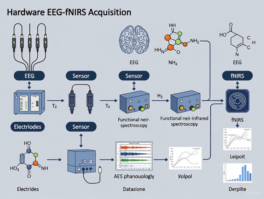
The integration of electroencephalography (EEG) and functional near-infrared spectroscopy (fNIRS) has emerged as a powerful multimodal neuroimaging approach, leveraging the complementary strengths of each modality. EEG provides millisecond-level temporal resolution of electrophysiological activity, while fNIRS offers better spatial resolution for hemodynamic responses [33]. The efficacy of this integration fundamentally depends on the synchronization architecture employed, which directly impacts data alignment precision, system complexity, and experimental flexibility. Within hardware integration for multimodal EEG-fNIRS acquisition research, two primary synchronization paradigms have emerged: unified (simultaneous) systems and separate (sequential) systems [6] [11].
The critical challenge in multimodal integration stems from the fundamentally different nature of the signals captured. EEG measures electrical potentials generated by synchronized neuronal firing, requiring microsecond-level temporal accuracy [34]. In contrast, fNIRS measures hemodynamic changes in blood oxygenation, a slow physiological process that unfolds over seconds [10]. This inherent discrepancy necessitates robust synchronization architectures to ensure meaningful data fusion and accurate interpretation of neurovascular coupling dynamics [35].
Unified System Integration
Unified integration employs a single hardware processor to acquire and process both EEG and fNIRS signals concurrently. This architecture achieves precise synchronization by generating drive signals for fNIRS light sources while simultaneously amplifying intensity signals from fNIRS and electrical potentials from EEG through a shared analog-to-digital conversion pathway [6] [11]. The synchronized data is then transmitted to a host computer for subsequent analysis, preprocessing, and fusion.
This approach typically utilizes integrated helmet designs where EEG electrodes and fNIRS optodes are mounted on a shared substrate. Advanced implementations employ 3D-printed or thermoplastic customized helmets that maintain consistent optode spacing and scalp contact pressure across subjects, which is crucial for data quality [6]. The unified architecture ensures microsecond-level temporal alignment between modalities, making it particularly valuable for investigating fast neural events and their corresponding hemodynamic correlates [35].
Separate System Integration
Separate system integration utilizes distinct, standalone EEG and fNIRS systems that are synchronized during post-processing analysis. In this architecture, the NIRScout (fNIRS) and BrainAMP (EEG) systems typically operate independently during data acquisition, with synchronization established retrospectively via a host computer [6] [11]. External triggers or shared clock systems, such as TTL pulses through parallel ports, facilitate this post-hoc alignment.
This approach often employs modified EEG caps with punctures to accommodate fNIRS probe fixtures, offering greater flexibility in experimental design and hardware selection [6]. However, the separate architecture faces significant challenges in achieving precise synchronization, particularly for analyzing EEG data with its inherent microsecond temporal resolution requirements. The inherent limitations of post-hoc synchronization may introduce temporal jitter between modalities, potentially obscuring fine-grained neurovascular coupling dynamics [6].
Table 1: Comparative Analysis of Synchronization Architectures
| Feature | Unified System Integration | Separate System Integration |
|---|---|---|
| Synchronization Precision | High (microsecond level) [6] | Moderate (limited by trigger alignment) [6] |
| System Complexity | High (requires specialized hardware) [11] | Lower (uses commercial off-the-shelf systems) [6] |
| Implementation Flexibility | Lower (fixed hardware configuration) [6] | Higher (modular component selection) [6] |
| Experimental Scalability | Moderate (constrained by integrated design) | High (adaptable to various experimental setups) [6] |
| Typical Artifact Handling | Coordinated noise rejection [36] | Separate artifact removal pipelines [14] |
| Optimal Application Scope | Research requiring precise temporal alignment of fast neural events [35] | Studies prioritizing flexibility over microsecond precision [6] |
Experimental Protocols and Methodologies
Protocol for Unified System Implementation
Objective: To implement a unified EEG-fNIRS system for investigating neural correlates during motor execution, observation, and imagery tasks.
Materials and Setup:
- Integrated EEG-fNIRS acquisition system (e.g., Hitachi ETG-4100 with embedded EEG) [35]
- Customized acquisition helmet with co-registered 128-electrode EEG and 24-channel fNIRS probe [35]
- 3D magnetic space digitizer (e.g., Fastrak, Polhemus) for optode localization [35]
- Stimulus presentation system with precision timing capabilities
Procedure:
- Helmet Fitting: Select appropriate helmet size based on participant head circumference. Ensure proper optode-scalp contact with consistent inter-optode spacing (typically 2.88±0.13 cm) [35].
- Optode Digitization: Digitize fNIRS optode positions relative to anatomical landmarks (nasion, inion, preauricular points) using the 3D digitizer to account for individual anatomical variability [35].
- System Calibration: Initialize the unified processor to simultaneously generate fNIRS drive signals and EEG amplification parameters. Verify synchronization lock between modalities.
- Data Acquisition: Present experimental paradigm (e.g., motor execution, observation, imagery tasks) while concurrently recording both modalities through the shared acquisition hardware [35].
- Quality Assessment: Monitor signal quality in real-time, ensuring >50% of data from both modalities meets quality thresholds for inclusion in analysis [35].
Data Processing Pipeline:
- Apply structured sparse multiset Canonical Correlation Analysis (ssmCCA) to fuse electrical and hemodynamic responses [35]
- Identify brain regions consistently activated across both modalities
- Extract fused features for subsequent statistical analysis and interpretation
Protocol for Separate System Integration
Objective: To implement separate but synchronized EEG-fNIRS systems for cognitive task classification.
Materials and Setup:
- Independent EEG system (e.g., BrainAMP) [6]
- Independent fNIRS system (e.g., NIRScout) [6]
- External synchronization unit (TTL pulse generator or shared clock)
- Standard EEG cap with fNIRS-compatible openings
- Host computer with synchronization software
Procedure:
- System Setup: Arrange EEG and fNIRS systems in parallel configuration. Establish master-slave relationship through TTL pulse synchronization [6].
- Sensor Placement: Mount fNIRS optodes through predefined openings in the standard EEG cap. Verify no physical interference between electrode and optode components [6].
- Synchronization Initialization: Implement trigger-based synchronization by sending simultaneous pulse markers to both systems at trial initiation.
- Data Acquisition: Conduct experimental tasks while both systems record independently but with shared event markers.
- Post-hoc Alignment: Align datasets during preprocessing using shared trigger timestamps. Account for potential clock drift between systems.
Data Fusion Approach:
- Preprocess modalities independently using modality-specific pipelines
- Apply decision-level fusion techniques such as Dempster-Shafer theory to combine classification outcomes [29]
- Implement uncertainty quantification through Dirichlet distribution parameter estimation [29]
Visualization of System Architectures
Unified System Architecture
Unified System Data Flow
Separate System Architecture
Separate System Data Flow
The Scientist's Toolkit: Research Reagent Solutions
Table 2: Essential Materials for EEG-fNIRS Multimodal Research
| Component | Function & Purpose | Implementation Example |
|---|---|---|
| Integrated Acquisition Helmets | Ensures stable optode/electrode placement and consistent scalp coupling [6] | 3D-printed custom helmets; Cryogenic thermoplastic sheets; Modified EEG caps with fNIRS openings [6] |
| Synchronization Interfaces | Enables temporal alignment of multimodal data streams [6] [11] | TTL pulse generators; Shared clock systems; Unified processors with simultaneous acquisition [6] |
| 3D Spatial Digitizers | Records precise sensor locations for co-registration with anatomical landmarks [35] | Fastrak (Polhemus) magnetic digitizers; Optical positioning systems [35] |
| Data Fusion Algorithms | Integrates electrophysiological and hemodynamic signals for comprehensive analysis [35] [29] | Structured sparse multiset CCA (ssmCCA) [35]; Dempster-Shafer theory [29]; Dual-decoder architectures [36] |
| Artifact Handling Tools | Mitigates motion artifacts and physiological confounders in both modalities [10] | Short-separation regression; Motion correction algorithms; Independent Component Analysis [10] |
| Deep Learning Frameworks | Enables end-to-end multimodal feature learning and classification [29] [37] | DeepSyncNet [37]; EEG-fNIRS data augmentation (EFDA-CDG) [14]; Feature decoupling networks [36] |
The selection between unified and separate synchronization architectures represents a critical decision point in multimodal EEG-fNIRS research design. Unified systems offer superior temporal precision essential for investigating neurovascular coupling dynamics, particularly in tasks requiring millisecond-level alignment of electrical and hemodynamic events [35]. Separate systems provide greater implementation flexibility and hardware selection options, suitable for experimental paradigms where modularity outweighs the need for microsecond synchronization accuracy [6].
Future developments in synchronization architectures will likely focus on hybrid approaches that balance precision with flexibility. Advancements in wearable technology, real-time processing capabilities, and adaptive fusion algorithms promise to further blur the boundaries between these architectural paradigms [10] [14]. As multimodal research progresses toward more naturalistic settings and clinical applications, synchronization architectures must evolve to support both high temporal fidelity and ecological validity, ultimately enhancing our understanding of complex brain functions through complementary neural signatures.
Co-registration Strategies for Spatial Alignment of EEG Electrodes and fNIRS Optodes
Hardware integration for multimodal electroencephalography (EEG) and functional near-infrared spectroscopy (fNIRS) acquisition presents a unique set of challenges, paramount among which is the spatial co-registration of EEG electrodes and fNIRS optodes. The complementary nature of EEG, which provides high temporal resolution of electrophysiological activity, and fNIRS, which tracks hemodynamic responses with better spatial localization, can only be fully leveraged when the two modalities are precisely aligned [11] [38]. Effective co-registration ensures that the recorded neural electrical activity and the associated hemodynamic changes originate from the same cortical region, thereby facilitating accurate data fusion and interpretation [11] [39]. This protocol outlines established and emerging strategies to achieve this critical spatial alignment, framed within the context of a broader thesis on multimodal hardware integration.
Fundamental Principles and Integration Approaches
The core challenge of co-registration stems from the fundamental differences in the biophysical principles and operational requirements of EEG and fNIRS. EEG measures electrical potentials on the scalp surface, with signal quality being paramount. fNIRS relies on the transmission of near-infrared light between a source and a detector placed on the scalp, where the source-detector distance is a critical parameter determining sensitivity and penetration depth [11] [40]. Precise co-registration is essential not only for accurate multimodal analysis but also for avoiding cross-talk; for instance, studies have shown that with careful design, fNIRS optodes do not cause observable interference in EEG spectral analysis [38].
There are two primary methodological paradigms for integrating EEG and fNIRS hardware, each with implications for co-registration and synchronization fidelity.
Table 1: Comparison of fNIRS-EEG Hardware Integration Methods
| Integration Method | Description | Synchronization Precision | System Complexity | Key Advantage |
|---|---|---|---|---|
| Separate Systems | EEG and fNIRS data are acquired on independent, commercially available systems (e.g., NIRScout and BrainAMP). A host computer synchronizes data during acquisition and analysis [11] [6]. | Lower; may be insufficient for microsecond EEG analysis [11]. | Relatively Simple | Ease of implementation using established hardware. |
| Unified Processor | A single, custom processor is used to acquire and process both EEG signals and fNIRS input/output simultaneously [11] [6]. | High; enables precise synchronization [11]. | Complex and Intricate | Streamlined analytical process and high-fidelity temporal alignment. |
The following diagram illustrates the logical workflow for selecting and implementing a co-registration strategy, connecting the fundamental principles with the specific approaches detailed in the subsequent sections.
Co-registration Hardware Strategies
The physical realization of co-registration is achieved through specialized helmet or cap designs. The choice of strategy involves a trade-off between precision, cost, comfort, and scalability.
Co-localized Optode-Electrode Design
This advanced strategy aims to minimize the physical distance between an EEG electrode and an fNIRS optode, allowing them to occupy nearly the same location on the scalp. A specific implementation involves designing custom fNIRS source optodes that attach directly to the external housing of active wet EEG electrodes [38]. The optode is mounted to leverage the electrode's access hole for conductive gel, allowing a 3-mm diameter light pipe to touch the scalp at a minimal center-to-center distance (e.g., ~4.87 mm) from the electrode's active contact area [38]. This method preserves standardized EEG layouts (e.g., 10-20 system) and high-density fNIRS arrays simultaneously, maximizing spatial correspondence and modularity.
Custom-Molded Rigid Helmets
To address issues of inconsistent probe placement and pressure on elastic caps, researchers have turned to custom-fitted rigid helmets.
- 3D-Printed Helmets: 3D printing technology allows for the creation of patient-specific helmets that perfectly conform to an individual's head geometry. This design allows for flexible and precise positioning of both EEG electrodes and fNIRS optodes, accommodating head-size variations and ensuring consistent probe-to-scalp distance and contact pressure [11]. A limitation is the relatively high cost of production.
- Cryogenic Thermoplastic Sheets: A more cost-effective alternative involves using composite polymer cryogenic thermoplastic sheets. This material becomes soft and malleable at approximately 60°C, allowing it to be shaped directly to a subject's head. It retains this shape upon cooling, providing a stable and customized platform for mounting hardware [11] [6]. A potential drawback is that the molded sheet may be slightly rigid and exert pressure on the head.
Modified Elastic Caps
The most common and straightforward approach utilizes standard flexible EEG electrode caps as a base. Holes are punctured at specific locations to accommodate fNIRS probe fixtures, often using plastic connectors to secure them in place [11] [6]. While this method is simple to implement and maintains satisfactory coupling, it has significant limitations: the high stretchability of the fabric can lead to uncontrollable variations in the distance between the fNIRS source and detector across subjects with different head shapes, and it can result in inconsistent probe-scalp contact pressure, especially during movement [11].
Table 2: Comparison of fNIRS-EEG Co-registration Hardware Strategies
| Strategy | Description | Advantages | Disadvantages | Ideal Use Case |
|---|---|---|---|---|
| Co-localized Design [38] | Custom optodes attached directly to EEG electrodes, sharing nearly the same scalp position. | Maximizes spatial correspondence; preserves high-density layouts; modular. | Requires custom hardware design and fabrication; potential for electronic cross-talk (mitigated by design). | High-precision research requiring maximal spatial alignment of signals. |
| Custom 3D-Printed Helmet [11] | Additively manufactured helmet customized to subject's head anatomy. | Excellent stability; consistent probe placement and pressure; high precision. | High cost and lead time for production; less adaptable for multi-subject use. | Long-term studies with a few subjects or clinical applications requiring extreme reliability. |
| Cryogenic Thermoplastic Helmet [11] [6] | Custom-molded helmet made from low-temperature thermoplastic material. | Cost-effective; lightweight; good stability and customization. | Can be rigid and exert pressure on the head; requires molding equipment and time. | Studies with a moderate number of subjects where a balance of cost and precision is needed. |
| Modified Elastic Cap [11] [6] | Standard EEG cap with holes punched for fNIRS probes. | Simple and fast to implement; low cost; utilizes existing equipment. | Unstable probe placement; variable source-detector distance; poor control over contact pressure. | Preliminary studies or environments where cost and simplicity are the primary concerns. |
Experimental Protocol for a Co-localized fNIRS-EEG Study
The following detailed protocol is adapted from a study that successfully implemented a co-localized, high-density fNIRS-EEG design for a cognitive task, providing a template for rigorous multimodal experimentation [38].
Aim
To measure simultaneous hemodynamic (fNIRS) and electrophysiological (EEG) responses in the prefrontal and temporal cortices during a cognitive task using a co-localized HD-fNIRS-EEG probe design.
Materials and Reagents
- EEG System: A high-density EEG acquisition system (e.g., BrainVision LiveAmp) with active wet electrodes.
- fNIRS System: A high-density, continuous-wave fNIRS system (e.g., NIRSport2).
- Custom Co-localized Optodes: 3D-printed optodes designed to mate with the specific EEG electrodes being used.
- Hybrid Cap: A flexible cap (e.g., made of NinjaFlex TPU) designed to hold the integrated probe array. The cap design should be created in a neuroimaging toolbox (e.g., AtlasViewer) and 3D-printed or fabricated to specification.
- Electrode Gel: Conductive electrolyte gel for EEG electrodes.
- Task Presentation Software: Software to present stimuli and record event markers (e.g., E-Prime, PsychoPy).
- Synchronization Module: Hardware or software to send simultaneous triggers from the stimulus computer to both the EEG and fNIRS acquisition units.
Procedure
Probe Design and Fabrication (Offline):
- Design the HD-fNIRS-EEG probe layout in a neuroimaging toolbox (e.g., AtlasViewer). Specify the positions of fNIRS sources (red) and detectors (blue), and EEG electrodes (e.g., using 10-05, 10-10, and 10-20 positions).
- For co-localized positions, model the custom optode attachments to ensure mechanical and electrical compatibility with the chosen EEG electrodes.
- Export the probe design and fabricate the custom cap and optodes.
System Setup and Synchronization:
- Connect the EEG and fNIRS systems to their respective acquisition computers.
- Establish a synchronization link between the stimulus presentation computer and both data acquisition systems. This is critical for aligning the data streams during analysis. Verify that event markers are reliably recorded in both systems.
Subject Preparation:
- Fit the custom hybrid cap onto the subject's head. Ensure it is snug and secure.
- For co-localized positions: Attach the custom fNIRS optodes to their corresponding EEG electrodes according to the design.
- For non-co-localized positions: Place EEG electrodes and fNIRS optodes in their designated positions on the cap.
- Apply conductive gel to all EEG electrodes via the access holes, ensuring good electrical contact.
Signal Quality Check:
- Initiate data streams from both EEG and fNIRS systems.
- Check the impedance for all EEG electrodes; aim for impedances below 20 kΩ.
- Check the raw light intensity or signal-to-noise ratio for all fNIRS channels; ensure values are within acceptable limits as per the fNIRS system manufacturer's guidelines.
Data Acquisition:
- Instruct the subject on the task procedure.
- Start recording on both EEG and fNIRS systems.
- Begin the experimental paradigm (e.g., a modified Stroop task). The stimulus software should send a trigger at the onset of each trial or condition block, which is recorded by both acquisition systems.
- Monitor data quality periodically throughout the recording session.
Data Completion:
- Stop recording on both systems after the task is complete.
- Export the data from both systems, ensuring event markers are included.
The experimental workflow, from preparation to analysis, is visualized below.
The Scientist's Toolkit: Essential Research Reagents and Materials
Table 3: Key Materials for fNIRS-EEG Co-registration Experiments
| Item Name | Function / Purpose | Example Specifications / Notes |
|---|---|---|
| Active Wet EEG Electrodes [38] | Records electrical brain activity with high temporal resolution. Integrated housing allows for co-localized fNIRS optode attachment. | e.g., LiveAmp electrodes (BrainProducts); include an access hole for gel and optode guide. |
| Custom 3D-Printed fNIRS Optode [38] | Delivers near-infrared light to the scalp. Custom design enables physical integration with EEG electrodes for co-localization. | Fabricated using SLS 3D-printing (e.g., Formlabs resin). Dielectric epoxy pots electronics to isolate from EEG gel. |
| Flexible Hybrid Cap Substrate [38] | Holds both EEG electrodes and fNIRS optodes in a stable, pre-defined geometry on the subject's head. | Made of flexible material like NinjaFlex TPU, designed in software (e.g., AtlasViewer) and 3D-printed. |
| Cryogenic Thermoplastic Sheet [11] [6] | Provides a cost-effective, customizable rigid substrate for creating subject-specific helmets. | Softens at ~60°C to be molded to head shape, retains stability when cool. |
| fNIRS System [38] [41] | A continuous-wave system to generate light and detect intensity changes for calculating hemodynamic responses. | e.g., NIRSport2 (NIRx); capable of high-density source-detector arrangements. |
| EEG Amplifier [41] | Amplifies and digitizes microvolt-level electrical signals from scalp electrodes. | Must be compatible with the chosen electrodes and support the required number of channels. |
| Conductive Electrolyte Gel | Ensures low-impedance electrical connection between the EEG electrode and the scalp. | Standard clinical or research-grade EEG gel. |
| Synchronization Trigger Box | Sends simultaneous electronic pulses (TTL) from the stimulus computer to both EEG and fNIRS systems to align data streams temporally. | Critical for any multimodal study. Can be a custom-built or commercial solution. |
The integration of electroencephalography (EEG) and functional near-infrared spectroscopy (fNIRS) represents a promising direction in multimodal neuroimaging, offering a pathway to study brain function with high spatiotemporal resolution in naturalistic scenarios [10]. This dual-modality approach leverages the complementary strengths of each technique: EEG captures neuro-electrical activity with millisecond temporal resolution, while fNIRS measures hemodynamic changes with better spatial localization [42] [11]. The fusion of these signals is particularly valuable for understanding neurovascular coupling and has demonstrated enhanced performance in brain-computer interfaces, clinical neurology, and neurorehabilitation applications [10] [43].
However, a significant challenge in designing effective fNIRS-EEG systems lies in optimizing channel configurations to balance data richness against practical constraints. The spatial arrangement and density of electrodes and optodes directly impact signal quality, spatial resolution, and participant comfort, while also influencing the system's portability and applicability in real-world settings [11]. This application note provides a structured framework for channel configuration optimization within the broader context of hardware integration for multimodal acquisition research.
Technical Specifications and Performance Metrics
Table 1: Comparative Technical Specifications of EEG and fNIRS Modalities
| Parameter | EEG | fNIRS | Integrated fNIRS-EEG |
|---|---|---|---|
| Temporal Resolution | Millisecond level [11] | Seconds [11] | High (leveraging EEG strength) |
| Spatial Resolution | Low [11] | Notable (due to exponential light attenuation in tissue) [11] | Enhanced through complementarity |
| Measured Phenomena | Neuronal electrical activity [42] | Hemodynamic fluctuations (HbO, HbR) [43] | Electrophysiological + hemodynamic |
| Key Quantitative Parameters | Power Spectral Density (PSD), Brain Symmetry Index (BSI), Phase Synchrony Index [42] | Oxygenated (HbO) & Deoxygenated (HbR) Hemoglobin concentrations [10] | Neurovascular-coupling related features [42] |
| Primary Artifact Sources | Ocular (EOG), muscle activity (EMG) [10] | Scalp hemodynamics, motion [10] | Combined physiological & motion artifacts |
Experimental Protocols for System Validation
Protocol for Motor Task Characterization
This protocol is adapted from studies investigating post-stroke motor recovery, leveraging well-established motor paradigms to elicit robust neural and hemodynamic responses [42].
- Objective: To validate the performance of an integrated fNIRS-EEG channel configuration during a controlled motor task.
- Subject Preparation: Position the subject in a comfortable chair. Apply EEG electrodes according to the international 10-20 system, focusing on the motor cortex (C3, C4, Cz). Arrange fNIRS optodes over the primary motor cortex and supplementary motor area to ensure coverage contralateral to the movement hand. Ensure proper scalp coupling and light shielding [11].
- Paradigm Design: Employ a block design consisting of:
- Rest Condition (30 seconds): Subject remains still with eyes open, focusing on a fixation cross.
- Activation Condition (20 seconds): Subject performs repetitive self-paced finger tapping or hand gripping.
- Repeat for a minimum of 10 blocks to ensure adequate signal-to-noise ratio.
- Data Acquisition: Record simultaneous EEG and fNIRS data throughout the experiment. Synchronize acquisition using a unified processor or precise external triggers [11].
- Analysis Metrics:
- EEG: Compute event-related desynchronization/synchronization (ERD/ERS) in the mu (8-13 Hz) and beta (13-30 Hz) frequency bands over the motor cortex [42].
- fNIRS: Calculate the mean concentration changes of HbO and HbR during activation blocks compared to rest using a General Linear Model (GLM) [10] [43].
- Integration: Assess the temporal correlation between EEG power band changes and the hemodynamic response.
Protocol for Cognitive Task Characterization with Music Stimuli
This protocol utilizes music-evoked brain responses to test the system's capability to capture complex cognitive and emotional processing [43].
- Objective: To evaluate channel configuration efficacy in distinguishing brain states evoked by different auditory stimuli.
- Subject Preparation: Configure the EEG cap and fNIRS helmet with a focus on the prefrontal cortex (PFC), a region critically involved in emotional and cognitive processing [43].
- Paradigm Design:
- Pre-Experiment: Conduct a questionnaire to identify each subject's personal preferred music. Select a neutral, unfamiliar piece of soft music as a control stimulus [43].
- Experimental Session: Present the two types of music in a randomized or counterbalanced order. Each trial should include a baseline period (quiet rest) followed by music listening.
- Data Acquisition: Record multimodal data continuously. Ensure the acoustic environment is controlled to minimize external noise.
- Analysis Metrics:
- EEG: Extract power spectral density (PSD) features from delta, theta, alpha, beta, and gamma bands [42] [43].
- fNIRS: Analyze the amplitude and spatial extent of HbO activation in the PFC.
- Integration: Employ feature-level fusion techniques, such as an improved Normalized-ReliefF algorithm, to classify the brain state associated with preferred vs. neutral music, using classification accuracy as a performance metric for the channel setup [43].
System Workflow and Signaling Pathways
The following diagram illustrates the end-to-end workflow for data acquisition, processing, and fusion in a multimodal fNIRS-EEG study.
Multimodal fNIRS-EEG Data Acquisition and Fusion Workflow
The Scientist's Toolkit: Research Reagent Solutions
Table 2: Essential Materials and Equipment for fNIRS-EEG Research
| Item Category | Specific Examples & Functions | Key Considerations |
|---|---|---|
| Acquisition Hardware | EEG Amplifier & Electrodes: Record electrical potentials. fNIRS System: Includes laser diodes/LEDs (sources) and photodetectors to measure light attenuation [11]. | Prefer systems with a unified processor for synchronized acquisition [11]. High-density arrays enable better spatial resolution (HD-DOT) [10]. |
| Integration Helmet | Custom 3D-printed helmet [11], Cryogenic thermoplastic sheet [11], or modified elastic EEG cap with integrated optodes. | Ensures stable and co-registered placement of EEG electrodes and fNIRS optodes. Custom helmets improve contact pressure consistency and data quality [11]. |
| Data Synchronization | Dedicated microcontroller, unified host computer software, or external trigger hardware (e.g, TTL pulse generators). | Critical for aligning high-temporal-resolution EEG with fNIRS. A unified processor offers the highest synchronization precision [11]. |
| Software & Algorithms | Artifact Removal Tools: ICA for EEG, PCA-based motion correction for fNIRS [10]. Fusion Algorithms: Data-level, feature-level (e.g., ReliefF), or decision-level fusion methods [10] [43]. | Robust artifact handling is essential. Data-driven fusion methods (e.g., unsupervised symmetric techniques) are promising for naturalistic studies [10]. |
| Validation Stimuli | Motor Paradigms: Finger tapping, hand gripping. Cognitive Paradigms: Personal preferred music, neutral music, n-back tasks. | Protocols should elicit robust and reproducible neural/hemodynamic responses in targeted brain regions for system validation [42] [43]. |
Channel Configuration Strategies and Optimization Framework
Optimizing channel configuration requires a principled approach that considers the specific research question, anatomical targets, and practical constraints. The table below summarizes key strategies and their trade-offs.
Table 3: Channel Configuration Strategies for fNIRS-EEG Integration
| Strategy | Description | Advantages | Challenges & Considerations |
|---|---|---|---|
| High-Density (HD) Mapping | Utilizing high-density arrays of multiple source-detector separations (e.g., for HD-DOT) with overlapping sensitivity profiles [10]. | Enables 3D image reconstruction of functional activation; achieves spatial resolution comparable to fMRI in some contexts [10]. | Increased system complexity, weight, and setup time; higher data throughput requirements; more susceptible to motion artifacts. |
| Targeted Sparse Configuration | Focusing a limited number of channels on specific regions of interest (ROI) known to be involved in the task (e.g., prefrontal cortex for music, motor cortex for movement) [42] [43]. | Practical and lightweight; suitable for portable and long-duration monitoring; simplifies data analysis. | Limited coverage outside ROIs; may miss distributed network activity; requires strong a priori hypothesis. |
| Hybrid Density Approach | Combining high-density coverage over a primary ROI with sparse coverage over secondary or associative areas. | Balances data richness and practicality; allows for focused investigation while retaining some whole-cortex exploratory capability. | Requires careful design to avoid excessive weight and complexity; channel placement must be justified by the experimental design. |
| Co-registration Precision | Precisely determining the spatial location of each EEG electrode and fNIRS channel on the scalp, often using 3D digitizers or structural MRI [11]. | Essential for accurate spatial alignment and interpretation of fused data; facilitates source localization and integration with anatomical atlases. | Adds an extra step to the experimental protocol; requires specialized equipment and software. |
| Auxiliary Signal Integration | Incorporating short-separation fNIRS channels and other auxiliary signals (e.g., EOG, ECG) for robust confounder correction [10]. | Significantly improves signal quality by separating cerebral from extracerebral physiological noise; enhances the robustness of fusion models. | Underutilized in current research; requires additional hardware channels and more complex processing pipelines [10]. |
The integration of Brain-Computer Interface (BCI) technology into clinical and research settings represents a paradigm shift in neurorehabilitation and cognitive assessment. A particularly promising development is the multimodal integration of electroencephalography (EEG) and functional near-infrared spectroscopy (fNIRS), which creates a synergistic system for monitoring brain activity by combining electrical and hemodynamic responses [6] [32]. This integration is foundational for hardware systems designed to probe the complex relationship between neuronal electrical activity and subsequent vascular changes, a process known as neurovascular coupling [19] [6]. For researchers and clinicians, these systems offer a portable, cost-effective solution with complementary strengths: EEG provides millisecond-level temporal resolution of electrical brain activity, while fNIRS offers superior spatial resolution for localizing hemodynamic changes in the cortex [6] [32]. This application note details the protocols and analytical frameworks for employing multimodal EEG-fNIRS systems in key areas of neurorehabilitation and mental workload monitoring, providing a practical guide for their implementation in research and clinical practice.
Multimodal EEG-fNIRS Acquisition: Core Principles and Hardware
Technical Synergy and Integration Methods
The efficacy of multimodal EEG-fNIRS stems from its ability to provide a more complete picture of brain function by measuring two distinct but related phenomena.
- EEG records electrical activity from populations of neurons, offering direct insight into neural processing with a high temporal resolution (on the order of milliseconds) [6] [32].
- fNIRS measures changes in concentrations of oxygenated (HbO) and deoxygenated hemoglobin (HbR) in the blood, serving as an indirect marker of neural activity with better spatial resolution than EEG [19] [6].
Two primary methods exist for integrating these modalities [6]:
- Synchronized Separate Systems: EEG and fNIRS data are acquired using separate, commercially available systems (e.g., NIRScout for fNIRS and BrainAMP for EEG) and synchronized during post-processing. This approach is simpler to implement but may lack the precision required for microsecond-level EEG analysis.
- Unified Hardware System: A single integrated processor is used for simultaneous acquisition and processing of both EEG and fNIRS signals. This method, though more complex, ensures precise temporal synchronization and streamlines the analytical workflow, making it the preferred approach for advanced applications.
Hardware Integration: The Joint Acquisition Helmet
A critical component for successful multimodal research is the design of the acquisition helmet. Current designs often integrate EEG electrodes and fNIRS optodes into a single cap [6]. Key design considerations and challenges include:
- Spatial Co-registration: The arrangement of EEG electrodes assists in co-registering the fNIRS channels, enabling precise spatial localization of the brain regions being probed by both modalities [6].
- Contact Pressure and Stability: The use of highly elastic fabrics in standard caps can lead to inconsistent probe-to-scalp contact pressure and variable distances between fNIRS sources and detectors, which negatively impacts data quality [6].
- Customized Solutions: To address these challenges, researchers are turning to 3D printing and cryogenic thermoplastic sheets to create custom-fitted helmets. These solutions offer a more secure and consistent fit, accommodating variations in head shape and size, which is crucial for data quality, especially in movement-oriented or long-duration studies [6].
Application Note 1: Motor Rehabilitation in Stroke
Clinical Background and Rationale
Stroke often leads to significant motor impairments, with upper limb function being a critical determinant of patient independence. BCI technology based on Motor Imagery (MI) and Motor Attempt (MA) has emerged as a promising tool for enhancing motor recovery. These tasks activate sensorimotor areas similar to actual movement, thereby promoting activity-dependent neuroplasticity [44]. Multimodal assessment is crucial for capturing the complex neurophysiological changes underlying recovery.
Detailed Experimental Protocol
Objective: To evaluate the effectiveness of a BCI system integrating MI and MA tasks in improving upper limb motor function in subcortical ischemic stroke patients using a multimodal assessment framework [44].
Study Design: Randomized, double-blind, placebo-controlled clinical trial.
Participants:
- Inclusion Criteria: Adults aged 35-79; first-onset ischemic stroke (2 weeks to 3 months prior); hemiplegia with proximal upper limb muscle strength grade 1-3; right-handed; sufficient sitting balance.
- Exclusion Criteria: Significant cognitive impairment (MMSE < 20); severe limb pain or limited mobility [44].
Intervention:
- BCI Group: Participants underwent 20-minute training sessions for both upper and lower limbs, twice daily for two weeks. The system comprised:
- An 8-electrode EEG acquisition system.
- A virtual reality training module that converted EEG signals into a quantitative "motor intention score" (Mscore).
- A pedaling training robot whose speed was controlled by the Mscore, providing real-time feedback.
- Control Group: Used identical equipment but received simulated feedback from pre-recorded EEG data instead of real-time signals. Both groups received standard rehabilitation (physiotherapy, occupational therapy) [44].
Multimodal Assessment Schedule: Assessments were conducted at baseline (T0) and immediately post-intervention (T1).
Table 1: Multimodal Assessment Protocol for Stroke Rehabilitation
| Assessment Domain | Specific Measure/Instrument | Key Parameters/Outcomes |
|---|---|---|
| Clinical Motor Function | Fugl-Meyer Assessment for Upper Extremity (FMA-UE) | Primary outcome: Change in FMA-UE score [44] |
| Neural Electrophysiology | Electroencephalography (EEG) | Delta/Alpha Ratio (DAR), Delta/Alpha Beta Ratio (DABR) [44] |
| Cortical Hemodynamics | Functional Near-Infrared Spectroscopy (fNIRS) | Functional connectivity & activation in PFC, SMA, M1 [44] |
| Muscle Activity | Electromyography (EMG) | Activity of deltoid and biceps muscles during flexion [44] |
Key Findings and Workflow
The BCI group demonstrated significantly greater improvement in upper extremity motor function compared to the control group (ΔFMA-UE: 4.0 vs. 2.0) [44]. The multimodal data revealed consistent neurophysiological improvements: a significant decrease in EEG power ratios (DAR and DABR), increased muscle activity on EMG, and enhanced functional connectivity in motor networks on fNIRS [44].
The following workflow diagram illustrates the closed-loop BCI system and multimodal assessment process:
Figure 1: Closed-Loop BCI Rehabilitation and Assessment Workflow.
Application Note 2: Differential Diagnosis of Disorders of Consciousness
Clinical Background and Rationale
Differentiating between patients with Unresponsive Wakefulness Syndrome (UWS) and those in a Minimally Conscious State (MCS) is critically important for prognosis and treatment but remains clinically challenging. Motor Imagery-based BCI (MI-BCI) offers a potential tool for objectively assessing residual cognitive function and responsiveness in these populations [45].
Detailed Experimental Protocol
Objective: To investigate the utility of an MI-BCI system for discriminating between UWS and MCS patients by analyzing EEG spectral features and BCI performance metrics [45].
Study Design: Observational, cross-sectional study.
Participants: 31 patients with prolonged Disorders of Consciousness (pDoC), including 12 with UWS and 19 with MCS, as diagnosed by standardized behavioral scales [45].
Procedure:
- EEG Recording: EEG data were collected during a resting state and during MI-BCI tasks.
- MI-BCI Task: Patients were instructed to imagine moving their hands. The system was calibrated to detect sensorimotor rhythms associated with this imagery.
- Data Analysis: Focused on the relative power spectral density across five frequency bands (delta, theta, alpha, beta, gamma) in motor imagery-related regions (frontal and parietal cortices). BCI performance was measured via classification accuracy and computed attention indices [45].
Outcome Measures:
- Primary: Patterns of neural oscillation modulation across frequency bands.
- Secondary: BCI classification accuracy and its correlation with clinical consciousness scores.
Table 2: Key Neural Oscillation Findings in DoC Diagnosis [45]
| Patient Group | Findings in Frontal & Parietal Lobes during MI-BCI |
|---|---|
| Minimally Conscious State (MCS) | Significant multiband modulation:- Enhancement in slow waves (Delta, Theta)- Suppression in fast waves (Alpha, Beta, Gamma) |
| Unresponsive Wakefulness Syndrome (UWS) | Only localized enhancement in parietal Gamma waves. |
Key Findings and Diagnostic Logic
The study found that MCS patients exhibited complex, multiband neural oscillation modulation during MI-BCI tasks, while UWS patients showed only minimal, localized changes [45]. Furthermore, the MCS group achieved significantly higher BCI classification accuracy (55% vs. 38%), and attention indices correlated moderately with clinical scores across all patients [45]. This suggests that BCI performance can serve as an objective auxiliary discriminator.
The diagnostic logic based on EEG spectral response is summarized below:
Figure 2: Diagnostic Logic for Disorders of Consciousness using MI-BCI.
Application Note 3: Mental Workload Monitoring
Background and Rationale
Mental workload reflects the interaction between the demands of a task and an individual's cognitive capacity to meet those demands. Excessive workload can lead to performance decline and errors, making its assessment vital in fields like aviation, surgery, and medical training [46] [47]. A neuroergonomic approach using objective neural and physiological measures is key to developing sensitive and reliable assessment tools.
Detailed Experimental Protocol
Objective: To comprehensively profile mental workload correlates across six foundational cognitive domains using a multi-modal, longitudinal design [46].
Study Design: Longitudinal study across four sessions over four weeks.
Participants: 23 healthy adults (ages 18-48) [46].
Cognitive Task Battery (Six Domains):
- Working Memory (e.g., N-back tasks)
- Vigilance (sustained attention tasks)
- Risk Assessment (decision-making under uncertainty)
- Shifting Attention (multitasking or task-switching)
- Situation Awareness (comprehension of dynamic environments)
- Inhibitory Control (e.g., Stop-Signal Task)
Each task included high and low difficulty levels to manipulate workload.
Multimodal Acquisition: Six biomedical modalities were recorded concurrently during task performance [46]:
- Brain Activity: EEG (electrical activity) and fNIRS (hemodynamic activity in prefrontal cortex).
- Cardiac Activity: ECG (electrocardiogram) and PPG (photoplethysmography).
- Ocular Activity: EOG (electrooculogram) and eye-tracking.
Data Analysis: Sensitivity of each modality to workload changes was assessed by comparing high vs. low difficulty conditions and tracking changes over sessions as expertise was acquired.
Key Insights and Measurement Framework
The study demonstrated that while all modalities showed some sensitivity to workload, neuroimaging modalities (EEG, fNIRS) were particularly effective at revealing differences between task conditions and cognitive domains [46]. The combination of central (brain) and peripheral (heart, eyes) nervous system measures provides a composite and more robust perspective on the operator's cognitive state.
Table 3: Multimodal Metrics for Mental Workload Assessment
| Modality | Measured Signal | Example Workload-Sensitive Metrics | Primary Advantage |
|---|---|---|---|
| EEG | Electrical brain activity | Power in Theta, Alpha bands; Event-Related Potentials (ERPs) | Excellent temporal resolution [46] |
| fNIRS | Hemodynamic (HbO/HbR) in PFC | Changes in HbO concentration during cognitive tasks | Good spatial resolution; motion robust [46] |
| ECG/PPG | Cardiac activity | Heart Rate (HR), Heart Rate Variability (HRV) | Indicates autonomic nervous system engagement [46] [47] |
| Eye-Tracking/EOG | Ocular activity | Pupil dilation, blink rate, saccadic patterns | Directly linked to visual attention & fatigue [46] |
The Scientist's Toolkit: Research Reagent Solutions
For researchers aiming to implement the protocols described, the following table lists essential materials and their functions based on the cited studies.
Table 4: Essential Research Materials and Equipment for Multimodal BCI Research
| Item Category | Specific Example / Function | Key Application / Rationale |
|---|---|---|
| EEG Amplifier & Electrodes | 8- to 32+ channel systems (e.g., BrainAMP); Ag/AgCl electrodes | Acquisition of electrical brain activity; critical for MI/MA decoding and workload assessment [45] [44]. |
| fNIRS System | Continuous-wave systems with sources & detectors (e.g., NIRScout) | Measures HbO/HbR concentration changes in cortex; provides hemodynamic correlate to EEG [6] [44]. |
| Integrated Headgear | Custom 3D-printed helmet or modified elastic cap with integrated holders | Ensures stable and co-registered placement of EEG electrodes and fNIRS optodes [6]. |
| Stimulation & Feedback Software | Virtual Reality (VR) environments, graphical feedback displays | Presents cognitive tasks, provides real-time BCI feedback, enhances user engagement and adherence [44]. |
| Actuation Devices | Rehabilitation robots (e.g., pedaling trainers, robotic orthoses) | Provides physical assistance or actuation based on decoded BCI commands, closing the loop in rehabilitation [44]. |
| Physiological Monitors | ECG/EMG sensors, eye-trackers, PPG sensors | Captures peripheral physiological correlates of mental workload for a comprehensive assessment [46]. |
| Data Analysis Suite | Software for signal processing (e.g., BCILAB, EEGLAB, NIRS-KIT), machine learning (e.g., for SVM, CNN) | Essential for preprocessing, feature extraction, and classification of multimodal data [48]. |
Overcoming Integration Hurdles: Artifacts, Placement, and Signal Quality
Mitigating Motion Artifacts and Scalp-Coupling Variability
The integration of electroencephalography (EEG) and functional near-infrared spectroscopy (fNIRS) offers a powerful, multimodal window into brain function, combining EEG's millisecond temporal resolution with fNIRS's robust hemodynamic measures [6]. However, the fidelity of this integrated signal is critically dependent on two interrelated hardware challenges: motion artifacts and scalp-coupling variability [6]. Motion artifacts introduce high-amplitude, non-neural noise into both EEG and fNIRS signals, while poor scalp-coupling, often exacerbated by movement, leads to signal degradation and loss [49] [6]. Within the context of hardware integration for multimodal acquisition, these issues are paramount. The design of the acquisition helmet, the stability of the optode and electrode interfaces, and the selection of real-time processing algorithms collectively determine the system's resilience. This document provides detailed application notes and experimental protocols to quantify, mitigate, and control for these artifacts, ensuring data quality for advanced research and drug development applications.
The performance of various motion artifact correction methods can be quantitatively evaluated using established metrics, primarily the improvement in Signal-to-Noise Ratio (ΔSNR) and the percentage reduction in motion artifacts (η) [49] [50]. The following tables summarize the efficacy of different techniques for EEG and fNIRS modalities, providing a benchmark for selection.
Table 1: Performance of Motion Artifact Correction Techniques for EEG Signals
| Correction Method | Key Details | Performance Metrics | Reference |
|---|---|---|---|
| WPD-CCA (Two-Stage) | Utilizes db1 wavelet packet | ΔSNR: 30.76 dB, η: 59.51% | [49] |
| WPD (Single-Stage) | Utilizes db2 wavelet packet | ΔSNR: 29.44 dB | [49] |
| Motion-Net (Deep Learning) | Subject-specific 1D-CNN | ΔSNR: 20 ±4.47 dB, η: 86% ±4.13 | [50] |
Table 2: Performance of Motion Artifact Correction Techniques for fNIRS Signals
| Correction Method | Key Details | Performance Metrics | Reference |
|---|---|---|---|
| WPD-CCA (Two-Stage) | Utilizes db1 & fk8 wavelet packets | ΔSNR: 16.55 dB, η: 41.40% | [49] |
| WPD (Single-Stage) | Utilizes fk4 wavelet packet | ΔSNR: 16.11 dB, η: 26.40% | [49] |
| Convolutional Neural Network | U-net based HRF reconstruction | Lowest Mean Squared Error vs. benchmarks | [51] |
Hardware Integration and Scalp-Coupling Solutions
A critical step in mitigating artifacts lies in the physical hardware design and its stable integration with the scalp. Inconsistent probe contact pressure, often caused by conventional elastic caps, is a primary source of scalp-coupling variability and motion artifacts [6].
1. Custom-Fit Helmets: The use of custom-fit helmets, fabricated via 3D printing or using cryogenic thermoplastic sheets, is highly recommended. These materials can be softened and molded to an individual subject's head shape, ensuring a precise and stable fit that maintains consistent optode and electrode placement and contact pressure across sessions [6].
2. Integrated Mounting Systems: Rather than simply puncturing an existing EEG cap, a more robust approach involves designing a unified substrate that integrates both EEG electrode and fNIRS optode fixtures. This co-registration is essential for precise spatial alignment of the measured electrophysiological and hemodynamic activity [6].
3. Stability Validation: Prior to data collection, it is crucial to validate the quality of the scalp coupling. This can be done by checking the signal strength on all fNIRS channels and the impedance on all EEG electrodes. Channels with poor signal quality or high impedance should be adjusted or noted for exclusion.
The relationship between hardware design and artifact mitigation is a systematic process, as shown in the workflow below.
Experimental Protocols for Artifact Mitigation
Protocol: Validating Motion Artifact Correction Algorithms
This protocol outlines the steps for quantitatively evaluating the performance of a motion artifact correction method (e.g., WPD-CCA or a deep learning model) using a benchmark dataset.
1. Objective: To quantify the efficacy of a motion artifact correction algorithm in improving signal quality for single-channel EEG or fNIRS signals.
2. Materials:
- A publicly available benchmark dataset containing motion-artifact-corrupted EEG/fNIRS signals with clean, ground-trtruth segments or a reliable method for synthesizing artifacts [49] [50].
- Computing environment (e.g., MATLAB, Python) with necessary toolboxes (e.g., Wavelet Toolbox).
- The algorithm to be validated (e.g., custom WPD-CCA script, pre-trained Motion-Net model).
3. Procedure:
- Data Preparation: Load the dataset. Segment the data into epochs containing clear motion artifacts and clean "ground truth" periods. If using a synthetic approach, add realistic motion artifacts to clean baseline recordings [49] [51].
- Algorithm Application: Apply the motion artifact correction algorithm to the corrupted signal segments. For deep learning models, ensure data is preprocessed to match the model's input requirements (e.g., normalization, segmentation) [50].
- Performance Calculation: Calculate the performance metrics for each processed epoch.
- ΔSNR (dB):
ΔSNR = SNR_corrected - SNR_uncorrected- Where SNR is calculated as the ratio of the variance of the ground-truth signal to the variance of the artifact noise.
- Artifact Reduction η (%):
η = (1 - (RMSE_corrected / RMSE_uncorrected)) * 100- Where RMSE is the Root Mean Square Error between the processed signal and the ground truth [49].
- ΔSNR (dB):
- Statistical Analysis: Report the average ΔSNR and η across all trials and subjects. Compare results against state-of-the-art methods using statistical tests (e.g., paired t-test).
Protocol: fNIRS-EEG Block Design with Motion Control
This protocol describes a block-design experiment optimized for simultaneous EEG-fNIRS studies, incorporating specific measures to minimize motion artifacts and habituation.
1. Objective: To acquire high-quality, simultaneous EEG-fNIRS data during a cognitive or motor task while controlling for motion artifacts and physiological confounds.
2. Materials:
- Simultaneous EEG-fNIRS system with a co-registered helmet.
- Stimulus presentation software.
- A standardized task (e.g., finger-tapping for motor cortex, working memory task for prefrontal cortex) [52].
3. Procedure:
- Helmet Fitting: Use a custom-fit helmet to ensure stable optode/electrode placement. Validate scalp coupling by checking fNIRS signal quality and EEG electrode impedance.
- Paradigm Design:
- Use a block design consisting of alternating task blocks and rest blocks.
- Task Block Duration: Set to 15-30 seconds to allow the hemodynamic response to evolve and plateau without saturation [52].
- Rest Block Duration: Set to 20-30 seconds, but apply jitter (e.g., ±2-4 seconds) to de-correlate the task timing from inherent physiological noise like Mayer waves (~0.1 Hz) [52].
- Number of Blocks: Repeat each condition/block type a minimum of 5-10 times to achieve a stable response and improve the signal-to-noise ratio through averaging.
- Task Instructions: Instruct participants to minimize head and body movement during both task and rest blocks. For computer-based tasks, display a fixation cross during rest periods to standardize visual input [52].
- Data Acquisition: Record EEG and fNIRS data simultaneously, ensuring precise hardware synchronization [6].
The logical structure of this optimized experimental design is summarized below.
The Scientist's Toolkit: Essential Research Reagents and Materials
Table 3: Key Materials and Tools for Multimodal EEG-fNIRS Research
| Item Name | Function/Benefit | Application Context |
|---|---|---|
| 3D-Printed or Thermoplastic Helmet | Provides a custom, rigid fit for stable optode/electrode placement, directly reducing scalp-coupling variability and motion artifacts. | Hardware Integration & Subject Setup [6] |
| Co-registered EEG-fNIRS Substrate | A unified cap or grid that ensures precise spatial alignment of EEG electrodes and fNIRS optodes, facilitating accurate multimodal data fusion. | Hardware Integration [6] |
| Wavelet Packet Decomposition (WPD) Software | Provides a mathematical basis for decomposing signals, serving as the core for single-stage (WPD) or two-stage (WPD-CCA) motion artifact correction. | Signal Processing [49] |
| Motion-Net or Similar Deep Learning Model | A subject-specific deep learning framework for removing complex motion artifacts from EEG, achieving high artifact reduction rates. | Signal Processing (Advanced) [50] |
| Block Design Paradigm with Jitter | An experimental design that optimizes the detection of the hemodynamic response while mitigating confounding physiological signals. | Experimental Design & Data Acquisition [52] |
Addressing Temporal Resolution Mismatch and Hemodynamic Delay
Multimodal integration of electroencephalography (EEG) and functional near-infrared spectroscopy (fNIRS) provides a powerful non-invasive approach for investigating human brain function by simultaneously capturing electrophysiological activity and hemodynamic responses [6]. However, a significant challenge in fNIRS-EEG research involves the inherent temporal resolution mismatch between the millisecond-range electrical signals from EEG and the slower, second-range hemodynamic responses measured by fNIRS. Furthermore, the hemodynamic delay—the temporal lag between neuronal firing and the subsequent blood flow change—complicates the precise alignment and interpretation of these complementary data streams [6] [53]. This application note provides detailed methodologies and protocols to address these challenges within the context of hardware integration for multimodal acquisition systems.
Quantitative Characterization of Temporal Mismatch
Temporal Resolution Profiles of Neuroimaging Modalities
Table 1: Temporal and Spatial Resolution of Neuroimaging Modalities [6]
| Technique | Temporal Resolution | Spatial Resolution | Primary Signal Source |
|---|---|---|---|
| Microelectrode Array (MEA) | Highest | Highest | Neuronal electrical activity |
| Cortical EEG (ECoG) | Very High | High | Neuronal electrical activity |
| Magnetoencephalography (MEG) | High (ms) | Low | Magnetic fields from neuronal currents |
| Electroencephalography (EEG) | High (ms) | Lowest | Scalp-recorded electrical potentials |
| functional Near-Infrared Spectroscopy (fNIRS) | Moderate (1-10s) | Moderate | Hemodynamic (blood oxygen) changes |
| functional MRI (fMRI) | Low (1-5s) | High (mm) | Hemodynamic (BOLD) response |
| Positron Emission Tomography (PET) | Lowest (minutes) | Moderate | Radioligand distribution |
Impact of Temporal Resolution and Delay on Quantification
Table 2: Impact of Heart Rate (Temporal Resolution) on Myocardial Blood Flow (MBF) Quantification Error [54]
| Heart Rate (bpm) | MBF Percentage Error (No Correction) | MBF Percentage Error (with Data Interpolation) |
|---|---|---|
| 30 | 55.4% | -14.2% |
| 60 | Baseline | Baseline |
| 90 | - | -14.2% |
| 120 | - | -14.2% |
| 150 | -62.7% | -14.2% |
Table 3: Hemodynamic Response Latency Ranges and Reconstruction Performance [53]
| Parameter | Range/Value | Notes |
|---|---|---|
| Typical HRF Latency Range | ± 5 s | Allowable range for reconstruction method |
| Reliable Absolute Latency Detection | ≥ 0.6 s | Best-case experimental conditions |
| Previous Method Limitations | ± 1 s | Range of asymmetric coefficient ratio-latency function |
| Common Latency Causes | Neurovascular coupling, reaction time, age, disease, genetics | [53] |
Experimental Protocols for Synchronized Acquisition
Hardware Integration and Synchronization Protocol
Objective: To achieve precise temporal synchronization of EEG and fNIRS hardware for concurrent data acquisition.
Materials:
- EEG system (e.g., BrainAMP, BioSemi, Neuroscan)
- fNIRS system (e.g., NIRScout)
- Integrated EEG-fNIRS helmet or cap
- Synchronization interface (e.g., TTL signal generator, Ethernet messaging)
- Host computer with acquisition software
Procedure:
System Configuration:
- Utilize a unified processor to simultaneously process and acquire both EEG and fNIRS signals. This method, while requiring more complex system design, achieves precise synchronization and streamlines analysis [6].
- Alternatively, if using separate acquisition systems (e.g., NIRScout for fNIRS and BrainAMP for EEG), synchronize them via the host computer for acquisition and analysis. Note that this method may not achieve microsecond-level precision required for some EEG analyses [6].
Helmet Integration:
- Integrate EEG electrodes and fNIRS optodes into a single acquisition helmet. Use customized solutions (e.g., 3D-printed helmets or cryogenic thermoplastic sheets) to ensure stable and consistent probe placement across subjects, minimizing variations in source-detector distance and scalp-coupling pressure [6].
Synchronization Signal Setup:
- Implement TTL signaling or Ethernet messaging to create common event markers in both the eye-tracking and EEG/fNIRS data streams. This allows for experimental event-based (e.g., stimulus onset) and eye event-based analysis [55].
- Configure the systems so that a single trigger initiates data collection on both devices simultaneously.
Validation:
- Conduct a validation experiment using a simple phantom or a known stimulus (e.g., a brief visual or auditory stimulus) to verify the temporal alignment of recorded signals across both modalities.
Protocol for Correcting Hemodynamic Delay and Temporal Mismatch
Objective: To align the fast EEG signals with the slower, delayed fNIRS hemodynamic responses for integrated analysis.
Materials:
- Acquired and synchronized EEG/fNIRS dataset
- Signal processing software (e.g., MATLAB, Python with SciPy)
- Computational resources for deconvolution and regression analysis
Procedure:
Data Preprocessing:
- EEG Preprocessing: Apply standard preprocessing steps: band-pass filtering, artifact removal (e.g., ocular, cardiac), and bad channel interpolation.
- fNIRS Preprocessing: Convert raw light intensities into optical densities. Perform motion artifact correction, band-pass filtering to remove physiological noise (e.g., cardiac, respiratory), and conversion to concentration changes in oxy-hemoglobin (HbO) and deoxy-hemoglobin (HbR).
Temporal Interpolation:
- To account for sparsity in sampling rate and enforce uniformity, interpolate the fNIRS signals to a fixed, higher temporal resolution (e.g., equivalent to a heart rate of 240 bpm) prior to quantification. This has been shown to reduce MBF estimation error to ≤10% across different heart rates [54].
- Use interpolation methods such as piecewise cubic spline or linear interpolation to upsample the fNIRS data.
Hemodynamic Response Function (HRF) Reconstruction:
- Employ a time-derivative approach to estimate and correct for the unknown hemodynamic latency in the fNIRS signal [53].
- Regress the measured fNIRS time course onto a superposition of a standard HRF (e.g., a double-gamma function) and its time derivative.
- Use the resulting regression coefficients for the HRF (( \beta1 )) and its derivative (( \beta2 )) in closed-form functional relations to reconstruct the true time-shifted HRF and its magnitude. This approach can handle hemodynamic shifts of up to ±5 s [53].
Data Alignment and Fusion:
- Align the EEG epochs or event-related potentials (ERPs) with the corrected, latency-adjusted fNIRS responses based on the shared stimulus markers.
- For integrated analysis, use the aligned data for methods such as joint ICA or multimodal general linear models (GLM) to identify coupled patterns of electrical and hemodynamic activity.
Visualization of Workflows and Signaling Pathways
Multimodal EEG-fNIRS Data Acquisition and Processing Workflow
Neurovascular Coupling and Hemodynamic Delay Pathway
The Scientist's Toolkit: Research Reagent Solutions
Table 4: Essential Hardware and Software for Multimodal EEG-fNIRS Research
| Item | Function / Application | Examples / Specifications |
|---|---|---|
| Integrated EEG-fNIRS Helmet | Co-registers electrodes and optodes; ensures stable placement and coupling. | 3D-printed custom helmets; Cryogenic thermoplastic sheets; Modified flexible EEG caps with fNIRS fixtures [6]. |
| Unified Acquisition Processor | Simultaneously processes EEG and fNIRS signals for precise synchronization. | Systems achieving microsecond-level precision by handling both inputs with a single processor [6]. |
| Synchronization Interface | Generates common event markers across all data streams (EEG, fNIRS, stimulus). | TTL signal generators; Ethernet messaging systems (e.g., from EyeLink systems) [55]. |
| Stimulus Presentation Software | Presents experimental tasks and sends synchronized triggers. | SR Research Experiment Builder; Presentation; PsychoPy; E-Prime [55]. |
| Signal Processing Toolboxes | For data preprocessing, HRF modeling, and deconvolution. | MATLAB; Python (SciPy, MNE-NIRS); Homer2; NIRS-KIT; SPM; FSL [54] [56] [53]. |
| High-Density Probe Layout | Provides comprehensive cortical coverage and improved spatial resolution. | Dense arrays of fNIRS sources and detectors; 32+ channel EEG setups [6]. |
| Time-Derivative Regression Algorithm | Corrects for hemodynamic latency in the fNIRS signal. | Custom scripts implementing the closed-form relations between regression coefficients and time shift [53]. |
Optode Placement Consistency and Its Impact on Signal Reproducibility
Within the advancement of multimodal EEG-fNIRS acquisition systems, the precise and consistent placement of optodes is a fundamental hardware integration challenge that directly dictates the quality, reliability, and interpretability of the collected physiological data. Functional Near-Infrared Spectroscopy (fNIRS) signals are exquisitely sensitive to the positioning of optodes on the scalp, as even minor shifts can alter the sensitivity profile of the measurement channels to the underlying cortical regions [57] [58]. The integration with Electroencephalography (EEG) introduces further complexity, necessitating co-localization strategies that optimize the arrangement for both modalities without compromising signal integrity [38] [6]. This application note details the critical impact of optode placement consistency on signal reproducibility and provides structured protocols to enhance the rigor of multimodal research hardware setup.
The Critical Link Between Placement and Reproducibility
Quantitative Evidence of Placement Impact
Evidence consistently demonstrates that inconsistencies in optode placement are a primary source of measurement variability in fNIRS studies. The relationship between placement shifts and signal quality can be summarized as follows:
Table 1: Documented Impacts of Optode Placement Variation on fNIRS Signals
| Impact Factor | Effect on Signal | Experimental Support |
|---|---|---|
| Spatial Overlap | Increased shifts in optode position correlate with less spatial overlap of activation maps across sessions [57]. | Multi-session visual and motor task data from 4 participants (≥10 sessions each) [57]. |
| Hemoglobin Reproducibility | Oxyhemoglobin (HbO) is significantly more reproducible over sessions than deoxyhemoglobin (HbR) [57]. | Repeated measures ANOVA: F(1, 66) = 5.03, p < 0.05 [57]. |
| Anatomical Specificity | Variations in cap placement coupled with limited anatomical information reduce spatial accuracy and targeting of Regions of Interest (ROIs) [58]. | Review of fNIRS real-time applications highlighting targeting challenges [58]. |
| Analysis Confidence | Higher self-reported analysis confidence, correlating with researcher experience, leads to greater inter-team agreement on results [59]. | fNIRS Reproducibility Study Hub (FRESH) initiative with 38 independent analysis teams [59]. |
Consequences for Multimodal Integration
In simultaneous EEG-fNIRS setups, the challenge of placement consistency is twofold. First, the mere addition of EEG electrodes competes for scalp real estate, potentially forcing suboptimal fNIRS optode arrangements [38]. Second, the complementary nature of the signals means that misalignment between the modalities can hinder the accurate interpretation of neurovascular coupling. A co-localized design, where fNIRS optodes are integrated directly with EEG electrodes, has been shown to mitigate these trade-offs between coverage, density, and portability, and demonstrates no observable interference in EEG spectral analysis [38].
Protocols for Ensuring Consistent Optode Placement
Achieving high reproducibility requires meticulous attention to the entire workflow, from pre-experiment planning to in-session verification. The following protocols are designed to be integrated into a standard multimodal experimental procedure.
Pre-Experimental Planning and Probe Design
Objective: To define an optode montage that maximizes sensitivity to the target cortical region and ensures a stable, reproducible configuration on the scalp.
Table 2: Strategies for Informed Optode Layout Design
| Approach | Description | Required Resources | Relative Benefit |
|---|---|---|---|
| Literature-Based (LIT) | Places optodes based on standardized systems (e.g., 10-20) and prior published work [60]. | No individual anatomy data. | Baseline; suboptimal for sparse layouts. |
| Probabilistic (PROB) | Uses individual anatomical MRI data with probabilistic fMRI activation maps from an independent group dataset [60]. | Individual T1 MRI; group fMRI map. | High improvement in signal quality and sensitivity vs. LIT. |
| Individual fMRI (iFMRI) | Uses individual anatomical and task-based fMRI data to guide placement [60]. | Individual T1 and task-based fMRI. | Similar high performance to PROB and fVASC. |
| fNIRS Optodes' Location Decider (fOLD) | A toolbox that automatically decides optode locations from a set of predefined 10-10/10-5 positions to maximize anatomical specificity to ROIs [61]. | Head atlases (e.g., Colin27, SPM12); photon transport simulation results. | Improves anatomical specificity of channel placement. |
Protocol Steps:
- Define the Region of Interest (ROI): Precisely identify the cortical target based on the research hypothesis.
- Select a Layout Design Approach: Choose an approach from Table 2 based on available resources. The PROB, iFMRI, and fVASC approaches have been shown to outperform the basic LIT approach in signal quality and sensitivity [60].
- Optimize the Montage: For targeted studies, use optimization algorithms (e.g., formulated as a linear integer programming problem) to compute an optimal source-detector montage that maximizes spatial sensitivity to the ROI with a constrained number of optodes [62].
- Select the Cap Infrastructure: Utilize custom 3D-printed caps made from flexible materials like NinjaFlex TPU, which provide stable and repeatable optode and electrode positioning superior to standard elastic caps [38] [6].
In-Session Placement and Co-Registration Protocol
Objective: To ensure the pre-defined optode montage is accurately and consistently implemented on the participant's scalp for every experimental session.
Protocol Steps:
- Fiducial Measurement and Cap Alignment: Precisely measure the nasion, inion, and preauricular points. Align the cap's reference system (e.g., Cz) with these anatomical landmarks.
- Implement Co-localized Mounting: For multimodal studies, use custom optodes designed to attach directly to active EEG electrodes. These should feature breakaway clips for safe and independent adjustment, ensuring both the electrode and the optical light pipe maintain optimal scalp contact without interference [38].
- Digitize Optode Positions: Use a neuronavigation system (e.g., a 3D digitizer) to record the real-world 3D coordinates of all optodes and electrodes after placement. This is critical for accounting for placement variances in subsequent analysis and for improving source localization [57].
- Verify Signal Quality: Before beginning the experiment, confirm good signal quality for both fNIRS (e.g., sufficient signal-to-noise ratio) and EEG (e.g., acceptable impedance levels).
The Scientist's Toolkit: Essential Research Reagents and Materials
The following table catalogues key hardware and software solutions critical for implementing the protocols described and achieving high placement consistency.
Table 3: Essential Research Reagents and Materials for Consistent Optode Placement
| Item Name | Type | Function and Application Notes |
|---|---|---|
| Custom 3D-Printed Cap | Hardware | Provides a rigid, subject-specific scaffold for stable optode and electrode positioning, overcoming the variability of elastic caps. Materials like NinjaFlex TPU are ideal [38] [6]. |
| Co-localized Optode-Electrode | Hardware | A custom fNIRS source optode designed to attach directly to a specific EEG electrode (e.g., BrainProducts LiveAmp), allowing both modalities to share the same scalp position and maximizing spatial correspondence [38]. |
| 3D Digitizer / Neuronavigation System | Hardware | Used to record the actual 3D spatial coordinates of placed optodes and electrodes relative to anatomical fiducials. This is essential for quantifying placement and for accurate source localization during analysis [57]. |
| fOLD Toolbox | Software | The "fNIRS Optodes' Location Decider" uses photon transport simulations on head atlases to recommend optode positions that maximize sensitivity to pre-defined brain ROIs [61]. |
| AtlasViewer Toolbox | Software | A software toolbox used for designing and visualizing HD-fNIRS-EEG probe layouts, converting the design into files compatible with 3D printing custom caps [38]. |
| Probabilistic Functional Maps | Data | Pre-existing fMRI activation maps from independent datasets. Used in the PROB approach to inform subject-specific optode placement when individual fMRI is unavailable [60]. |
The pursuit of reproducible findings in multimodal EEG-fNIRS research is inextricably linked to the physical hardware setup. Inconsistent optode placement is a significant, yet addressable, source of variability that can obscure true brain signals and compromise the validity of scientific conclusions. By adopting the detailed protocols and tools outlined in this document—ranging from informed, optimized probe design and the use of custom 3D-printed headgear to rigorous in-session placement and digitization—researchers can significantly enhance the spatial specificity, signal quality, and overall reliability of their multimodal brain imaging data.
Advanced Materials for Customized, Subject-Specific Helmets
The integration of electroencephalography (EEG) and functional near-infrared spectroscopy (fNIRS) represents a frontier in multimodal neuroimaging, combining EEG's millisecond-scale temporal resolution with fNIRS's superior spatial localization of hemodynamic responses [32]. The physical interface for these technologies—the acquisition helmet—is a critical determinant of data quality. Traditional, one-size-fits-all helmets with elastic substrates often result in poor fit, inconsistent sensor-scalp contact, and variable source-detector distances, leading to significant data artifacts [11]. This application note details the advanced materials and methodologies enabling the creation of customized, subject-specific helmets, which are fundamental to achieving the high-fidelity data acquisition required for robust hardware integration in multimodal EEG-fNIRS research.
Advanced Materials and Manufacturing for Customization
The shift towards customized helmets is propelled by innovations in materials science and additive manufacturing. These advancements facilitate the creation of helmets that offer unparalleled fit, precise sensor placement, and enhanced user comfort, which are essential for long-term or naturalistic studies.
Table 1: Advanced Materials for Subject-Specific Helmets
| Material / Technology | Key Properties | Application in Helmet Design | Impact on Performance |
|---|---|---|---|
| 3D Printable Polymers | High dimensional stability, low weight, design flexibility [63] | Fabrication of helmet shell and integrated sensor holders [64] [63] | Enables precise, repeatable sensor placement (2.2±1.5 mm accuracy); improves signal stability [63] |
| Cryogenic Thermoplastic Sheets | Softens at ~60°C, moldable, retains shape upon cooling [11] | Low-cost, custom-fit helmet substrate formed directly to subject's head [11] | Ensures uniform scalp contact pressure; reduces motion artifacts |
| Flexible/Conductive Polymers | Directly conductive, flexibility, biocompatibility [64] | 3D printing of dry EEG electrodes [64] | Eliminates need for gel/Ag/AgCl coatings; enhances comfort for long-term use [64] |
| Statistical Shape Modeling | Digital modeling of population-wide anatomical variation [64] | Informs design of a single helmet that fits a diverse range of head shapes and sizes [64] | Improves overall fit, usability, and consistency of electrode placement across a cohort [64] |
Experimental Protocol for Fabrication and Validation
This protocol outlines the end-to-end process for creating and validating a customized, subject-specific fNIRS-EEG helmet, leveraging the ninjaCap methodology [63] and recent innovations in integrated sensor design.
Protocol: Creation of a 3D-Printed, Customized fNIRS-EEG Helmet
Objective: To fabricate a subject-specific helmet that ensures precise co-registration of fNIRS optodes and EEG electrodes, thereby maximizing signal quality for multimodal acquisition.
Materials and Equipment:
- Software: MRI/CT imaging system, 3D modeling software (e.g., Blender, FreeCAD), ninjaCap cloud-based pipeline (openfnirs.org) [63], slicer software for 3D printing.
- Hardware: 3D printer (FDM or SLA capable of high-detail prints).
- Materials: PLA or ABS filament, cryogenic thermoplastic sheet (alternative low-cost method) [11], fNIRS optodes, dry or wet EEG electrodes [64] [32].
Procedure:
- Obtain Anatomical Data: Acquire a high-resolution T1-weighted MRI or 3D surface scan of the subject's head. For population-based designs, use statistical shape modeling from multiple scans [64].
- Generate Head Model: Segment the scalp and brain surfaces from the MRI data to create a 3D digital model of the subject's head.
- Design Sensor Layout: Co-register the international 10-5 EEG system positions onto the 3D head model. Determine the desired locations for fNIRS sources and detectors, ensuring optimal sensitivity to the cortical regions of interest.
- Flatten 3D Model to 2D Panels: Utilize a spring-relaxation algorithm (as implemented in the ninjaCap pipeline) to computationally "flatten" the 3D head coordinates surrounding the sensor locations into 2D, printable panels. This step is critical for maintaining geometrical fidelity [63].
- Design and Integrate Holders: Model custom holders for both EEG electrodes and fNIRS optodes directly into the 2D panels. These holders should ensure stable sensor placement and correct optode orientation. For integrated systems, use specially designed holders that allow both sensor types to occupy the same location without interference [32].
- 3D Print the Helmet: Print the designed panels and structural components using a suitable polymer. Post-process as needed (e.g., support removal, smoothing).
- Assemble and Fit: Attach the fNIRS optodes and EEG electrodes to their respective holders. Fit the assembled helmet onto the subject's head. For thermoplastic sheets, heat the material, mold it directly to the subject's head, and allow it to cool and set [11].
Workflow Visualization
The following diagram illustrates the streamlined workflow from anatomical data to a functional, custom-fitted helmet.
The Scientist's Toolkit: Research Reagent Solutions
Table 2: Essential Materials for Custom Helmet Research & Development
| Item | Function/Application in Research | Example/Note |
|---|---|---|
| ninjaCap Generation Pipeline | Cloud-based, open-source platform for generating customizable, 3D-printable cap designs from head models [63]. | Available at openfnirs.org; supports various probe types and manufacturer-agnostic. |
| Integrated EEG-fNIRS Holders | Specialized fixtures that allow EEG electrodes and fNIRS optodes to be placed at the same scalp location, enabling precise co-registration [32]. | Critical for minimizing spatial error in multimodal data fusion. |
| Dry EEG Electrodes | Electrodes that operate without conductive gel, simplifying setup and improving comfort for long-duration studies [64]. | Can be 3D-printed with conductive polymers [64]. |
| Flexible Printed Circuit Boards (Flex PCBs) | Enable the integration of electronic components directly into the helmet structure while conforming to the head's curvature [64]. | Enhances system portability and robustness. |
| Statistical Shape Modeling Software | Software to analyze anatomical variability across a population for designing a single, well-fitting "one-size-fits-all" helmet [64]. | An alternative to fully individualized printing for larger cohort studies. |
Data Acquisition and Multimodal Fusion Protocol
With a custom helmet in place, the following protocol ensures high-quality, synchronized data acquisition, which is the foundation for effective multimodal fusion.
Objective: To acquire synchronized EEG and fNIRS data during a cognitive or sensory task, leveraging the improved signal quality afforded by the custom helmet.
Materials and Equipment:
- Custom-fitted fNIRS-EEG helmet.
- Synchronized fNIRS-EEG acquisition system (e.g., TMSi/Artinis integrated system) [32].
- Stimulus presentation software.
Procedure:
- System Synchronization: Employ a hardware-triggered or unified processor system to achieve microsecond-level synchronization between EEG and fNIRS data streams. This is superior to post-hoc software synchronization [11].
- Signal Quality Check: Verify impedance for EEG electrodes and scalp coupling for fNIRS optodes. The custom helmet should provide consistently good signal quality across all channels.
- Task Paradigm: Execute the experimental paradigm (e.g., motor execution, auditory stimulation, or cognitive task). The stability of the helmet allows for more naturalistic movement and longer recording times [10].
- Data Pre-processing:
- EEG: Apply band-pass filtering, and remove artifacts (e.g., ocular, muscle) using techniques like Independent Component Analysis (ICA).
- fNIRS: Convert raw light intensity to optical density, then to concentration changes in oxygenated (HbO) and deoxygenated hemoglobin (HbR). Perform motion artifact correction and band-pass filtering to isolate hemodynamic responses [10].
- Multimodal Data Fusion: Implement data-driven fusion algorithms to extract complementary information.
- Data-Level Fusion: Analyze the neurovascular coupling relationship by temporally aligning the pre-processed EEG and fNIRS (HbO) signals [43].
- Feature-Level Fusion: Extract temporal and spectral features from EEG (e.g., power in alpha band) and temporal features from fNIRS (e.g., mean HbO). Normalize features and use advanced feature selection algorithms (e.g., Improved Normalized-ReliefF) to create an optimized, fused feature vector for classification or regression [43].
- Asymmetric Integration: Use EEG information (e.g., specific rhythmic modulations) to inform and improve the General Linear Model (GLM) analysis of fNIRS data [43].
Multimodal Fusion Pathway
The relationship between the acquired signals and the fusion strategies is depicted below.
Customized, subject-specific helmets, fabricated using advanced materials and 3D printing technologies, are no longer a conceptual ideal but a practical and powerful tool in multimodal EEG-fNIRS research. By ensuring precise sensor placement, stable scalp coupling, and enhanced comfort, these helmets directly address critical sources of noise and artifact that have long plagued neuroimaging studies. The resulting high-quality, co-registered data provides a solid foundation for applying sophisticated, data-driven fusion algorithms. This integrated hardware-and-software approach is pivotal for unlocking the full potential of multimodal imaging, paving the way for more reliable discoveries in cognitive neuroscience, clinical diagnostics, and neurorehabilitation.
Hardware and Software Strategies for Robust Artifact Removal
In multimodal EEG-fNIRS acquisition research, the synergistic integration of hardware and software is paramount for achieving robust artifact removal. These artifacts, if not adequately addressed, can severely compromise data quality and the validity of neuroscientific findings. Effective artifact mitigation begins at the point of acquisition with thoughtful hardware design and extends through sophisticated software processing pipelines. This document details the core strategies and protocols for tackling these challenges, providing a framework for obtaining clean, reliable neural signals in complex experimental settings, including applications in clinical drug development.
Hardware Integration and Artifact Challenges
The physical integration of EEG and fNIRS systems presents the first line of defense against artifacts. A well-designed acquisition platform can significantly reduce the ingress of noise before software processing begins.
Helmet and Probe Design
The design of the headgear used for simultaneous EEG-fNIRS acquisition is a critical hardware factor influencing artifact susceptibility. Current approaches each present distinct trade-offs:
- Elastic EEG Caps with Integrated Fixtures: A common method involves making punctures in standard elastic EEG caps to accommodate fNIRS probe fixtures, using plastic connectors to maintain positioning [6]. While straightforward to implement, this approach can lead to inconsistent probe-to-scalp contact pressure due to the high stretchability of the fabric. This variability can cause motion artifacts and fluctuations in data quality, especially during movement or long-duration experiments [6].
- Customized Rigid Helmets: To overcome the limitations of elastic caps, researchers have turned to customized helmets fabricated via 3D printing or using cryogenic thermoplastic sheets [6]. These can be tailored to individual head shapes, ensuring stable positioning of both EEG electrodes and fNIRS optodes. This stability minimizes motion-induced artifacts and improves the reproducibility of measurements. The primary drawback is the higher cost and potential for discomfort due to rigidity [6].
System Synchronization
Precise temporal synchronization of EEG and fNIRS data streams is a fundamental hardware and software requirement for effective multimodal fusion and artifact correction. Two primary methods are employed:
- Synchronized Separate Systems: Here, separate commercial systems (e.g., NIRScout for fNIRS and BrainAMP for EEG) are synchronized during acquisition and analysis via host computer software [6]. This method offers flexibility but may lack the microsecond-level precision required for analyzing high-temporal-resolution EEG data alongside fNIRS.
- Unified Processor Systems: A more integrated approach uses a unified processor to acquire and process EEG and fNIRS signals simultaneously [6]. This architecture, though more complex to design, achieves highly precise synchronization, simplifies the analytical process, and is the most robust method for concurrent data collection.
Software Processing and Data-Driven Fusion
Once data is acquired, software strategies are deployed to identify and remove artifacts that originate from both physiological and non-physiological sources.
Understanding the nature of artifacts is essential for selecting the appropriate removal strategy. The key sources are:
- Motion Artifacts: Particularly problematic in mobile EEG (mo-EEG), these are caused by electrode displacement, head movements, and muscle twitches. They can manifest as sharp transients, baseline shifts, and periodic oscillations that mimic neural activity [65].
- Physiological Artifacts: These non-neural biological signals include:
- Electrooculographic (EOG): from eye movements.
- Electromyographic (EMG): from muscle activity in the head and neck.
- Electrocardiographic (ECG): from heartbeats.
- Cardiac and Respiratory Activities: which also introduce systemic physiological noise in fNIRS signals by affecting cerebral blood flow [10] [65].
- Technical Artifacts: Arising from environmental noise or limitations of the recording equipment itself [65].
Artifact Removal Algorithms
A range of algorithms, from classical signal processing to modern deep learning, are used for artifact removal.
- Signal Processing-based Methods:
- Filtering: Basic low-pass and high-pass filters are used but are limited when artifact frequencies overlap with the neural signal of interest [65].
- Blind Source Separation (e.g., ICA): Methods like Independent Component Analysis (ICA) separate mixed signals into statistically independent components, allowing for the manual or automated removal of components identified as artifacts [65].
- Deep Learning-based Methods:
- Motion-Net: A subject-specific, CNN-based deep learning model designed for removing motion artifacts from EEG. Its U-Net architecture is trained on individual subject data and can incorporate visibility graph features to enhance performance with smaller datasets. Reported performance includes an artifact reduction percentage of 86% ±4.13 and an SNR improvement of 20 ±4.47 dB [65].
- Multimodal Representation Learning (EFRM): This model learns both shared and modality-specific representations from EEG and fNIRS using a Masked Autoencoder (MAE) and contrastive learning. Pre-trained on ~1250 hours of data, it enables effective classification with minimal labeled data, demonstrating how shared domain learning can improve robustness [66].
Table 1: Quantitative Performance of Featured Artifact Removal Algorithms
| Algorithm Name | Modality | Artifact Target | Key Metric | Reported Performance |
|---|---|---|---|---|
| Motion-Net [65] | EEG | Motion Artifacts | Artifact Reduction (η) | 86% ±4.13 |
| SNR Improvement | 20 ±4.47 dB | |||
| Mean Absolute Error | 0.20 ±0.16 | |||
| EFRM [66] | EEG-fNIRS | General / Feature Learning | Classification Accuracy (Few-shot) | Competitive with state-of-the-art supervised models |
Experimental Protocols for Validation
To validate the efficacy of any artifact removal strategy, controlled experiments and quantitative metrics are essential. The following protocols provide a template for rigorous testing.
Protocol for Validating Motion Artifact Removal in EEG
This protocol is adapted from the validation methodology for the Motion-Net algorithm [65].
- Objective: To quantitatively evaluate the performance of a motion artifact removal algorithm on EEG data collected during structured movements.
- Equipment:
- Mobile EEG system with a sufficient number of electrodes.
- Synchronized motion capture system or accelerometer.
- Standard PC for data processing.
- Procedure:
- Participant Preparation: Apply the EEG cap according to standard 10-20 system procedures. Ensure accelerometers are securely attached to the head.
- Data Acquisition:
- Baseline Recording (2 mins): Record EEG while the participant is at rest with eyes open.
- Ground-Truth Clean EEG (5 mins): Record EEG while the participant remains perfectly still.
- Motion-Artifact Recording (10 mins): Instruct the participant to perform a series of predefined movements (e.g., walking in place, head tilts, jaw clenches) while EEG and accelerometer data are recorded.
- Data Preprocessing:
- Synchronize EEG and accelerometer data streams based on trigger pulses.
- Resample all data to a uniform sampling rate.
- Apply a basic band-pass filter (e.g., 0.5-45 Hz) to the "Ground-Truth Clean EEG."
- Algorithm Application & Analysis:
- Process the "Motion-Artifact Recording" through the target artifact removal algorithm (e.g., Motion-Net).
- Calculate performance metrics (Artifact Reduction %, SNR Improvement, MAE) by comparing the algorithm's output to the "Ground-Truth Clean EEG" segments.
Protocol for fNIRS Confounder Correction Assessment
This protocol assesses the ability to distinguish and remove systemic physiological confounders from fNIRS signals [10].
- Objective: To evaluate the effectiveness of software filters and multimodal regression in removing cardiac, respiratory, and blood pressure-related artifacts from fNIRS data.
- Equipment:
- fNIRS system with short-separation channels.
- Synchronized physiological monitors (Pulse oximeter, respiratory belt, blood pressure monitor).
- Procedure:
- Setup: Place standard fNIRS optodes over the region of interest (e.g., prefrontal cortex). Ensure short-separation detectors (< 1 cm) are placed nearby to capture systemic artifacts.
- Data Collection (15 mins): Record fNIRS and physiological data during both a resting-state period and a task period (e.g., a cognitive task known to evoke a hemodynamic response).
- Data Processing & Analysis:
- Convert raw light intensity to optical density and then to HbO and HbR concentrations using the Modified Beer-Lambert Law.
- Apply Standard Filtering: Use band-pass or high-pass filtering to remove slow drifts and cardiac pulsations.
- Apply Advanced Correction: Use the data from the short-separation channels and physiological monitors as regressors in a General Linear Model (GLM) or blind source separation algorithm to subtract the systemic physiological component.
- Compare: Quantify the signal quality (e.g., contrast-to-noise ratio) of the task-evoked hemodynamic response before and after the advanced correction.
The Scientist's Toolkit: Essential Research Reagents and Materials
Table 2: Key Hardware and Software for a Multimodal EEG-fNIRS Acquisition Setup
| Item | Function/Description | Example/Note |
|---|---|---|
| Elastic EEG Cap with Integrated Fixtures | Base headgear for holding EEG electrodes and fNIRS probes. | Commonly used but may lead to variable probe pressure [6]. |
| Custom 3D-Printed Helmet | Rigid, subject-specific headgear for stable optode/electrode placement. | Improves stability and reduces motion artifacts; higher cost [6]. |
| Unified EEG-fNIRS Acquisition System | Hardware with a single processor for simultaneous data acquisition. | Ensures precise temporal synchronization of multimodal data [6]. |
| Short-Separation fNIRS Channels | fNIRS source-detector pairs with very short distances (<1.5 cm). | Critical for measuring and regressing out systemic physiological artifacts from the scalp [10]. |
| Accelerometer | A sensor to measure head motion. | Used to inform motion artifact removal algorithms, e.g., for Motion-Net validation [65]. |
| Structured Sparse Multiset CCA (ssmCCA) | A data fusion algorithm. | Fuses EEG and fNIRS data to find brain regions consistently activated in both modalities [35]. |
| Masked Autoencoder (MAE) | A self-supervised learning model. | Used in pre-training frameworks (e.g., EFRM) to learn modality-specific features by reconstructing masked signal segments [66]. |
Visualization of Workflows
Below are diagrams illustrating the core hardware integration challenge and a modern software processing pipeline for artifact removal.
Hardware Integration Challenge
Multimodal Artifact Removal Pipeline
Benchmarking Performance: Accuracy, Reproducibility, and Clinical Efficacy
Classification Accuracy Metrics in BCI and Motor Imagery Tasks
Brain-Computer Interfaces (BCIs) harness neural signals to create direct communication pathways between the brain and external devices, offering significant potential for neurorehabilitation and assistive technologies [67]. For individuals with motor impairments resulting from conditions such as stroke, spinal cord injury, or amyotrophic lateral sclerosis, Motor Imagery (MI)-based BCIs present a promising non-invasive approach to restore function and facilitate recovery [68]. The classification of electroencephalography (EEG) signals during motor imagery tasks represents a core technological challenge in BCI systems, as the accuracy of this decoding process directly determines the system's efficacy and usability in real-world applications.
The pursuit of enhanced classification accuracy faces substantial obstacles due to the inherent characteristics of EEG signals, which typically exhibit low signal-to-noise ratio, high dimensionality, and non-stationarity [67] [69]. These challenges are further compounded by significant inter-subject and inter-session variability, necessitating robust algorithms capable of generalizing across diverse populations and usage contexts [70]. Within this framework, researchers are increasingly focusing on the development of sophisticated deep learning architectures and multimodal approaches that can improve both the reliability and practicality of BCI systems for clinical deployment.
Table 1: Key Performance Metrics for BCI Motor Imagery Classification
| Metric | Definition | Importance in BCI Evaluation |
|---|---|---|
| Classification Accuracy | Percentage of correctly classified trials | Primary indicator of system reliability and performance |
| Information Transfer Rate (ITR) | Bits per unit of time transmitted by the system | Measures communication speed and practical utility |
| Subject-Dependent Accuracy | Accuracy when model is trained and tested on the same subject | Assesses personalized model performance |
| Subject-Independent Accuracy | Accuracy when model is trained and tested on different subjects | Evaluates model generalizability across populations |
| Spatial Resolution | Ability to distinguish between adjacent neural sources | Critical for precise motor imagery discrimination |
| Temporal Resolution | Precision in capturing neural activity timing | Essential for real-time BCI control applications |
Current State of Classification Accuracy in MI-BCI
Benchmark Performance Across Methodologies
Classification accuracy in MI-BCI systems varies considerably based on the algorithmic approach, dataset characteristics, and experimental paradigm. Recent advances in deep learning have demonstrated remarkable improvements over traditional machine learning methods, with several studies reporting accuracy exceeding 90% for well-defined classification tasks.
Transformers and hybrid deep learning models represent the current state-of-the-art in EEG classification. The EEGEncoder model, which integrates modified transformers with Temporal Convolutional Networks (TCNs), has achieved an average accuracy of 86.46% for subject-dependent classification and 74.48% for subject-independent evaluation on the BCI Competition IV-2a dataset [67]. This architecture employs a Dual-Stream Temporal-Spatial Block (DSTS) to capture both temporal and spatial features, significantly enhancing classification performance compared to conventional approaches.
Even more impressive results have been reported with attention-enhanced architectures. One study utilizing a hierarchical deep learning framework combining convolutional layers, long short-term memory networks, and attention mechanisms achieved exceptional accuracy of 97.25% on a custom four-class motor imagery dataset comprising 4,320 trials from 15 participants [69]. This approach demonstrates that selective attention mechanisms, which mirror the brain's own information processing strategies, can dramatically improve the identification of task-relevant neural patterns within high-dimensional EEG data.
Source localization techniques coupled with deep learning have also shown tremendous promise. By transforming EEG signals into cortical activity maps using beamforming and analyzing them with custom ResNet convolutional neural networks, researchers have reached remarkable accuracy levels of 99.15% for motor imagery tasks involving left hand, right hand, both feet, and tongue movements [71]. This represents a substantial improvement over traditional sensor-domain approaches and highlights the value of incorporating neuroanatomical information into the classification pipeline.
Table 2: Reported Classification Accuracies Across Studies and Paradigms
| Study | Methodology | Dataset | Classes | Accuracy |
|---|---|---|---|---|
| EEGEncoder [67] | Transformer + TCN | BCI Competition IV-2a | 4-class MI | 86.46% (subject-dependent) |
| Hierarchical Attention [69] | CNN-LSTM with Attention | Custom Dataset (15 subjects) | 4-class MI | 97.25% |
| Source Localization [71] | Beamforming + ResNet | Custom Dataset | 4-class MI | 99.15% |
| Feature Reweighting [72] | Temporal/Channel Feature Reweighting | BCI Competition IV-2a | 4-class MI | 3.82% improvement over baseline |
| CSP + SVM/LDA [68] | Traditional Machine Learning | Custom Dataset (Post-stroke) | 2-class (Left vs Right Hand) | 96.25% (experienced users) |
| Same-Limb Classification [70] | Various Traditional Methods | NeuroSCP Dataset | 3-class (Same Limb) | 53% (average) |
The Challenge of Same-Limb Motor Imagery Classification
While high accuracy rates are achievable for distinguishing movements between different limbs (e.g., left hand vs. right hand vs. feet), classifying different motor imagery tasks within the same limb presents a significantly greater challenge. Research indicates that techniques commonly applied to classify different limb MI based on EEG features perform poorly when classifying different MI tasks with the same limb [70].
The BCI Competition IV-2a dataset, which involves MI tasks of different limbs, can achieve mean classification accuracy of 76% with best-case performance reaching 94%. In contrast, the NeuroSCP EEG-MI dataset, consisting of EEG recordings of different movements with the same limb (right dominant arm), yields substantially lower average accuracy of 53%, with best-case and worst-case performances of 71% and 35%, respectively [70]. This performance接近随机水平(33%)突显了在BCI系统中实现高维控制的固有限制。
This performance gap接近随机水平(33%)突显了在BCI系统中实现高维控制的固有限制。 underscores the fundamental difficulty in discriminating between more subtle neural patterns associated with different movements of the same limb, presenting a significant obstacle for applications requiring fine motor control, such as neuroprosthetics or complex robotic manipulation.
Experimental Protocols for MI-BCI Classification
EEG Data Acquisition and Preprocessing
Standardized experimental protocols are essential for obtaining reliable and comparable results in MI-BCI research. The data acquisition process typically follows well-established guidelines to ensure signal quality and consistency across subjects and sessions.
For motor imagery experiments, EEG signals are typically recorded using multi-channel systems with electrodes positioned over sensorimotor areas according to the international 10-20 system. Common montages include electrodes at FC3, FCz, FC4, C5, C3, C1, Cz, C2, C4, C6, CP3, CP1, CPz, CP2, CP4, and Pz, providing comprehensive coverage of the motor cortex [68]. Data is usually sampled at 250 Hz or higher to adequately capture relevant neural dynamics, with a reference electrode placed on the right earlobe and a ground electrode at AFz.
Preprocessing pipelines generally include band-pass filtering (typically 0.5-40 Hz) to remove slow drifts and high-frequency noise, followed by notch filtering to eliminate line interference. Artifact removal techniques, such as Independent Component Analysis (ICA), are commonly employed to identify and remove ocular, cardiac, and muscular artifacts. The preprocessed signals are then segmented into epochs time-locked to the presentation of motor imagery cues, with common epoch ranges being from -0.5 to 4 seconds relative to cue onset.
Diagram 1: EEG Preprocessing Workflow
Motor Imagery Paradigm Design
Effective motor imagery paradigms are crucial for eliciting robust and discriminative brain patterns. Recent research has investigated various cueing methodologies to enhance subject engagement and task performance, particularly for naive BCI users.
The traditional arrow paradigm presents left- or right-pointing arrows to indicate which hand to imagine moving. While widely used, this abstract cueing method may not optimally engage motor imagery processes. Alternative approaches include:
- Picture paradigms: Displaying images of hands to facilitate mental visualization of the movement
- Video paradigms: Showing videos of hand movements to provide dynamic visual guidance for imagery [68]
These enhanced paradigms have demonstrated significant improvements in classification accuracy, particularly for naive subjects. Studies report that picture and video paradigms can achieve accuracy up to 97.5% in naive subjects, compared to lower performance with traditional arrow cues [68]. This suggests that more concrete and engaging visual representations can enhance the quality and consistency of motor imagery, leading to more discriminative EEG patterns.
The experimental timing parameters typically follow a structured sequence: each trial begins with a fixation cross displayed for 1-2 seconds, followed by the imagery cue presented for 2-5 seconds during which the subject performs the motor imagery task. A rest period of 2-4 seconds separates consecutive trials, allowing the EEG signals to return to baseline. Each experimental session typically includes 40-80 trials per class to ensure adequate data for model training and validation.
Advanced Architectures for Enhanced Classification
Deep Learning Approaches
Modern MI-BCI systems increasingly leverage sophisticated deep learning architectures that automatically learn relevant features from raw or minimally processed EEG data, overcoming limitations of traditional hand-crafted feature extraction methods.
The EEGEncoder framework exemplifies this trend by integrating multiple advanced components in a unified architecture. The model begins with a Downsampling Projector module that employs convolutional layers to reduce input dimensionality and noise while preserving discriminative information [67]. The core feature extraction occurs through multiple parallel Dual-Stream Temporal-Spatial Blocks (DSTS), each integrating Temporal Convolutional Networks (TCNs) and stabilized transformers to capture both local temporal patterns and global dependencies [67]. This hybrid approach effectively addresses the challenge of modeling both short-term and long-range temporal dynamics in EEG signals.
Attention mechanisms have emerged as particularly powerful components in MI classification pipelines. Hierarchical attention architectures combine spatial feature extraction through convolutional layers, temporal dynamics modeling via long short-term memory networks, and selective attention mechanisms for adaptive feature weighting [69]. This tripartite approach mirrors the brain's own selective processing strategies, enabling the model to focus computational resources on the most discriminative spatial locations and temporal segments within the high-dimensional EEG data.
Diagram 2: Deep Learning Architecture for MI-BCI
Feature Reweighting and Selection
The feature reweighting approach represents another significant advancement in MI-EEG classification. This method addresses the challenge of irrelevant information in feature maps by automatically learning relevance scores across temporal and channel dimensions, then using these scores to emphasize informative features while suppressing noise [72].
The feature reweighting module consists of two subcomponents: the Temporal Feature Score (TFS) module, which assesses the importance of different time points, and the Channel Feature Score (CFS) module, which evaluates the relevance of different EEG channels [72]. A Score Fusion (SF) module then combines these temporal and channel scores to generate comprehensive reweighting coefficients that are applied to the feature maps before classification.
This approach has demonstrated significant performance improvements, achieving accuracy gains of 3.82% on the BCI Competition IV-2a dataset and 9.34% on the Physionet motor imagery dataset over conventional CNN-based methods [72]. Furthermore, the method has shown excellent generalizability, performing competitively on both speech imagery and motor movement tasks, indicating its robustness across different BCI paradigms.
The Scientist's Toolkit: Research Reagent Solutions
Table 3: Essential Research Materials and Computational Tools for MI-BCI Research
| Resource Category | Specific Tools/Solutions | Function/Purpose |
|---|---|---|
| EEG Acquisition Systems | g.Nautilus PRO, recoveriX | High-quality multi-channel EEG data acquisition with precise timing |
| Deep Learning Frameworks | TensorFlow, PyTorch | Implementation of complex neural architectures for EEG classification |
| Signal Processing Libraries | MNE-Python, EEGLAB | Preprocessing, artifact removal, and feature extraction from raw EEG |
| Spatial Filtering Algorithms | Common Spatial Patterns (CSP), Filter Bank CSP | Enhancement of discriminative spatial patterns in motor imagery |
| Source Localization Tools | Minimum Norm Estimation (MNE), Beamforming | Transformation of sensor-level EEG to cortical activity maps |
| Performance Metrics | Classification Accuracy, Information Transfer Rate (ITR) | Quantitative evaluation of BCI system performance and efficiency |
| Experimental Paradigm Software | Psychtoolbox, OpenSesame | Precise presentation of visual cues and collection of behavioral responses |
Hardware Integration and Multimodal Approaches
EEG-fNIRS Integration for Enhanced Classification
The integration of multiple neuroimaging modalities represents a promising direction for improving classification accuracy in MI-BCI systems. Combined EEG-functional Near-Infrared Spectroscopy (fNIRS) systems leverage complementary information from electrical and hemodynamic brain activities to provide a more comprehensive characterization of neural processes underlying motor imagery.
EEG offers excellent temporal resolution in the millisecond range, directly measuring electrical neural activity, while fNIRS provides better spatial localization of cortical activation areas, measuring hemodynamic responses correlated with neural activity [32]. This complementary relationship enables multimodal systems to overcome limitations of either modality alone, potentially yielding more robust and accurate classification, particularly for complex tasks like same-limb motor imagery.
Technical implementation of integrated EEG-fNIRS systems requires careful consideration of sensor placement, synchronization, and potential interference. Recent advances include the development of specialized holders that accommodate both EEG electrodes and fNIRS optodes at the same location, enabling truly simultaneous acquisition [32]. Additionally, hardware and software solutions for precise time synchronization between modalities are essential for meaningful data fusion and analysis.
Hardware Considerations for Practical Deployment
The translation of high-accuracy classification algorithms to practical BCI applications necessitates careful consideration of hardware constraints and optimization. For battery-powered or implantable devices, power efficiency becomes a critical design parameter alongside classification performance [73].
Counterintuitively, research suggests that increasing the number of recording channels can simultaneously reduce power consumption per channel through hardware sharing while increasing the Information Transfer Rate by providing more input data [73]. This relationship highlights the importance of holistic system design that considers both algorithmic performance and hardware implementation constraints.
Custom hardware solutions for BCI applications have demonstrated significant advantages over general-purpose processors in terms of power efficiency. These specialized circuits implement optimized signal processing and classification algorithms tailored to the specific characteristics of neural signals, enabling high-performance decoding with minimal power consumption [73]. Such hardware-aware approaches are essential for the development of practical, clinically viable BCI systems suitable for long-term use outside laboratory environments.
Classification accuracy remains the paramount metric for evaluating MI-BCI system performance, with recent advances in deep learning architectures pushing accuracy boundaries beyond 95% for well-defined tasks. The integration of transformer networks, temporal convolutional networks, and attention mechanisms has demonstrated remarkable success in capturing the complex spatiotemporal patterns inherent in motor imagery EEG signals.
Nevertheless, significant challenges persist, particularly in the domain of same-limb motor imagery classification, where current methods achieve only approximately 53% accuracy – barely above chance levels. This performance gap underscores the need for continued innovation in feature extraction, multimodal integration, and hardware-efficient algorithms.
The future of MI-BCI classification accuracy enhancement lies in the synergistic combination of algorithmic advances and hardware optimizations. Integrated EEG-fNIRS systems, attention-based deep learning architectures, and power-efficient custom circuits represent promising directions that may ultimately bridge the gap between laboratory demonstrations and clinically impactful BCI technologies for motor rehabilitation and assistive applications.
In the advancing field of multimodal functional neuroimaging, the hardware integration of electroencephalography (EEG) and functional near-infrared spectroscopy (fNIRS) presents a unique opportunity to concurrently capture electrical and hemodynamic brain activities [6] [31]. For these integrated systems to yield reliable data for both basic neuroscience and clinical drug development, the reproducibility of their derived signals is paramount. This application note focuses on a critical aspect of this reproducibility: the comparative reliability across sessions of two primary fNIRS signals—oxygenated hemoglobin (HbO) and deoxygenated hemoglobin (HbR). A clear understanding of their reliability profiles is essential for designing robust experiments, interpreting longitudinal studies, and validating the performance of integrated neuroimaging hardware.
Quantitative Reliability Profile of HbO and HbR
The reliability of neuroimaging metrics is typically quantified using the Intraclass Correlation Coefficient (ICC), where values below 0.4 are considered poor, 0.4-0.59 fair, 0.6-0.74 good, and 0.75-1.0 excellent [74]. The table below summarizes findings from key studies investigating the test-retest reliability of HbO and HbR signals.
Table 1: Test-Retest Reliability of fNIRS Signals Across Sessions
| Study / Paradigm | Brain Region(s) | ICC Range for HbO | ICC Range for HbR | Key Findings / Notes |
|---|---|---|---|---|
| Vergence Eye Movements [74] | Frontal Eye Fields (FEF) | 0.60 - 0.70 (Good) | 0.32 - 0.51 (Poor-Fair) | HbO and HbT showed good reliability, while HbR reliability was significantly lower. |
| Resting-State Functional Networks [75] | Whole-Brain (Frontal, Temporal, Parietal, Occipital) | ~0.90 for functional connections [75] | Similar to HbO for functional connections [75] | Global metrics like clustering coefficient showed excellent reliability (HbO: 0.76; HbR: 0.78). |
| Resting-State Functional Networks [75] | Whole-Brain (Frontal, Temporal, Parietal, Occipital) | Global Efficiency: 0.76 [75] | Global Efficiency: 0.70 [75] | Nodal degree and efficiency were highly reliable for both signals. |
| Cerebral TD-NIRS [76] | Frontal, Central, Parietal | Reproducibility: ~1.8 - 6.9% (CV) for all hemoglobin species [76] | Short-term reproducibility assessed by coefficient of variation (CV). Mean baseline HbO: 33.3 ± 9.5 μM [76] |
The evidence consistently demonstrates that the HbO signal generally exhibits superior and more robust test-retest reliability compared to the HbR signal, particularly during task-based paradigms [74]. This has critical implications for hardware integration, where ensuring signal quality for HbO should be a primary design goal.
Detailed Experimental Protocols for Reliability Assessment
To ensure the findings on signal reliability can be effectively replicated and applied in evaluating integrated systems, the following detailed experimental protocols are provided.
Protocol for Task-Based Reliability Assessment (e.g., Vergence Eye Movements)
This protocol is adapted from a study that established good reliability for HbO signals in the Frontal Eye Fields [74].
Subject Preparation & Hardware Setup:
- Recruit subjects with normal binocular vision. Perform a full binocular vision workup.
- Use a custom high-strength neoprene rubber head cap to ensure stable optode-scalp contact.
- Position fNIRS optodes over the FEF regions bilaterally, based on the international 10-10 or 10-20 system.
- For integrated EEG-fNIRS systems, ensure the EEG electrodes and fNIRS optodes are co-registered on the same substrate to minimize crosstalk and motion artifacts [6] [31].
Stimulus Presentation & Data Acquisition:
- Paradigm: Implement a block design. A single block consists of a 16-second "REST" phase (sustained gaze fixation on a central target) followed by a 24-second "TASK" phase (vergence eye movements to a symmetrically stepping target).
- Session Structure: Repeat the block 10 times per session. Conduct two identical scanning sessions with a short break in-between to assess test-retest reliability.
- Data Recording: Collect continuous fNIRS data at a sampling rate ≥ 10 Hz. Monitor and record eye movements to ensure task compliance.
Data Pre-processing & Analysis:
- Convert raw light intensity signals into HbO and HbR concentration changes using the Modified Beer-Lambert Law [77].
- Apply band-pass filtering (e.g., 0.01 - 0.2 Hz) to remove physiological noise (cardiac, respiratory) and slow drifts.
- For each channel over the FEF, extract the beta values or peak amplitudes of the HbO and HbR responses during task blocks relative to baseline.
- Reliability Calculation: Use a two-way mixed-effects model to calculate the ICC for absolute agreement for both HbO and HbR signals across the two sessions [74].
The workflow for this reliability assessment is outlined below.
Protocol for Resting-State Functional Connectivity Reliability
This protocol assesses the reliability of functional brain networks derived from spontaneous HbO and HbR fluctuations [75].
Subject Preparation & Hardware Setup:
Data Acquisition:
- Paradigm: Instruct subjects to remain still with eyes closed without falling asleep for the duration of the scan (e.g., 11 minutes).
- Session Structure: Conduct two resting-state scans separated by a short interval (e.g., 20 minutes). Check signal quality before the second session and readjust the probe holder if necessary [75].
Data Pre-processing & Network Analysis:
- Pre-process the data as in the task-based protocol. A band-pass filter with cutoff frequencies of 0.009 and 0.08 Hz is recommended to isolate low-frequency oscillations relevant for resting-state networks [75].
- Network Construction: For each subject and session, construct a functional brain network. Define channels as nodes. Calculate the temporal correlation (Pearson's r) between the pre-processed HbO (or HbR) time series of every pair of channels to form the edges of the network.
- Graph Metrics Calculation: Apply graph-theoretical analysis to extract global and nodal metrics, such as:
- Global Efficiency: The average inverse shortest path length in the network, reflecting integration capacity.
- Clustering Coefficient: The likelihood that neighbors of a node are connected, reflecting functional segregation.
- Reliability Calculation: Calculate ICC for each graph metric (e.g., global efficiency, clustering coefficient) derived from both HbO and HbR signals across the two sessions [75].
The Scientist's Toolkit: Essential Reagents & Materials
The table below lists key materials and their functions for conducting reproducibility studies with integrated EEG-fNIRS systems.
Table 2: Essential Research Reagents and Materials for EEG-fNIRS Reproducibility Studies
| Item Name | Function / Application | Specification Notes |
|---|---|---|
| Integrated EEG-fNIRS Head Cap | Mechanically integrates electrodes and optodes on a single substrate [6] [31]. | Custom designs via 3D printing or cryogenic thermoplastic are optimal for stability [6]. |
| fNIRS System | Measures cortical hemodynamic changes via near-infrared light [77]. | Time-Domain (TD) systems can provide absolute hemoglobin concentrations [76]. |
| EEG System | Records electrical potentials from the scalp [6] [31]. | Integrated systems with a shared ADC minimize synchronization issues [31]. |
| Electrode Gel (Wet EEG) | Facilitates ionic conduction for EEG electrodes [31]. | Ag/AgCl electrodes with electrolyte gel standard for high-quality signals [31]. |
| 3D Digitizer | Records precise 3D locations of optodes and electrodes [40]. | Critical for co-registration with anatomical scans and reproducibility across sessions [40]. |
| Anatomical Landmark Kit | Marks standard reference points (e.g., Nz, Iz, Cz) on the scalp. | Ensures consistent cap placement across multiple sessions based on the 10-20 system [76]. |
This application note establishes that HbO signals generally provide more reliable measurements across sessions than HbR, a critical consideration for the design and validation of multimodal EEG-fNIRS hardware. The provided detailed protocols and toolkit empower researchers and drug development professionals to systematically evaluate the performance of their integrated systems, ensuring that longitudinal and interventional studies are built upon a foundation of reproducible and robust hemodynamic data.
The integration of electroencephalography (EEG) and functional near-infrared spectroscopy (fNIRS) represents a paradigm shift in neuroimaging, leveraging the complementary strengths of each modality to overcome their individual limitations. EEG provides millisecond-level temporal resolution crucial for capturing rapid neural dynamics but suffers from limited spatial resolution and susceptibility to motion artifacts [78]. In contrast, fNIRS measures hemodynamic responses with superior spatial localization and resistance to motion artifacts but offers lower temporal resolution due to the slow nature of blood flow changes [41]. This complementary relationship forms the foundation for multimodal systems that simultaneously capture both electrophysiological and hemodynamic aspects of brain activity.
Multimodal integration addresses a critical challenge in neuroscience and clinical practice: the inherent limitations of single imaging modalities that have hindered the translation of brain-computer interfaces (BCIs) into robust clinical applications [79]. By combining electrical and hemodynamic activity measurements, researchers can achieve a more comprehensive characterization of brain responses, potentially leading to improved system accuracy and reliability [80]. The hardware integration of these systems presents significant technical challenges, including synchronization of data acquisition streams and physical sensor integration, but offers substantial rewards in terms of decoding performance and clinical utility [79] [41].
This application note examines the comparative performance of unimodal versus multimodal EEG-fNIRS systems, focusing on quantitative accuracy metrics across various applications. We present structured experimental protocols and data demonstrating how multimodal integration enhances system performance beyond the capabilities of either modality alone.
Performance Data Comparison
Table 1: Classification Accuracy Across Applications
| Application Domain | EEG-only | fNIRS-only | EEG-fNIRS Multimodal | Improvement vs Best Unimodal | Citation |
|---|---|---|---|---|---|
| Motor Imagery (General) | 79.48% | - | 83.26% | +3.78% | [29] |
| Emotion Recognition | 70.09% | 63.71% | 76.15% | +6.06% | [81] |
| Cognitive Task Decoding | - | - | ~90% (Est.) | +5-10% (Est.) | [78] [41] |
Table 2: Complementary Characteristics of EEG and fNIRS
| Parameter | EEG | fNIRS | Multimodal Advantage |
|---|---|---|---|
| Temporal Resolution | Millisecond level | Seconds (hemodynamic response) | Comprehensive temporal coverage |
| Spatial Resolution | Limited (~10-20 mm) | Better (5-10 mm) | Improved spatial localization |
| Signal Type | Electrical neural activity | Hemodynamic (HbO/HbR) | Electrophysiological + metabolic |
| Artifact Resistance | Sensitive to EMG/EOG | Resistant to electrical artifacts | Cross-validation and artifact rejection |
| Depth Sensitivity | Cortical surface | Superficial cortex (2-3 cm) | Enhanced surface layer coverage |
| Portability | High | High | Full mobility maintained |
The quantitative evidence consistently demonstrates that multimodal EEG-fNIRS systems outperform unimodal approaches across diverse applications. In motor imagery tasks, deep learning approaches combining EEG and fNIRS with evidence theory have achieved 83.26% classification accuracy, representing a 3.78% improvement over state-of-the-art unimodal methods [29]. For affective BCIs performing cross-subject emotion recognition, multimodal integration achieved 76.15% accuracy, surpassing EEG-only (70.09%) and fNIRS-only (63.71%) approaches by 6.06% and 12.44%, respectively [81]. These improvements, while seemingly modest in percentage terms, represent significant advancements in reliability for BCI systems where consistent performance is critical for clinical adoption.
The performance advantage stems from the fundamental complementarity of electrical and hemodynamic signals. EEG captures rapid neural oscillations and event-related potentials with millisecond precision, while fNIRS tracks the slower hemodynamic responses associated with neural metabolic demands with better spatial specificity [10] [41]. This temporal-spatial synergy allows multimodal systems to capture a more complete picture of brain activity, enabling more robust decoding algorithms that are less vulnerable to the limitations of either modality alone.
Experimental Protocols
Multimodal Neurofeedback for Motor Imagery
Objective: To assess the benefits of combined EEG-fNIRS neurofeedback (NF) for motor imagery (MI) tasks in comparison to unimodal approaches [79] [80].
Hardware Setup:
- Integrated cap with 32-channel EEG system (ActiCHamp, Brain Products GmbH) and continuous-wave NIRS system (NIRScout XP, NIRx) with 16 detectors, 16 LED sources (λ₁=760 nm, λ₂=850 nm), and 8 short channels
- EEG electrodes and fNIRS channels positioned over sensorimotor cortices according to 10-10 international system
- Custom software for real-time signal processing and NF presentation
Experimental Design:
- Participants: 30 right-handed healthy adults
- Conditions: Three randomized NF conditions (EEG-only, fNIRS-only, EEG-fNIRS)
- Task: Left-hand motor imagery (kinesthetic imagination of grasping movement)
- Feedback: Visual representation of a ball moving along a one-dimensional gauge corresponding to brain activity level
- Trial Structure: 2-second visual cue, 10-second execution phase, 15-second inter-trial interval
Data Processing:
- Real-time computation of NF score from right primary motor cortex activity
- For EEG: Analysis of event-related desynchronization (ERD) in sensorimotor rhythms
- For fNIRS: Calculation of oxygenated hemoglobin (HbO) concentration changes
- Multimodal integration: Combined score based on both EEG and fNIRS features
This protocol enables direct comparison of unimodal versus multimodal NF effectiveness by measuring the specificity of task-related brain activity in sensorimotor cortices across the three conditions [79].
Hybrid EEG-fNIRS for Intracerebral Hemorrhage Rehabilitation
Objective: To develop and validate a hybrid BCI system for motor imagery decoding in intracerebral hemorrhage (ICH) patients [41].
Participant Cohort:
- 17 normal subjects (12 males, 5 females; mean age 23.6 ± 1.8 years)
- 20 ICH patients (17 males, 3 females; mean age 50.8 ± 10.3 years)
- Clinical assessments: Fugl-Meyer Assessment for Upper Extremities, Modified Barthel Index, modified Rankin Scale
Hardware Configuration:
- Synchronized g.HIamp amplifier (g.tec) for EEG and NirScan system (Danyang Huichuang) for fNIRS
- Custom hybrid cap with 32 EEG electrodes, 32 optical sources, and 30 photodetectors (90 fNIRS channels)
- Sampling rates: 256 Hz for EEG, 11 Hz for fNIRS
- Temporal synchronization via E-Prime 3.0 event markers
Motor Imagery Paradigm:
- Preparatory phase: Grip strength calibration with dynamometer and stress ball to enhance MI vividness
- Baseline recording: 1-minute eyes-closed followed by 1-minute eyes-open states
- Trial structure: 2-second visual cue (directional arrow), 10-second execution phase (kinesthetic MI of grasping at 1 Hz), 15-second inter-trial interval
- Session design: Minimum of 2 sessions, 15 trials per hand per session
Data Analysis:
- Unified deep learning framework for neural signal classification
- Feature-level fusion with adaptive weighting mechanisms
- Performance comparison between healthy subjects and ICH patients
This protocol addresses the critical need for ICH-specific multimodal datasets and algorithms, as neurovascular uncoupling in ICH patients alters signal dynamics compared to healthy individuals [41].
Signaling Pathways and Workflows
Diagram 1: Multimodal EEG-fNIRS Signaling and Processing Pathway. This workflow illustrates the parallel acquisition of electrical and hemodynamic signals, their respective feature extraction processes, and the three primary fusion strategies employed in multimodal integration.
Diagram 2: Experimental Workflow for Multimodal System Validation. This protocol outlines the comprehensive workflow from hardware integration through data analysis specifically designed for comparing unimodal versus multimodal system performance.
The Scientist's Toolkit
Table 3: Essential Research Reagents and Hardware Solutions
| Component | Example Products/Specifications | Function in Multimodal Research |
|---|---|---|
| Integrated EEG-fNIRS Caps | EasyCap with EEG electrodes + fNIRS optodes; Custom designs (M54-58cm) [79] [41] | Simultaneous spatial co-registration of electrical and optical sensors |
| EEG Acquisition Systems | g.HIamp (g.tec), ActiCHamp (Brain Products); 32+ channels, 256+ Hz sampling [79] [41] | High-temporal resolution recording of electrical brain activity |
| fNIRS Acquisition Systems | NIRScout (NIRx), NirScan (Danyang Huichuang); 16+ sources, 16+ detectors [79] [41] | Hemodynamic response measurement via light absorption at 760/850 nm |
| Synchronization Solutions | E-Prime 3.0, LabStreamingLayer, hardware triggers [41] [35] | Temporal alignment of multimodal data streams with precision <100ms |
| Real-time Processing Software | Custom MATLAB/Python platforms; OpenVIBE; BCILAB [79] [80] | Online signal processing, feature extraction, and neurofeedback |
| Multimodal Fusion Algorithms | Structured Sparse Multiset CCA (ssmCCA); Cross-Modal Attention; Deep Learning [29] [81] [35] | Integration of complementary information from both modalities |
| 3D Digitization Systems | Fastrak (Polhemus), optical digitizers [35] | Precise spatial localization of sensors for source reconstruction |
The implementation of successful multimodal EEG-fNIRS research requires careful selection of compatible hardware components specifically designed for simultaneous operation. Integrated caps must balance the competing demands of EEG electrode contact pressure and fNIRS optode coupling, often requiring custom designs that maintain proper source-detector distances (typically 3cm for fNIRS) while avoiding electromagnetic interference between systems [79] [41].
Synchronization solutions represent a critical component often overlooked in initial experimental design. Hardware triggers combined with software platforms like LabStreamingLayer ensure temporal alignment of data streams, which is essential for analyzing the relationship between fast electrical signals and slower hemodynamic responses [41] [35]. Advanced fusion algorithms like structured sparse multiset Canonical Correlation Analysis (ssmCCA) enable researchers to identify neural activity consistently detected by both modalities, validating findings across complementary neurophysiological processes [35].
The evidence consistently demonstrates that multimodal EEG-fNIRS systems achieve superior accuracy compared to unimodal approaches across diverse applications including motor imagery, emotion recognition, and cognitive state decoding. The performance improvements of 3-12% classification accuracy represent significant advancements for brain-computer interface systems where reliability directly impacts clinical utility [29] [81]. These gains stem from the fundamental complementarity of electrical and hemodynamic signals, which provide temporally and spatially rich information respectively.
Future developments in multimodal hardware integration will focus on enhancing wearability, reducing setup complexity, and improving real-time processing capabilities. The growing availability of public datasets, such as the HEFMI-ICH dataset containing recordings from both healthy individuals and intracerebral hemorrhage patients, will accelerate algorithm development and validation [41]. As multimodal systems become more accessible and standardized, their implementation in clinical settings—particularly for stroke rehabilitation and neurological disorder assessment—is expected to expand significantly, ultimately translating laboratory demonstrations into practical therapeutic applications.
For researchers implementing multimodal systems, priority considerations should include rigorous synchronization methods, artifact management strategies that leverage cross-modal information, and validation against unimodal baselines to properly quantify performance improvements. The experimental protocols and technical insights provided in this document serve as a foundation for developing robust multimodal EEG-fNIRS systems that leverage the complementary strengths of both modalities to achieve superior accuracy and reliability.
Validation Through Public Datasets and Clinical Trial Outcomes
The integration of electroencephalography (EEG) and functional near-infrared spectroscopy (fNIRS) presents a powerful multimodal approach for studying brain activity, combining EEG's millisecond-level temporal resolution with fNIRS's superior spatial localization of hemodynamic responses [10] [35]. However, the path to widespread adoption of this technology in both neuroscience research and clinical drug development hinges on robust validation frameworks. This application note details protocols for validating multimodal EEG-fNIRS systems through public datasets and clinical trial outcomes, providing researchers and drug development professionals with standardized methodologies for establishing system credibility. By framing this within a broader hardware integration thesis, we address critical gaps in current validation practices, including the scarcity of multimodal public datasets and the underutilization of clinical outcomes data for neurotechnology assessment [10] [82].
The complementary nature of EEG and fNIRS signals makes them particularly valuable for understanding complex brain processes. EEG captures synchronous neuro-electrical activity with high temporal resolution, while fNIRS measures slow changes in cerebral blood flow with better spatial localization, with both measures linked via neurovascular coupling [10]. This multimodality enables more comprehensive brain decoding with high spatiotemporal resolution in naturalistic scenarios, but requires sophisticated validation approaches to establish reliability across diverse applications from basic research to clinical trials [10] [35].
Public Datasets Availability and Challenges
A significant challenge in multimodal EEG-fNIRS research is the scarcity of curated public datasets. As noted in a recent systematic review, there is a "scarcity of multimodal public datasets," which hampers method development and validation [10]. Some research groups have begun addressing this gap by generating realistic synthetic datasets that simulate specific tasks, such as finger tapping motor tasks, with concurrent suppression of EEG alpha-band power and an increase in hemoglobin in fNIRS from a shared neuronal source [10]. These synthetic datasets provide valuable ground truth for validating analytical approaches when real-world data is limited.
The table below summarizes the current landscape of data resources available for validating EEG-fNIRS systems:
Table 1: Data Resources for EEG-fNIRS System Validation
| Resource Type | Availability Status | Key Characteristics | Example Use Cases |
|---|---|---|---|
| Public Multimodal Datasets | Limited availability; synthetic datasets emerging | Simulated ground truth with known neural sources; some real task data (e.g., motor execution, observation, imagery) | Algorithm validation; method comparison; signal processing development |
| Clinical Outcomes Databases | Commercially available (e.g., CODEX); extensive coverage | 65+ therapeutic indications across 8 areas; 13,500 studies; 4.8M patients [82] | Trial design optimization; endpoint selection; competitive landscape assessment |
| Proprietary Research Data | Available through collaborations and publications | Live-action paradigms; ecologically valid tasks; varied participant populations [35] | Hypothesis testing; clinical validation; biomarker discovery |
Clinical Trial Outcomes Databases
For drug development professionals, clinical outcomes databases such as Certara's CODEX platform provide valuable resources for understanding therapeutic contexts where EEG-fNIRS might serve as valuable biomarkers [82]. These databases capture key safety and efficacy endpoints across multiple therapeutic areas, facilitating comparison of treatments even in the absence of head-to-head trials [82]. The CODEX platform alone spans eight therapeutic categories including oncology, immunology, cardiovascular, metabolic, and CNS disorders, containing data from 13,500 studies across 65 highly-curated, indication-specific databases [82].
Such resources enable researchers to contextualize EEG-fNIRS findings within established clinical endpoints and disease progression patterns, potentially identifying opportunities where multimodal neuroimaging could provide earlier or more sensitive biomarkers of treatment response than traditional clinical measures alone.
Experimental Validation Protocols
Protocol 1: Motor Task Validation Paradigm
Objective: To validate EEG-fNIRS system performance during motor execution, observation, and imagery tasks, targeting the Action Observation Network (AON).
Participants: 20-60 healthy adult participants (18-65 years), with screening for recent neurological conditions or concussion history [35].
Equipment Specifications:
- fNIRS System: 24-channel continuous-wave system (e.g., Hitachi ETG-4100) measuring oxygenated hemoglobin (HbO) and deoxyhemoglobin (HbR) at 695 nm and 830 nm wavelengths, sampling at 10 Hz [35]
- EEG System: 128-electrode system (e.g., Electrical Geodesics) embedded within elastic EEG cap
- Integration: fNIRS probes embedded within EEG cap with digitization of optode positions relative to nasion, inion, and preauricular landmarks using 3D magnetic space digitizer [35]
Procedure:
- Setup: Position participant face-to-face with experimenter across a table. Apply integrated EEG-fNIRS cap, ensuring proper optode-scalp contact and electrode impedance checks.
- Task Conditions:
- Motor Execution (ME): Participant grasps, lifts, and moves a cup approximately two feet toward themselves using their right hand upon audio cue "Your turn" [35]
- Motor Observation (MO): Participant observes experimenter performing the same cup movement upon audio cue "My turn" [35]
- Motor Imagery (MI): Participant mentally rehearses the cup movement without physical execution upon audio cue [35]
- Data Collection: Record simultaneous EEG-fNIRS data throughout task blocks with appropriate baseline periods and randomized condition presentation.
Validation Metrics:
- Hemodynamic response in bilateral sensorimotor and parietal cortices (fNIRS)
- Event-related desynchronization in mu and beta bands over central electrodes (EEG)
- Cross-modal concordance using structured sparse multiset Canonical Correlation Analysis (ssmCCA) [35]
Table 2: Key Research Reagent Solutions for EEG-fNIRS Validation
| Item | Specification | Function |
|---|---|---|
| Integrated EEG-fNIRS Cap | Elastic cap with embedded electrodes and optodes; customizable using 3D printing or thermoplastic sheets [11] | Ensures precise co-registration of electrophysiological and hemodynamic measurements |
| 3D Magnetic Digitizer | Fastrak, Polhemus or equivalent spatial digitization system [35] | Records precise optode and electrode positions relative to cranial landmarks |
| Structured Sparse Multiset CCA | MATLAB/Python implementation for multimodal data fusion [35] | Identifies brain regions consistently detected by both fNIRS and EEG modalities |
| CW fNIRS System | Dual-wavelength (695±830nm); 10Hz sampling; 24+ channels [35] | Measures hemodynamic changes via HbO and HbR concentration changes |
| High-Density EEG System | 128+ electrodes; compatible with fNIRS integration [35] | Captures electrical neural activity with high temporal resolution |
Protocol 2: Clinical Population Validation
Objective: To validate EEG-fNIRS system sensitivity to clinical state changes in neurological or psychiatric disorders.
Participants: Patient populations relevant to therapeutic area (e.g., ADHD, epilepsy, stroke rehabilitation) with matched healthy controls.
Equipment Specifications: As in Protocol 1, with potential for portable systems for bedside or natural environment monitoring [11].
Procedure:
- Baseline Assessment: Record resting-state EEG-fNIRS data for 10 minutes with eyes open and closed conditions.
- Task Paradigm: Implement disorder-relevant cognitive or motor tasks (e.g., Go/No-Go for ADHD, motor imagery for stroke recovery).
- Intervention Monitoring: Record during therapeutic interventions (medication administration, rehabilitation exercises) where appropriate.
- Clinical Correlation: Collect standard clinical assessment scores concurrent with neuroimaging sessions.
Validation Metrics:
- Effect size differences between patient and control groups
- Correlation between neural signals and clinical assessment scores
- Test-retest reliability in stable patients
- Sensitivity to change following known effective interventions
Data Analysis Workflow
The validation of EEG-fNIRS systems requires a structured analytical approach that handles both unimodal and fused data streams. The following diagram illustrates the core analytical workflow:
The workflow emphasizes robust artifact handling, which is particularly important given the different artifact profiles of each modality. EEG requires handling of ocular, muscle, and cardiac artifacts, while fNIRS needs correction for motion, scalp hemodynamics, and systemic physiological fluctuations [10]. The fusion step employs methods like structured sparse multiset Canonical Correlation Analysis (ssmCCA) to identify neural activity consistently detected by both modalities [35].
Hardware Integration Considerations
Successful validation of EEG-fNIRS systems depends critically on proper hardware integration. The complementary nature of these modalities creates specific technical challenges that must be addressed:
Helmet Design and Probe Placement
Effective integration requires careful consideration of helmet design to ensure proper probe placement and signal quality. Current approaches include:
- Shared Substrate Integration: EEG electrodes and NIR probes integrated on a common material
- Separate Arrangement: EEG electrodes arranged separately from NIR fiber-optic components to assist co-registration [11]
- Customized Helmets: 3D-printed or thermoplastic-shaped helmets for improved fit and consistent probe placement [11]
The integration approach significantly impacts data quality, as variations in head shapes and insufficient probe-to-scalp contact can introduce artifacts and signal quality issues [11]. Customized helmets using thermoplastic materials that soften at approximately 60°C and retain their shape when cooled have shown promise in addressing these challenges [11].
Signal Acquisition Synchronization
Two primary methods exist for synchronizing EEG and fNIRS data acquisition:
- Separate System Synchronization: Using separate systems (e.g., NIRScout and BrainAMP) synchronized during post-processing [11]
- Unified Processor Approach: Employing a single processor for simultaneous acquisition and processing of both modalities [11]
While the separate system approach is simpler to implement, the unified processor method provides more precise synchronization, which is particularly important for analyzing fast EEG signals in relation to slower hemodynamic responses [11].
Clinical Trial Applications
Validated EEG-fNIRS systems have significant potential applications throughout the clinical trial process, from early target engagement studies to Phase III clinical outcomes assessment.
Application Across Trial Phases
Table 3: EEG-fNIRS Applications in Clinical Trial Development
| Trial Phase | Primary Application | Key Validation Metrics | Regulatory Considerations |
|---|---|---|---|
| Phase I | Target engagement biomarkers; safety profiling | Signal stability; test-retest reliability; dose-response relationships | Biomarker qualification not typically required; focus on technical validation |
| Phase II | Proof-of-concept; dose optimization; patient stratification | Effect size vs. standard measures; sensitivity to change; predictive validity | Exploratory biomarker status with plans for prospective validation |
| Phase III | Secondary endpoints; companion diagnostics | Correlation with clinical outcomes; multicenter reliability; standardization | Companion diagnostic requirements; full biomarker qualification |
The integration of multimodal neuroimaging data with clinical outcomes databases enables richer insights into treatment mechanisms and effects. For example, Certara's CODEX platform allows researchers to "perform a simple network meta-analysis to compare key safety and efficacy endpoints for multiple treatments" [82], which could be enhanced through the addition of EEG-fNIRS derived biomarkers.
Protocol 3: Clinical Trial Implementation
Objective: To implement validated EEG-fNIRS protocols in multicenter clinical trials for CNS drug development.
Equipment Validation:
- Conduct phantom testing and harmonization across sites
- Establish standardized operator training and certification
- Implement quality control metrics for data acquisition
Data Collection:
- Standardized pre-processing pipelines across sites
- Centralized data management with quality monitoring
- Blind analysis procedures with pre-specified analytical plans
Endpoint Validation:
- Pre-specified primary and secondary neural endpoints
- Analysis of relationship to traditional clinical endpoints
- Assessment of sensitivity to detect treatment effects
Robust validation of multimodal EEG-fNIRS systems through public datasets and clinical trial outcomes is essential for advancing both neuroscience research and clinical drug development. The protocols outlined in this application note provide a framework for establishing the reliability and validity of these integrated systems, addressing current limitations in multimodal data resources and validation methodologies. As hardware integration techniques continue to improve and more comprehensive datasets become available, EEG-fNIRS systems are poised to make significant contributions to our understanding of brain function and the development of novel therapeutics for neurological and psychiatric disorders.
Cost-Benefit Analysis of Integrated Systems for Widespread Adoption
Quantitative Market and Performance Data
The following tables consolidate key quantitative data from the market and scientific literature to provide a grounded basis for the cost-benefit analysis of EEG-fNIRS multi-modal integration systems.
Table 1: Global Market Analysis & Financial Projections for EEG-fNIRS Systems
| Metric | Value / Projection | Context & Timeline |
|---|---|---|
| Estimated Market Size (2025) | USD 450 Million | Base year for projections [25] |
| Projected Market Size (2033) | > USD 1,500 Million | Forecast for the end of the period [25] |
| Compound Annual Growth Rate (CAGR) | 18% | Projected from 2025 to 2033 [25] |
| Projected CAGR for "Others" Application Segment | ~14% | Highest among application segments; includes emerging uses [25] |
| Regional Dominance | North America (especially the United States) | Attributed to robust research ecosystem and high R&D spending [25] |
Table 2: System Performance and Technical Characteristics
| Aspect | EEG | fNIRS | Integrated EEG-fNIRS |
|---|---|---|---|
| Temporal Resolution | High (millisecond scale) [6] [83] [19] | Lower (constrained by hemodynamics) [6] [83] | High (inherits EEG's temporal strength) [83] |
| Spatial Resolution | Relatively Low [6] [19] | Better than EEG [6] [83] | Enhanced (improves upon single-modality limits) [6] |
| Reported Classification Accuracy | — | — | Up to 98.38% for distinguishing brain activity [43] |
| Typical BCI Performance Improvement | — | — | ~5% average increase over single modality [19] |
Experimental Protocols for EEG-fNIRS Research
This section outlines detailed methodologies for key experimental paradigms utilizing integrated EEG-fNIRS systems.
Protocol: Investigating Brain Activity Evoked by Preferred Music
This protocol is adapted from a study exploring the neural correlates of music perception, which successfully demonstrated the utility of multi-modal feature fusion [43].
- 1. Objective: To characterize and distinguish brain activity evoked by personal preferred music versus neutral music using synchronized EEG and fNIRS signals.
- 2. Subject Preparation:
- Recruit right-handed volunteers with no history of neurological or psychiatric disorders.
- Obtain written informed consent.
- Conduct a pre-experiment questionnaire for subjects to provide one piece of their favorite music (with lyrics). Select one piece of unfamous, soft relaxation music as the neutral stimulus [43].
- 3. Equipment Setup & Data Acquisition:
- Integrated System: Use a system capable of concurrent EEG and fNIRS recording with precise synchronization [6] [83].
- EEG Setup: Apply standard scalp electrodes.
- fNIRS Setup: Position optodes over the prefrontal cortex and other regions of interest.
- Stimulus Delivery: Play music externally via a mobile phone, maintaining a consistent volume and quiet environment [43].
- 4. Experimental Paradigm:
- Instruct the subject to close their eyes and remain seated and awake.
- The trial structure is as follows, repeated for both music types:
- Baseline/Rest (30-60s): No music.
- Auditory Cue: A short beep signals the start.
- Music Stimulus (e.g., 2-5 min): Play either preferred or neutral music.
- Post-stimulus Rest (30-60s).
- 5. Data Analysis:
- Preprocessing: Apply standard filtering and artifact removal (e.g., for motion, heartbeat) to both EEG and fNIRS data [10].
- Feature Extraction:
- EEG Features: Extract band powers (e.g., Alpha, Beta, Theta) from specific channels.
- fNIRS Features: Calculate concentration changes of oxygenated (HbO) and deoxygenated hemoglobin (HbR).
- Feature Fusion & Classification:
- Normalize the multi-modal feature set.
- Use a feature selection algorithm (e.g., an improved Normalized-ReliefF method) to optimize the feature vector [43].
- Feed the fused features into a classifier (e.g., Support Vector Machine, Linear Discriminant Analysis) to distinguish between the two music conditions.
Protocol: Motor Imagery for Brain-Computer Interface (BCI)
This protocol is common in BCI research, where EEG-fNIRS integration has been shown to improve classification accuracy and address the inherent delay of the hemodynamic response [19].
- 1. Objective: To decode user intention (e.g., left-hand vs. right-hand motor imagery) for BCI control using combined EEG and fNIRS features.
- 2. Subject Preparation: Similar to Protocol 2.1, with sensors placed over the sensorimotor cortex [19].
- 3. Equipment Setup: As in Protocol 2.1, ensure coverage of the sensorimotor cortex.
- 4. Experimental Paradigm (Cued Motor Imagery):
- A single trial typically consists of:
- Fixation/Rest (e.g., 2s): A cross-hair is displayed on screen.
- Cue (e.g., 3s): A visual cue (e.g., an arrow pointing left or right) indicates the required motor imagery task.
- Motor Imagery (e.g., 4s): The subject performs the kinesthetic imagination of the movement without executing it.
- Rest (e.g., 5-10s): Break period.
- A single trial typically consists of:
- 5. Data Analysis:
- Preprocessing: As above.
- Feature Extraction:
- EEG Features: Compute event-related desynchronization/synchroniation (ERD/ERS) in the mu (8-12 Hz) and beta (13-30 Hz) rhythms [19].
- fNIRS Features: Extract the mean or slope of HbO and HbR during the imagery period. The "slope indicator" feature has been shown to reduce latency to peak classification performance [19].
- Fusion & Classification:
The experimental workflow for these protocols is summarized in the diagram below.
The Scientist's Toolkit: Essential Research Reagents & Materials
Table 3: Key Components of an Integrated EEG-fNIRS Research System
| Item / "Reagent" | Function & Specification in the Experimental Context |
|---|---|
| Integrated EEG-fNIRS Helmet | A customized cap or helmet that holds both EEG electrodes and fNIRS optodes in a stable, co-registered spatial configuration. Designs include modified elastic caps, 3D-printed helmets, or those made from cryogenic thermoplastic sheets for a customized fit [6]. |
| Concurrent Data Acquisition System | The core hardware that synchronously records EEG electrical potentials and fNIRS optical signals. It typically includes a microcontroller for signal generation, amplification, and analog-to-digital conversion [6]. |
| fNIRS Light Sources & Detectors | Components that emit near-infrared light (typically at two wavelengths, e.g., 760 nm and 850 nm) and detect the intensity of light that has scattered through the tissue. This allows for the calculation of HbO and HbR concentration changes [6] [19]. |
| EEG Electrodes | Scalp sensors (e.g., Ag/AgCl) that record electrical potentials generated by neuronal activity. The number and placement (e.g., according to the 10-20 system) are determined by the research question [6] [19]. |
| Data Fusion & Analysis Software | Computational tools and algorithms for processing the multi-modal data. This includes pipelines for artifact removal, feature extraction, and the implementation of fusion strategies (e.g., data-level, feature-level, or decision-level fusion) [43] [10]. |
| Stimulus Presentation Software | Software (e.g., PsychoPy, E-Prime) used to deliver precisely timed sensory stimuli (visual, auditory) and record event markers that are synchronized with the neurophysiological data acquisition. |
Analysis of Benefits, Costs, and Adoption Challenges
The decision to adopt an integrated EEG-fNIRS system is guided by a balance of its significant technical benefits against substantial costs and technical hurdles.
Core Benefits and Technical Synergy
The primary benefit of integration is the synergistic combination of complementary information, providing a more comprehensive picture of brain activity than either modality alone [6] [83]. This synergy is visually summarized below.
Key benefits include:
- Enhanced Data Completeness: The system captures both the fast electrophysiological processes (via EEG) and the slower, metabolically coupled hemodynamic responses (via fNIRS) [6] [83] [10].
- Improved BCI and Classification Performance: The complementary features lead to higher accuracy and reliability in decoding brain states. Studies report accuracy improvements of around 5% in BCI tasks and up to 98.38% in distinguishing cognitive states like music preference [43] [19].
- Cross-Validation of Signals: fNIRS can help interpret certain EEG patterns, and vice versa, leading to more robust conclusions [6].
- Portability and Ecological Validity: Both technologies are relatively portable, allowing for studies in more naturalistic settings outside the MRI scanner [10].
Cost and Adoption Barrier Analysis
Despite the benefits, widespread adoption faces several challenges:
- High Initial Financial Outlay: Integrated systems represent a significant capital investment. While more cost-effective than fMRI or MEG, advanced wireless or high-density systems can be prohibitively expensive for some labs [25] [84].
- Technical and Analytical Complexity: The integration poses substantial challenges.
- Data Fusion: Combining signals with different temporal resolutions and physiological origins is non-trivial. While simple concatenation is common, more sophisticated methods (e.g., data-driven, source-decomposition) are needed to fully exploit synergies [10] [19].
- Artifact Removal: Both modalities are susceptible to artifacts (e.g., motion, physiological confounders like heartbeat and blood pressure changes), requiring robust and often complex preprocessing pipelines [10].
- Hardware Integration: Designing a comfortable, stable helmet that provides good scalp coupling for both electrodes and optodes without signal crosstalk is an engineering challenge [6].
- Regulatory and Training Hurdles: Gaining regulatory approval (e.g., from the FDA) for clinical use can be a lengthy process. Furthermore, a shortage of personnel trained to operate these integrated systems and interpret the multi-modal data limits their deployment [25] [84].
Conclusion
The hardware integration of EEG and fNIRS represents a transformative advancement in neuroimaging, successfully leveraging the complementary strengths of both modalities to provide a more comprehensive window into brain function. While significant progress has been made in helmet design, synchronization techniques, and signal processing, challenges remain in optimizing system portability, cost-effectiveness, and robustness for real-world applications. Future directions should focus on developing more sophisticated data-driven fusion algorithms, creating standardized integration protocols, and advancing personalized, subject-specific hardware solutions. The continued evolution of these integrated systems holds immense promise for revolutionizing clinical diagnostics, personalized neurorehabilitation, and drug development by providing rich, multimodal biomarkers of brain health and disease progression in naturalistic settings.
References
- 1. Concurrent fNIRS and EEG for Brain Function Investigation [https://pmc.ncbi.nlm.nih.gov/articles/PMC9371171/]
- 2. fNIRS vs EEG: Comparing Non-Invasive Brain Imaging ... [https://imotions.com/blog/learning/research-fundamentals/fnirs-vs-eeg/?srsltid=AfmBOoqvH9Kkl0aIJBAW2bOk8FuKQyAwJhLhOXlpFAo_IsSozdTY4zTD]
- 3. Functional near-infrared spectroscopy for the assessment ... [https://www.frontiersin.org/journals/neurology/articles/10.3389/fneur.2025.1524806/full]
- 4. How fNIRS technology enables hemodynamic response [https://www.thinkiesystem.com/blog/how-fnirs-technology-enables-hemodynamic-response?srsltid=AfmBOooChjQ7a6Hs_kPkTof0RkgxHzU0YdYXrPKxJ3En0cJxS6UHI4F0]
- 5. Optimal hemodynamic response model for functional near- ... [https://pmc.ncbi.nlm.nih.gov/articles/PMC4468613/]
- 6. A Cross-Disciplinary Review of the fNIRS-EEG Dual-Modality ... [https://pmc.ncbi.nlm.nih.gov/articles/PMC11506513/]
- 7. EEG-Controlled Functional Electrical Stimulation Therapy With ... [https://pmc.ncbi.nlm.nih.gov/articles/PMC6037320/]
- 8. The role of fNIRS in assessing motor function: A bibliometric ... [https://pmc.ncbi.nlm.nih.gov/articles/PMC12338178/]
- 9. Advances in brain-computer interface controlled functional ... [https://www.sciencedirect.com/science/article/pii/S0361923025001662]
- 10. Multimodal fNIRS–EEG sensor fusion: Review of data-driven ... [https://pmc.ncbi.nlm.nih.gov/articles/PMC12592382/]
- 11. A Cross-Disciplinary Review of the fNIRS-EEG Dual ... [https://www.mdpi.com/2076-3425/14/10/1022]
- 12. Applications and advances of combined fMRI-fNIRs ... [https://www.frontiersin.org/journals/neurology/articles/10.3389/fneur.2025.1542075/full]
- 13. Comparison of fNIRS with other neuroimaging modalities [https://www.artinis.com/blogpost-all/comparison-fnirs-vs-fmri]
- 14. Neurophysiological data augmentation for EEG-fNIRS ... [https://www.sciencedirect.com/science/article/abs/pii/S0169260725000112]
- 15. Ensuring high-quality EEG recordings in integrated fNIRS ... [https://www.artinis.com/blogpost-all/ensuring-high-quality-eeg-recordings-in-integrated-fnirs-eeg]
- 16. NIRS-BIDS: Brain Imaging Data Structure Extended to ... [https://www.nature.com/articles/s41597-024-04136-9]
- 17. Neurovascular Coupling: Scientometric Analysis of 30 Years ... [https://pmc.ncbi.nlm.nih.gov/articles/PMC12606022/]
- 18. Neurovascular coupling in humans: Physiology ... [https://pmc.ncbi.nlm.nih.gov/articles/PMC4821024/]
- 19. Multi-Modal Integration of EEG-fNIRS for Brain-Computer ... [https://www.frontiersin.org/journals/human-neuroscience/articles/10.3389/fnhum.2017.00503/full]
- 20. An analysis framework for the integration of broadband ... [https://www.nature.com/articles/s41598-021-83420-9]
- 21. Toolbox for studying neurovascular coupling in vivo, with a ... [https://pmc.ncbi.nlm.nih.gov/articles/PMC8920490/]
- 22. A quantitative model for human neurovascular coupling with ... [https://journals.plos.org/ploscompbiol/article?id=10.1371/journal.pcbi.1010818]
- 23. A multimodal EEG-fNIRS-TCD investigation [https://www.sciencedirect.com/science/article/pii/S1053811924004075]
- 24. Differences in Neurovascular Coupling in Patients with ... [https://arxiv.org/abs/2506.11634]
- 25. EEG-fNIRS Multi-Modal Integration System to Grow at XX ... [https://www.datainsightsmarket.com/reports/eeg-fnirs-multi-modal-integration-system-213598]
- 26. Neurotech Devices Market Size & Forecast 2025-2035 [https://www.futuremarketinsights.com/reports/neurotech-devices-market]
- 27. Demand Patterns in Functional Near-Infrared Spectroscopy ... [https://www.marketreportanalytics.com/reports/functional-near-infrared-spectroscopy-fnirs-279460]
- 28. Neurotechnology Market Size, Share & Industry Forecast ... [https://www.mordorintelligence.com/industry-reports/neurotechnology-market]
- 29. EEG–fNIRS signal integration for motor imagery ... [https://www.sciencedirect.com/science/article/pii/S2772528625000299]
- 30. EEG Acquisition System 2025 Trends and Forecasts 2033 [https://www.archivemarketresearch.com/reports/eeg-acquisition-system-541032]
- 31. Wearable, Integrated EEG–fNIRS Technologies: A Review [https://www.mdpi.com/1424-8220/21/18/6106?src=1499473]
- 32. Multimodal Measurement: Integration of EEG & fNIRS - TMSi [https://www.tmsi.artinis.com/blog/eeg-fnirs-integration]
- 33. fNIRS vs EEG: Comparing Non-Invasive Brain Imaging ... [https://imotions.com/blog/learning/research-fundamentals/fnirs-vs-eeg/?srsltid=AfmBOoqdoV7KxTN_mdXdqrhANc_zyPSlRd1DzzuGeFsM_MTR_HYTkIa5]
- 34. Exploring Imagined Movement for Brain–Computer Interface ... [https://pmc.ncbi.nlm.nih.gov/articles/PMC12468679/]
- 35. Simultaneous multimodal fNIRS-EEG recordings reveal ... [https://www.nature.com/articles/s41598-023-31609-5]
- 36. Towards effective joint learning for EEG-fNIRS integration [https://www.sciencedirect.com/science/article/abs/pii/S0925231225008008]
- 37. DeepSyncNet:Deep synchronized fusion network for EEG- ... [https://www.sciencedirect.com/science/article/abs/pii/S1746809425016118]
- 38. Co-localized optode-electrode design for multimodal ... [https://pmc.ncbi.nlm.nih.gov/articles/PMC11978466/]
- 39. Multi-Modal Integration of EEG-fNIRS for Brain-Computer ... [https://pmc.ncbi.nlm.nih.gov/articles/PMC5651279/]
- 40. Optimizing spatial specificity and signal quality in fNIRS [https://www.frontiersin.org/journals/neuroergonomics/articles/10.3389/fnrgo.2024.1286586/full]
- 41. HEFMI-ICH: a hybrid EEG-fNIRS motor imagery dataset for ... [https://www.nature.com/articles/s41597-025-06100-7]
- 42. Current implications of EEG and fNIRS as functional ... [https://pmc.ncbi.nlm.nih.gov/articles/PMC11629311/]
- 43. Multi-Modal Integration of EEG-fNIRS for Characterization ... [https://www.frontiersin.org/journals/neurorobotics/articles/10.3389/fnbot.2022.823435/full]
- 44. Multimodal assessment of a BCI system for stroke ... [https://jneuroengrehab.biomedcentral.com/articles/10.1186/s12984-025-01723-8]
- 45. Motor Imagery-Based Brain-Computer Interface for ... [https://www.frontiersin.org/journals/human-neuroscience/articles/10.3389/fnhum.2025.1695730/full]
- 46. Mental workload assessment by monitoring brain, heart ... [https://www.frontiersin.org/journals/neuroergonomics/articles/10.3389/fnrgo.2024.1345507/full]
- 47. Measurement of Mental Workload in Clinical Medicine [https://brieflands.com/journals/aapm/articles/17295]
- 48. Advancing Brain-Computer Interface Closed-Loop Systems for ... [https://pmc.ncbi.nlm.nih.gov/articles/PMC12588595/]
- 49. Motion Artifacts Correction from Single-Channel EEG and ... [https://pmc.ncbi.nlm.nih.gov/articles/PMC9102309/]
- 50. Motion Artifact Removal from EEG Signals Using the ... [https://www.sciencedirect.com/science/article/pii/S1746809425013886]
- 51. Learning based motion artifacts processing in fNIRS: a mini ... [https://www.frontiersin.org/journals/neuroscience/articles/10.3389/fnins.2023.1280590/full]
- 52. Which experimental designs to use in fNIRS — Block design [https://www.artinis.com/blogpost-all/fnirs-block-design]
- 53. Reconstruction of time-shifted hemodynamic response | Scientific Reports [https://www.nature.com/articles/s41598-022-17601-5]
- 54. Impact of Temporal Resolution and Methods for Correction on Cardiac Magnetic Resonance Perfusion Quantification - PMC [https://pmc.ncbi.nlm.nih.gov/articles/PMC9790572/]
- 55. EEG and fNIRS Eye-Tracking Research Solutions [https://www.sr-research.com/solutions/eeg-fnirs-solutions/]
- 56. Multimodal neuroimaging computing: the workflows ... [https://pmc.ncbi.nlm.nih.gov/articles/PMC4737665/]
- 57. Reproducibility of fNIRS within Subject for Visual and Motor ... [https://www.sciencedirect.com/science/article/pii/S1053811925004951]
- 58. Optimizing spatial specificity and signal quality in fNIRS [https://pmc.ncbi.nlm.nih.gov/articles/PMC11188482/]
- 59. fNIRS reproducibility varies with data quality, analysis ... [https://www.nature.com/articles/s42003-025-08412-1]
- 60. Guiding functional near-infrared spectroscopy optode- ... [https://pmc.ncbi.nlm.nih.gov/articles/PMC8211086/]
- 61. fNIRS Optodes' Location Decider (fOLD): a toolbox for ... [https://www.nature.com/articles/s41598-018-21716-z]
- 62. Optimal Optode Montage on electroencephalography ... [https://pubmed.ncbi.nlm.nih.gov/24525860/]
- 63. ninjaCap: A fully customizable and 3D printable headgear ... [https://pmc.ncbi.nlm.nih.gov/articles/PMC11118375/]
- 64. Design, Manufacturing, and Electroencephalography of the ... [https://www.mdpi.com/2571-5577/8/2/56]
- 65. Motion Artifact Removal from EEG Signals Using the ... [https://www.sciencedirect.com/science/article/abs/pii/S1746809425013886]
- 66. EFRM: A Multimodal EEG–fNIRS Representation-learning ... [https://www.sciencedirect.com/science/article/abs/pii/S0010482525016464]
- 67. Advancing BCI with a transformer-based model for motor ... [https://www.nature.com/articles/s41598-025-06364-4]
- 68. Motor Imagery Acquisition Paradigms: In the Search to ... [https://pmc.ncbi.nlm.nih.gov/articles/PMC12526967/]
- 69. Hierarchical attention enhanced deep learning achieves ... [https://www.nature.com/articles/s41598-025-17922-1]
- 70. Classification of Different Motor Imagery Tasks with the Same ... [https://pmc.ncbi.nlm.nih.gov/articles/PMC12431285/]
- 71. Decoding motor execution and motor imagery from EEG ... [https://www.sciencedirect.com/science/article/pii/S266709922500012X]
- 72. Feature reweighting for EEG-based motor imagery ... [https://www.sciencedirect.com/science/article/abs/pii/S1746809425007268]
- 73. Comparison metrics and power trade-offs for BCI motor ... [https://www.frontiersin.org/journals/human-neuroscience/articles/10.3389/fnhum.2025.1547074/full]
- 74. Reliability of Frontal Eye Fields Activation and Very Low- ... [https://www.nature.com/articles/s41598-020-57597-4]
- 75. Test-Retest Reliability of Graph Metrics in Functional Brain ... [https://journals.plos.org/plosone/article?id=10.1371/journal.pone.0072425]
- 76. Cerebral time domain-NIRS: reproducibility analysis, optical ... [https://pmc.ncbi.nlm.nih.gov/articles/PMC5695946/]
- 77. Cortical Signal Analysis and Advances in Functional Near ... [https://pmc.ncbi.nlm.nih.gov/articles/PMC4899446/]
- 78. Frontiers | Multimodal MBC-ATT: cross-modality attentional fusion of... [https://www.frontiersin.org/journals/human-neuroscience/articles/10.3389/fnhum.2025.1660532/full]
- 79. Evaluating the effects of multimodal EEG-fNIRS ... [https://pmc.ncbi.nlm.nih.gov/articles/PMC12425317/]
- 80. Evaluating the effects of multimodal - EEG ... | PLOS One fNIRS [https://journals.plos.org/plosone/article?id=10.1371/journal.pone.0331177]
- 81. A bidirectional cross-modal transformer representation ... [https://www.sciencedirect.com/science/article/abs/pii/S0957417424029488]
- 82. CODEX - Clinical Trial Outcomes Databases [https://www.certara.com/codex-clinical-trial-outcomes-databases/]
- 83. Concurrent fNIRS and EEG for Brain Function Investigation... [https://pubmed.ncbi.nlm.nih.gov/35957421/]
- 84. North America EEG-fNIRS Multi-Modal Integration System ... [https://www.linkedin.com/pulse/north-america-eeg-fnirs-multi-modal-integration-tqq0c]