Spatial Extent Replicability: Overcoming Model Fit Challenges in Biomedical Research and Drug Development
This article addresses the critical challenge of ensuring the replicability of model fit across varying spatial extents, a pivotal concern for researchers and drug development professionals.
Spatial Extent Replicability: Overcoming Model Fit Challenges in Biomedical Research and Drug Development
Abstract
This article addresses the critical challenge of ensuring the replicability of model fit across varying spatial extents, a pivotal concern for researchers and drug development professionals. We explore the foundational principles defining spatial extent and its impact on result validity, drawing parallels from geospatial and environmental modeling. The piece systematically reviews methodological frameworks for defining spatial parameters, highlights common pitfalls that compromise replicability, and presents robust validation techniques. By integrating case studies from neuroimaging and clinical trial design, we provide a comprehensive guide for achieving reliable, generalizable spatial models in biomedical research, ultimately aiming to enhance the rigor and predictive power of quantitative analyses in drug development.
Defining the Problem: Why Spatial Extent is a Foundational Pillar for Replicable Model Fits
The Critical Impact of Spatial Extent on Model Output Accuracy and Completeness
Troubleshooting Guides
Why does my model's performance vary drastically when applied to a new geographic area?
Issue: A model trained and validated in one region performs poorly when applied to a different spatial extent, showing inaccurate predictions and unreliable species richness estimates.
Explanation: The reliability of geospatial models is highly dependent on the spatial extent used for training and validation [1]. Models trained on limited environmental variability often fail to generalize to new areas with different environmental conditions. Furthermore, the problem of spatial autocorrelation (SAC) can create deceptively high performance metrics during training that don't hold up in new locations [2].
Solution:
- Expand Training Diversity: Incorporate data from multiple spatial extents that collectively capture the full range of environmental conditions your model may encounter [1].
- Spatial Cross-Validation: Implement validation techniques that account for spatial clustering rather than using random train-test splits [2].
- Uncertainty Estimation: Always quantify and report prediction uncertainty, especially when applying models to new spatial extents [2].
How can I improve species richness predictions from stacked ecological niche models (ENMs) at different spatial scales?
Issue: Stacked ENMs consistently overpredict or underpredict species richness, particularly at smaller spatial extents.
Explanation: Stacked Ecological Niche Models tend to be poor predictors of species richness at smaller spatial extents because species interactions and dispersal limitations become more influential at local scales [1]. The accuracy generally improves with larger spatial extents that incorporate more environmental variability [1].
Solution:
- Extent Matching: Ensure the spatial extent of your analysis matches the ecological processes you're modeling. For reliable richness estimates, consider using coarser extents (75-100 km) [1].
- Taxonomic Group Consideration: Account for biological differences between taxonomic groups. Cactaceae models showed different error patterns (overprediction) compared to Pinaceae models (both over- and underprediction) [1].
- Environmental Heterogeneity: Include sufficient environmental variability in your training data to better discriminate between suitable and unsuitable habitats [1].
Frequently Asked Questions
What is the minimum spatial extent required for reliable species richness modeling?
Research indicates that for reliable species richness estimates from stacked ENMs, spatial extents of approximately 75-100 kilometers provide more reliable results than smaller extents [1]. The relationship between observed and predicted richness improves noticeably with the size of spatial extents, though this varies by taxonomic group [1].
How does spatial autocorrelation affect my model's reported accuracy?
Spatial autocorrelation can create deceptively high predictive power during validation if not properly accounted for [2]. When training and test data are spatially clustered, traditional validation methods may indicate good performance that doesn't generalize to new areas. Proper spatial cross-validation techniques that separate spatially clustered data are essential for accurate performance assessment [2].
Why do models for different taxonomic groups perform differently at the same spatial extents?
Taxonomic groups with narrow environmental limits (like Cactaceae) often yield more accurate models than groups with wider environmental tolerances (like Pinaceae) [1]. Species with broad environmental limits are more difficult to model accurately due to partial knowledge of species presence and the limited number of environmental variables used as parameters [1].
Model Performance vs. Spatial Extent
| Spatial Extent Range | Cactaceae Model Reliability | Pinaceae Model Reliability | Key Limitations |
|---|---|---|---|
| 10² - 10³ ha | Poor predictors | Poor predictors | Strong overprediction for Cactaceae; both over- and underprediction for Pinaceae [1] |
| 10³ - 10⁴ ha | Low correlation with observed richness | Low correlation with observed richness | High influence of species interactions [1] |
| 10⁴ - 10⁵ ha | Improving correlation | Improving correlation | Decreasing effect of local species interactions [1] |
| 10⁵ - 10⁶ ha | Better reliability | Better reliability | Incorporation of more environmental variability [1] |
| 10⁶ - 10⁷ ha | Most reliable for richness estimates | Most reliable for richness estimates | Best environmental discrimination capacity [1] |
Taxonomic Group Modeling Characteristics
| Characteristic | Cactaceae | Pinaceae |
|---|---|---|
| Environmental Niche | Narrow, warm arid regions [1] | Broad, subarctic to tropics [1] |
| Typical Modeling Error | Overprediction [1] | Both over- and underprediction [1] |
| Model Sensitivity | Higher [1] | Lower [1] |
| Model Specificity | Lower [1] | Higher [1] |
| Range Size Effect | More accurate for limited ranges [1] | Less accurate for broad ranges [1] |
Experimental Protocols
Methodology for Assessing Spatial Extent Impact on Stacked ENMs
Objective: Evaluate how spatial extent influences the reliability of species richness predictions from stacked ecological niche models for different taxonomic groups [1].
Data Collection:
- Floral Data: Collect species lists from published floras across a stratified random sample of spatial extents (10¹ to 10⁷ hectares) [1]
- Occurrence Data: Gather species occurrence records from biodiversity databases (e.g., GBIF) for target taxonomic groups [1]
- Environmental Predictors: Compile relevant environmental variables covering the study region [1]
Modeling Procedure:
- ENM Development: Generate ecological niche models for each species using occurrence data and environmental predictors [1]
- Model Stacking: Overlay potential distribution estimates for all species within each flora's bounding box [1]
- Richness Prediction: Sum binary or probabilistic predictions to generate species richness estimates [1]
- Performance Validation: Compare predicted richness against observed richness from published floras [1]
Analysis:
- Calculate correlation coefficients between observed and predicted richness across different spatial extents [1]
- Assess sensitivity and specificity of predictions for each taxonomic group [1]
- Evaluate systematic overprediction or underprediction trends [1]
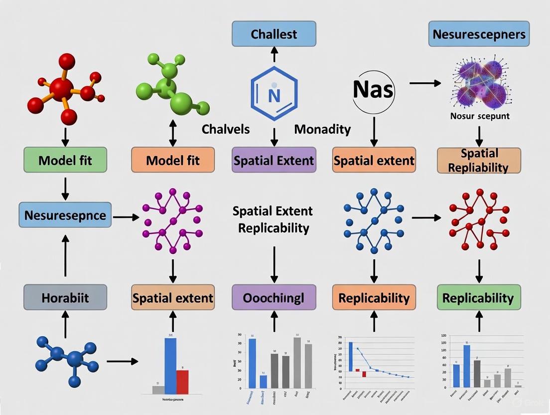
Workflow Visualization
Spatial Modeling Workflow: This diagram outlines the key steps in assessing spatial extent impacts on model accuracy, highlighting critical phases where spatial considerations must be addressed.
Data Challenges & Solutions: This diagram illustrates common spatial data challenges and their corresponding solutions, showing the relationship between problems and mitigation strategies.
Research Reagent Solutions
| Research Tool | Function | Application Notes |
|---|---|---|
| GBIF Data | Global biodiversity occurrence records [1] | Primary source for species occurrence points; requires quality filtering |
| Environmental Predictors | Bioclimatic and topographic variables [1] | Should represent relevant ecological gradients; resolution should match study extent |
| Ecological Niche Modeling Algorithms | MaxEnt, Random Forest, etc. [1] | Choice affects model transferability; multiple algorithms should be compared |
| Spatial Cross-Validation | Account for spatial autocorrelation [2] | Essential for realistic error estimation; use spatial blocking instead of random splits |
| Uncertainty Estimation Methods | Quantify prediction reliability [2] | Critical for model interpretation and application; should be reported for all predictions |
Understanding Spatial Autocorrelation and its Effect on Model Generalization
FAQ: Core Concepts
What is Spatial Autocorrelation? Spatial autocorrelation describes how the value of a variable at one location is similar to the values of the same variable at nearby locations. It is a mathematical expression of Tobler's First Law of Geography: "everything is related to everything else, but nearby things are more related than distant things" [3] [4]. Positive spatial autocorrelation occurs when similar values cluster together in space, while negative spatial autocorrelation occurs when dissimilar values are near each other [3] [4].
Why should researchers be concerned about its effect on model generalization? Spatial autocorrelation (SAC) violates the fundamental statistical assumption of independence among observations [3] [5]. When unaccounted for, it can lead to over-optimistic estimates of model performance, inappropriate model selection, and poor predictive power when the model is applied to new, independent locations [6]. This compromises the replicability of findings across different spatial extents [7] [6].
How does spatial autocorrelation lead to an inflated perception of model performance? In cross-validation, if the training and testing sets are spatially dependent, the model appears to perform well because it is essentially being tested on data that is similar to what it was trained on. One study on transfer functions demonstrated that when a spatially independent test set was used, the true root mean square error of prediction (RMSEP) was approximately double the previously published, over-optimistic estimates [6]. This inflation occurs because the model internalizes the spatial structure rather than learning the true underlying relationship [6].
Troubleshooting Guide: Diagnosing and Mitigating SAC
Problem: Model performs well in cross-validation but fails on new spatial data.
This is a classic symptom of a model that has overfit to the spatial structure of the training data rather than learning the generalizable process of interest [6].
Diagnostic Steps:
Quantify SAC in your response variable and model residuals.
- Action: Calculate Global Moran's I for your original data and for the residuals of your fitted model.
- Interpretation: A significant Moran's I value in the residuals indicates that your model has not adequately captured the spatial pattern, leaving behind structured noise that harms generalization [5] [6]. The presence of SAC in the response variable alone does not necessarily indicate a problem, but its presence in the residuals does [5].
- Tools: Use the
Spatial Autocorrelation (Global Moran's I)tool in ArcGIS [8] or themoran.test()function in R with thespdeppackage [4].
Assess the scale of autocorrelation.
- Action: Create a spatial correlogram or a smoothed distance scatter plot to visualize how autocorrelation changes with distance [9] [5].
- Interpretation: This helps you understand the distance range over which observations influence each other, which is critical for designing appropriate mitigation strategies [5].
Test model scalability with a spatially independent hold-out set.
- Action: Instead of a random train-test split, hold out entire geographic regions (e.g., all data from a specific fire, watershed, or administrative district) for validation [7] [6].
- Interpretation: This provides a more realistic estimate of how your model will perform in a true generalization context. A study on wildfire prediction found this method crucial for evaluating real-world applicability [7].
Mitigation Strategies:
Increase Sample Spacing:
- Protocol: Systematically increase the distance between your training data points. This reduces the redundancy caused by SAC and provides a more robust test of the model's ability to capture the underlying process [7].
- Evidence: A wildfire prediction study showed that model accuracy declined with increased sample spacing, but the relationships identified by feature importance remained consistent, indicating the model was capturing real processes rather than just spatial noise [7].
Incorporate Spatial Structure Explicitly:
- Protocol: Use spatial regression models that directly model the dependency.
- Protocol: Use spatial filtering methods like Moran Eigenvector Spatial Filtering to create synthetic covariates that capture the spatial structure, which can then be included in a standard linear model [3].
Account for Spatial Heterogeneity:
- Protocol: Apply Geographically Weighted Regression (GWR). This technique allows the relationships between predictors and the response variable to vary across the study area, accounting for spatial non-stationarity [3].
Problem: Computational failure when calculating spatial autocorrelation for large datasets.
Large datasets can cause memory errors during spatial weights matrix creation, especially if the distance band results in features having tens of thousands of neighbors [10].
Solutions:
- Reduce the number of neighbors: Lower the
Distance Band or Threshold Distanceparameter so that no feature has an excessively large number of neighbors (e.g., aim for a maximum of a few hundred, not thousands) [10]. - Remove spatial outliers: A few distant features can force a very large default distance band. Create a selection set that excludes these outliers for the initial analysis [10].
- Use a spatial weights matrix file: For very large analyses (e.g., >5,000 features), pre-calculating a spatial weights matrix file (
.swm) can be more memory-efficient than using an ASCII file [8] [10].
Experimental Protocols for Assessing SAC's Impact
Protocol 1: Evaluating Model Scalability Across Spatial Regions
- Objective: To test whether a model trained in one region can generalize to a different, spatially independent region.
- Methodology:
- Partition your dataset into two or more distinct geographic regions (e.g., Region A and Region B).
- Train your model exclusively on data from Region A.
- Use the trained model to predict outcomes in Region B.
- Compare the performance metrics (e.g., R², RMSE) from the Region B prediction with the cross-validation performance from Region A.
- Interpretation: A significant drop in performance when predicting in Region B indicates that the model may have overfit to the local spatial context of Region A and lacks generalizability [7] [6].
Protocol 2: Incremental Sample Spacing Test
- Objective: To determine if model performance is driven by fine-scale spatial autocorrelation or by the underlying ecological/process-based relationships.
- Methodology:
- Start with your full, finely-spaced dataset and calculate a baseline model performance.
- Systematically thin your dataset by increasing the minimum allowable distance between any two sample points.
- Re-train and re-evaluate the model (using a consistent method like cross-validation) at each level of sample spacing.
- Monitor changes in overall performance and in the feature importance rankings.
- Interpretation: If performance declines sharply with increased spacing, the model is highly dependent on fine-scale SAC. If feature importance remains stable even as performance declines modestly, it suggests the model is capturing meaningful processes [7].
The Researcher's Toolkit: Key Reagents & Solutions
Table 1: Essential Tools for Spatial Autocorrelation Analysis
| Item Name | Function / Purpose | Key Considerations |
|---|---|---|
| Global Moran's I | A global metric to test for the presence and sign (positive/negative) of spatial autocorrelation across the entire dataset [3] [4]. | Values range from -1 to 1. Significance is tested via z-score/ p-value or permutation [8] [9]. |
| Spatial Weights Matrix (W) | Defines the neighborhood relationships between spatial units, which is fundamental to all SAC calculations [3] [4]. | Can be contiguity-based (e.g., Queen, Rook) or distance-based. The choice of conceptualization critically impacts results [3] [8]. |
| LISA (Local Indicators of Spatial Association) | A local statistic (e.g., Local Moran's I) to identify specific clusters of high or low values and spatial outliers [3] [9]. | Helps pinpoint where significant spatial clustering is occurring, decomposing the global pattern [3]. |
| Spatial Correlogram | A graph plotting autocorrelation (e.g., Moran's I) against increasing distance intervals [9] [5]. | Reveals the scale or range at which spatial dependence operates, informing an appropriate distance threshold [5]. |
| Spatial Regression Models (SAR, CAR) | Statistical models that incorporate spatial dependence directly into the regression framework, either in the dependent variable (SAR) or the errors (CAR) [3]. | Corrects for the bias in parameter estimates and standard errors that arises from ignoring SAC [3] [5]. |
Workflow Diagram for Diagnosis and Mitigation
The following diagram outlines a logical workflow for diagnosing issues related to spatial autocorrelation and selecting an appropriate mitigation strategy.
SAC Diagnosis and Mitigation Workflow
FAQs: Spatial Extent and Model Replicability
Q1: What is the core problem with using a user-defined Area of Interest (AOI) as the spatial extent for all model inputs?
A1: The core problem is a fundamental mismatch between user-defined boundaries and the natural processes being modeled. Spatial processes are not bounded by user-assigned areas [11]. Using the AOI for all inputs ignores the spatial context required for accurate modeling. A classic example is extracting a stream network: using a Digital Elevation Model (DEM) clipped only to the AOI, rather than the entire upstream catchment, will produce incorrect or incomplete results because it ignores the contributing area from upstream [11]. This introduces cascading errors, especially in workflows chaining multiple models.
Q2: How can improper spatial extents impact the replicability of my research findings?
A2: Improper spatial extents directly undermine replicability—the ability to obtain similar results using similar data and methods in a different spatial context [12]. This occurs due to spatial heterogeneity, where the expected value of a variable and the performance of models vary across the Earth's surface [12]. If a model's spatial extent does not properly account for this heterogeneity, findings become place-specific and cannot be reliably reproduced in other study areas, limiting their scientific and practical value.
Q3: What are the common symptoms of a cascading error caused by an improper spatial extent?
A3: You may encounter one or more of the following issues:
- Incomplete Results: The model runs but produces a physically implausible or truncated output, such as a river network that ends abruptly within the AOI [11].
- Biased Predictions: The model appears to work but shows systematically poor performance in certain areas because key driving variables are missing their full contextual scope [13].
- Poor Generalization: A model that is highly accurate in one region performs poorly when applied to a new region, even if the environmental conditions seem similar, due to unaccounted spatial heterogeneity [12] [13].
- Irreplicable Findings: Results from one study area cannot be reproduced in another, leading to a "replicability crisis" for spatial studies [12].
Q4: Beyond the DEM, what other data types commonly require a spatial extent different from the AOI?
A4:
- Meteorological Data: For spatial interpolation of variables like temperature, the extent must include not just stations within the AOI, but also nearby stations outside it to create a reliable interpolation field [11].
- Species Distribution Data: Modeling habitat suitability requires data from a region large enough to capture the full range of environmental conditions the species tolerates [13].
- Data for Distance Calculations: Inputs for deriving variables like "distance-to-river" require the source data (e.g., for river network extraction) to cover the entire upstream watershed that hydrologically influences the AOI [11].
Troubleshooting Guide: Resolving Spatial Extent Errors
Problem: My geographical model workflow executes without crashing, but the output is physically implausible or cannot be replicated in a different study area.
| Step | Action | Key Questions to Ask | Expected Outcome |
|---|---|---|---|
| 1. Diagnosis | Identify the specific model in your workflow producing suspicious output. Trace its inputs back to their source data. | Is the output incomplete (e.g., rivers don't flow)? Does it ignore clear edge-influences? | Pinpoint the model and data layer where the error first manifests. [11] |
| 2. Input Analysis | For the identified model input, determine its required spatial context. | Does this input represent a process that extends beyond the AOI (e.g., water flow, species dispersal, atmospheric transport)? | A formalized rule defining the necessary spatial extent for the specific input. [11] |
| 3. Workflow Correction | Apply knowledge rules to automatically adjust the input's spatial extent during workflow preparation. | Should the extent be a watershed, a buffer zone, a minimum bounding polygon, or a different ecological region? | An execution-ready workflow where inputs are automatically fetched at their correct, process-based extents. [11] |
| 4. Validation | Use a resampling and spatial smoothing framework (e.g., MESS) to test the sensitivity of your results to zoning and scale. | How consistent are my results if the analysis grain or boundary placement changes slightly? | A robust understanding of how spatial context affects your findings, improving the interpretability and potential replicability of your results. [14] |
Detailed Experimental Protocol for Spatial Validation
The following protocol, based on the Macro-Ecological Spatial Smoothing (MESS) framework, helps diagnose and overcome the Modifiable Areal Unit Problem (MAUP), a core challenge for replicability [14].
Objective: To standardize the analysis of spatial data from different sources or regions to facilitate valid comparison and synthesis, thereby assessing the replicability of patterns.
Methodology:
- Define the Spatial Grain (
s): Select the size for the sampling regions (moving windows) that will slide across your landscape. - Set Sampling Parameters:
ss: Specify the sample size (number of local sites) to be randomly drawn within each window.n: Specify the number of random subsamples to be drawn with replacement in each window.mn: Set the minimum number of local sites a window must contain to be included.
- Sliding Window Execution: Slide the moving window of size
sacross the entire landscape. - Resampling and Metric Calculation: For each window location that meets the minimum site requirement (
mn):- Draw
nrandom subsamples of sizess. - For each subsample, calculate your metrics of interest (e.g., β-diversity, γ-richness, mean α-richness, environmental heterogeneity).
- Retain the average value of these metrics across the
nsubsamples for that window.
- Draw
- Synthesis and Mapping: The averaged metric values for each window create a continuous surface of spatial pattern, sidestepping arbitrary zonation. This allows for direct, standardized comparison between different studies or regions [14].
The Spatial Workflow Logic: Problem vs. Solution
The following diagram illustrates the critical difference between a flawed, common approach and a robust, knowledge-driven methodology for handling spatial extents.
The Scientist's Toolkit: Key Research Reagents & Solutions
| Item / Solution | Function in Addressing Spatial Extent & Replicability |
|---|---|
| Knowledge Rule Base | A systematic formalization of how the spatial extent for a model input should be determined based on its semantic relation to the output and its data type. This is the core of intelligent spatial workflow systems [11]. |
| Macro-Ecological Spatial Smoothing (MESS) | A flexible R-based framework that uses a moving window and resampling to standardize datasets, allowing for inferential comparisons across landscapes and mitigating the Modifiable Areal Unit Problem (MAUP) [14]. |
| Place-Based (Idiographic) Analysis | An analytical approach focused on the distinct nature of places. It acknowledges spatial heterogeneity and is used when searching for universal, replicable laws (nomothetic science) is confounded by local context [12]. |
| Convolutional Neural Networks (CNNs) | A class of deep learning algorithms particularly adept at learning from spatial data. They can inherently capture spatial patterns and contexts, but their application must still consciously account for spatial heterogeneity to ensure replicability [12]. |
| Spatial Cross-Validation | A validation technique where data is split based on spatial location or clusters (rather than randomly). It is crucial for obtaining realistic performance estimates and testing a model's ability to generalize to new, unseen locations [13]. |
| Cloud-Based Data Platform (e.g., S3) | Provides the necessary processing capabilities and scalability to handle large geospatial file sizes and the computational demands of resampling, smoothing, and running complex spatial models [15]. |
| Uncertainty Estimation Metrics | Tools and techniques to quantify the certainty of model predictions. This is especially important when a model is applied in areas where the input data distribution differs from the training data (out-of-distribution problem) [13]. |
Frequently Asked Questions (FAQs)
FAQ 1: Why does my spatial model perform well in one geographic area but fails in another, even for the same phenomenon?
This is a classic sign of the replicability challenge in spatial modeling, primarily driven by spatial heterogeneity. Spatial processes and the relationships between variables are not uniform across a landscape; they change from one location to another due to local environmental, social, or biological factors [16] [13]. A model trained in one region learns the specific relationships present in that data. When applied to a new area where these underlying relationships differ, the model's performance degrades because the fundamental rules it learned are no longer fully applicable.
FAQ 2: What is spatial autocorrelation, and how can it mislead my model's performance evaluation?
Spatial autocorrelation (SAC) is the concept that near things are more related than distant things, a principle often referred to as Tobler's First Law of Geography [17]. In modeling, SAC causes a violation of the common assumption that data points are independent. When training and test datasets are split randomly across a study area, they may not be truly independent if they are located near each other. This can lead to deceptively high predictive performance during validation because the model is effectively tested on data that is very similar to its training data, a problem known as spatial overfitting. Properly evaluating a model requires spatial validation techniques, such as splitting data by distinct spatial clusters or geographic regions, to ensure a realistic assessment of its performance on truly new, unseen locations [13] [16].
FAQ 3: My dataset has significant 'holes' or missing data in certain areas. How can I fill these gaps without biasing my results?
Filling missing data, or geoimputation, should be done with extreme caution. The best practice is to use the values of spatial neighbors, as guided by Tobler's Law [17]. However, this can introduce bias. Key considerations include:
- Amount of Missing Data: A common rule of thumb is to fill no more than 5% of the values in a dataset [17].
- Fill Method: The choice depends on your data and goal:
- Average of Neighbors: Use when values are expected to be similar to surroundings.
- Median of Neighbors: Preferred if outliers are suspected locally.
- Maximum of Neighbors: Use when you want to avoid underestimation (e.g., in public health risk mapping).
- Minimum of Neighbors: Use when you want a conservative estimate.
- Neighborhood Definition: You can define neighbors by a fixed distance, a fixed number of nearest features, or by contiguity (sharing a border) [17]. Always compare the data distribution (e.g., mean, standard deviation, histogram) before and after imputation to understand how your actions have altered the dataset.
FAQ 4: What is a "threshold parameter" in a spatial context, and why is it different from non-spatial models?
In classic non-spatial epidemic models, the basic reproductive number ( R_0 ) has a critical threshold of 1. However, in spatial models, this threshold is often higher. For example, in nearest-neighbour lattice models, the threshold value lies between 2 and 2.4 [18]. This is because spatial constraints and the local clustering of contacts change the dynamics of spread. An infected individual in a spatial model cannot contact all susceptible individuals in the population, only nearby ones, which reduces the efficiency of transmission and raises the transmissibility required for a large-scale outbreak [18].
Troubleshooting Guides
Problem: Model Fails to Generalize Across Geographic Regions
Symptoms:
- High accuracy in the original study area, poor accuracy in a new area.
- The spatial pattern of prediction errors is not random but clustered in the new region.
Investigation & Resolution Protocol:
Step 1: Diagnose Spatial Heterogeneity
- Action: Conduct an exploratory spatial data analysis (ESDA) for both the original and new study areas.
- Method: Calculate local statistics (e.g., Local Indicators of Spatial Association - LISA) to map local relationships between your dependent and independent variables. If the relationships are visually and statistically different between the two regions, spatial heterogeneity is confirmed as a primary cause [16] [13].
- Toolkit: Software with ESDA capabilities (e.g., GeoDa, ArcGIS Pro, R with
spdeppackage).
Step 2: Assess and Account for Spatial Autocorrelation
- Action: Validate your model using a spatially-aware method.
- Method: Instead of a random train-test split, use a spatial cross-validation or block cross-validation approach. This involves training and testing the model on distinct geographic clusters or tiles, ensuring the model is evaluated on spatially independent data [13].
- Toolkit: The
blockCVR package or custom scripting in Python withscikit-learn.
Step 3: Quantify Replicability
- Action: Generate a "replicability map" [16].
- Method:
- Train your model on the original region.
- Apply it to a moving window across the entire domain of interest (e.g., a continent).
- For each window, calculate a performance metric (e.g., accuracy, F1-score).
- Map this performance metric. The resulting replicability map visually reveals which geographic areas the model generalizes well to and where it fails, directly illustrating the challenge of user-defined bounds [16].
Problem: Unrealistic Model Predictions at the Edges of the Study Area
Symptoms:
- Sharp, unrealistic transitions in predicted values at the boundary of the area of interest.
- High prediction uncertainty for locations near the edge.
Investigation & Resolution Protocol:
Step 1: Check for Edge Effects
- Action: Confirm the problem is an edge effect.
- Method: Visually inspect prediction maps. High uncertainty or clearly artificial patterns that align with the study boundary are key indicators. This happens because the model lacks contextual information from beyond the user-defined area [13].
Step 2: Incorporate a Spatial Buffer
- Action: Expand the data collection area to create a buffer.
- Method: When defining your study area, include a generous buffer zone around your core area of interest. Gather predictor variable data for this larger area. This provides the model with the necessary spatial context to make more realistic predictions near the edges of your core area. The buffer should be large enough to capture the spatial process's scale of influence.
Step 3: Model with Spatial Context
- Action: Use modeling techniques that explicitly incorporate spatial context.
- Method: Implement models that use spatial lag variables or deep learning models that can process the spatial context around each pixel or polygon (e.g., Convolutional Neural Networks). This allows the model to "see" the neighborhood around a location, mitigating the edge effect.
Key Experimental Protocols
Protocol: Spatial Cross-Validation for Robust Model Evaluation
Objective: To accurately evaluate a spatial model's predictive performance and its potential to generalize to new, unseen locations.
Methodology:
- Define Spatial Blocks: Instead of randomly assigning data points to training and test sets, partition the study area into distinct spatial blocks or clusters. These can be regular tiles (e.g., quadrats) or irregular clusters based on k-means or spatial proximity [13].
- Iterative Training and Testing: Iteratively hold out one block as the test set and use all other blocks as the training set. This is repeated until each block has been used as the test set once.
- Aggregate Performance: Calculate the performance metric (e.g., R², RMSE, Accuracy) for each fold and then aggregate these (e.g., average) to get a final, spatially-robust performance estimate.
Interpretation: A model that performs well under spatial cross-validation is more likely to have captured the true underlying spatial process rather than just memorizing local spatial structure, giving greater confidence in its application to new areas.
Protocol: Conducting a Spatial Extension (Likelihood Ratio) Test
Objective: To statistically determine if an observed spatial pattern is better described by an extended source model (e.g., a spreading process) or a point-source model.
Methodology (as implemented in Fermipy software) [19]:
- Define Models: Fit two models to the data: a null model representing a point source, and an alternative model representing an extended source (e.g., a 2D Gaussian or Disk).
- Likelihood Scan: Perform a one-dimensional scan of the spatial extension parameter (e.g., width or radius of the source) over a predefined range.
- Calculate Test Statistic: For each value of the extension parameter, compute the log-likelihood of the data. The test statistic for spatial extension (( TS{ext} )) is derived from the difference in log-likelihoods between the best-fit extended model and the point-source model: ( TS{ext} = 2(\log L{ext} - \log L{ptsrc}) ) [19].
- Significance Testing: A large ( TS_{ext} ) value provides evidence against the point-source hypothesis, suggesting the process is spatially extended beyond a single point.
Data Presentation
Table 1: Impact of Spatial Validation on Model Performance Metrics
This table illustrates how using a naive random validation approach can severely overestimate model performance compared to a spatially robust method. The following data is synthesized from common findings in spatial literature [13] [16].
| Model Type | Study Area | Validation Method | Reported Accuracy (F1-Score) | Inference on Generalizability |
|---|---|---|---|---|
| Land Cover Classification | North Carolina, USA | Random Split | 0.92 | Overly Optimistic - Model performance is inflated due to spatial autocorrelation. |
| Land Cover Classification | North Carolina, USA | Spatial Block CV | 0.75 | Realistic - Better represents performance on truly new geographic areas. |
| Species Distribution Model | Amazon Basin | Random Split | 0.88 | Overly Optimistic - Fails to account for spatial heterogeneity in species-environment relationships. |
| Species Distribution Model | Amazon Basin | Spatial Cluster CV | 0.62 | Realistic - Highlights model's limitations when transferred across regions. |
Table 2: Guidelines for Selecting Geoimputation Methods for Missing Spatial Data
This table provides a structured approach to filling missing data based on the nature of the research question and data structure, following best practices [17].
| Research Context | Goal of Imputation | Recommended Fill Method | Recommended Neighborhood Definition | Rationale & Caution |
|---|---|---|---|---|
| Public Health Risk Mapping (e.g., lead poisoning) | Avoid underestimation of risk | Maximum of neighbor values | Contiguous neighbors (share a border) | Overestimation is safer than underestimation for public safety. Assumes similar risk factors in adjacent areas. |
| Cartography / Visualization | Create an aesthetically complete map | Average of neighbor values | Fixed number of nearest neighbors | Smooths data and fills "holes." Less concerned with statistical bias. |
| Environmental Sensing (e.g., soil moisture) | Avoid influence of local outliers | Median of neighbor values | Neighbors within a fixed distance | Robust to sensor errors or extreme local values. Distance should reflect process scale. |
| Socio-economic Analysis | Preserve local distribution | Average of neighbor values | Spatial and attribute-based neighbors | If data is Missing Not At Random (MNAR), all methods can introduce significant bias. |
The Scientist's Toolkit: Key Research Reagent Solutions
| Item / Concept | Function in Spatial Analysis |
|---|---|
| Spatial Cross-Validation | A validation technique that partitions data by location to provide a realistic estimate of a model's performance when applied to new geographic areas [13]. |
| Replicability Map | A visualization tool that maps the geographic performance of a model, highlighting regions where it generalizes well and where it fails, thus quantifying spatial replicability [16]. |
| Geoimputation | The process of filling in missing data values in a spatial dataset using the values from neighboring features in space or time, guided by Tobler's First Law of Geography [17]. |
| Spatial Autocorrelation (SAC) | A measure of the degree to which data points near each other in space have similar values. It is a fundamental property of spatial data that must be accounted for to avoid biased models [13]. |
| Spatial Heterogeneity | The non-stationarity of underlying processes and relationships across a landscape. It is a primary reason why models trained in one area may not work in another [16] [13]. |
| Gravity / Radiation Models | Mathematical models used to describe and predict human movement patterns between locations (e.g., between census tracts), which are crucial for building accurate spatial epidemic models [18]. |
| Threshold Parameters (R₀) | In spatial models, the critical value for the basic reproductive number is often greater than 1 (e.g., 2.0-2.4 for lattice models), reflecting the constrained nature of local contacts [18]. |
Frequently Asked Questions (FAQs) & Troubleshooting
Q1: Why does my spatial heterogeneity model fail to replicate when I change the spatial extent of the study area? A1: This is a common replicability challenge. The model's parameters might be over-fitted to the specific spatial scale of the initial experiment. To troubleshoot:
- Action: Re-run the core model on the original and new spatial extents, but only using a subset (e.g., 70%) of the data from each.
- Validation: Compare the model's performance on the withheld 30% of data for both extents. A significant performance drop on the new extent indicates over-fitting.
- Solution: Implement multi-scale spatial cross-validation to ensure model parameters are robust across different spatial scales [20].
Q2: How can I visually diagnose fitting errors in my spatial model's output?
A2: Use the Graphviz diagrams in the "Mandatory Visualization" section below. Compare your experimental workflow and results logic against the provided diagrams. Inconsistencies often reveal errors in data integration or result interpretation. The fixedsize attribute in Graphviz is crucial for preventing node overlap, which can misrepresent data relationships [21].
Q3: What is the most critical step in preparing single-cell RNA sequencing data for spatial heterogeneity analysis? A3: Ensuring batch effect correction and spatial normalization. Technical variations between sequencing batches can be misinterpreted as spatial heterogeneity.
- Protocol: Apply harmonization methods (e.g., Harmony, ComBat) before integrating data from multiple samples or regions. Validate by checking if known, ubiquitous cell markers show uniform expression across batches [20].
Q4: My Graphviz diagrams have poor readability. How can I improve the color contrast?
A4: Adhere to the WCAG (Web Content Accessibility Guidelines) for enhanced contrast. For text within nodes, explicitly set the fontcolor to contrast highly with the fillcolor [22]. Use the provided color palette and the following principles:
- For Dark Text: Use light backgrounds (e.g.,
fillcolor="#FBBC05",fontcolor="#202124"). - For Light Text: Use dark backgrounds (e.g.,
fillcolor="#4285F4",fontcolor="#FFFFFF"). - Avoid combinations like
#F1F3F4(light gray) fill with#FFFFFF(white) text, which has insufficient contrast [22] [23].
Mandatory Visualization
Diagram 1: Experimental Workflow for Spatial Heterogeneity Analysis
Diagram 2: Logic of Model Fit Replicability Challenges
Research Reagent Solutions
| Reagent / Material | Function in Spatial Heterogeneity Research |
|---|---|
| Single-Cell RNA-Seq Kits (e.g., 10x Genomics) | Enables profiling of gene expression at the individual cell level, fundamental for identifying cellular subpopulations within a tissue [20]. |
| Spatial Transcriptomics Slides (e.g., Visium) | Provides a grid-based system to capture and map gene expression data directly onto a tissue section, linking molecular data to spatial context. |
| Cell Type-Specific Antibodies | Used for immunohistochemistry (IHC) or immunofluorescence (IF) to validate the presence and location of specific cell types identified by computational models. |
| Spatial Cross-Validation Software (e.g., custom R/Python scripts) | Computational tool for rigorously testing model performance across different spatial partitions of the data, crucial for assessing replicability [20]. |
Experimental Protocols
Protocol 1: Multi-Scale Spatial Cross-Validation
- Objective: To evaluate the robustness of a spatial heterogeneity model across varying spatial extents and prevent over-fitting.
- Methodology:
- Define a set of spatial extents (e.g., 1x1 mm, 2x2 mm, 5x5 mm) from your tissue sample.
- For each spatial extent, partition the data into k-folds (e.g., k=5). Ensure folds are spatially contiguous to avoid data leakage.
- Iteratively train the model on k-1 folds and validate on the held-out spatial fold.
- Repeat the process for all defined spatial extents.
- Quantitative Measures: Calculate the mean and standard deviation of the model's performance metric (e.g., R², AUC) across all folds and extents. A low standard deviation indicates high replicability [20].
Protocol 2: Validation of Spatial Clusters via IHC
- Objective: To biologically validate computationally derived spatial clusters of cell types.
- Methodology:
- Based on single-cell and spatial transcriptomics data, computationally identify regions (clusters) enriched with a specific cell type.
- Select tissue sections corresponding to these computationally identified regions.
- Perform immunohistochemistry (IHC) staining using a validated antibody for a marker gene highly expressed in that cell cluster.
- Quantify the density of marker-positive cells in the predicted regions versus control regions.
- Quantitative Measures: Use a statistical test (e.g., t-test) to compare cell density between the computationally predicted cluster and a randomly selected area of the same size. A p-value < 0.05 confirms the cluster's biological validity.
Methodological Frameworks for Defining and Applying Spatially Replicable Models
Knowledge-Based and Modeling-Goal-Driven Approaches for Intelligent Spatial Extent Determination
Frequently Asked Questions (FAQs)
FAQ 1: Why can't I simply use my output Area of Interest (AOI) as the spatial extent for all my input data? Using the user-defined AOI for all inputs ignores that natural spatial processes extend beyond human-defined boundaries. For example, when extracting a stream network, the required Digital Elevation Model (DEM) must cover the entire upstream catchment area of the AOI; using only the AOI itself will ignore contributing upstream areas and produce incorrect or incomplete results due to the missing context of the spatial process [11].
FAQ 2: What is the core difference between execution-procedure-driven and modeling-goal-driven approaches?
- Execution-procedure-driven approaches require users to manually search for raw data and customize workflows based on the model's input requirements, relying heavily on user expertise and understanding of the complete modeling procedure [11].
- Modeling-goal-driven approaches automate the selection of suitable model input data and preparation methods, iteratively extending workflows starting from the user's modeling goal. This reduces reliance on the user's modeling expertise [11].
FAQ 3: What are the main types of knowledge used in modeling-goal-driven approaches for input preparation?
- Basic Input Data Descriptions: Formalizes basic data semantics (e.g., DEM, road networks) and data types (e.g., raster, polygon) to discover raw data and define input-output relationships [11].
- Application Context Knowledge: Incorporates knowledge about preparing proper input data based on specific application contexts, including factors like application purpose, study area characteristics, and data characteristics [11].
FAQ 4: How does improper spatial extent determination create problems in geographical model workflows? In workflows combining multiple models, an individual error in input data preparation can raise a chain effect, leading to cascading errors and ultimately incorrect final outputs. Each model often requires input data with different spatial extents due to distinct model and input characteristics [11].
Troubleshooting Guides
Problem: Inaccurate Model Results Due to Incorrect Spatial Extents
Symptoms:
- Model outputs appear truncated or contain unexpected boundary artifacts.
- Results for flow accumulation, watershed delineation, or other process-based models are illogical or incomplete.
- Model performance degrades when applied to different geographical areas.
Solutions:
- Implement Knowledge Rules: Formalize and apply knowledge rules that systematically consider the semantic relation between spatial extents of input and output and the data type of the corresponding input [11].
- Adopt a Modeling-Goal-Driven Workflow: Use an approach that automatically determines the proper spatial extent for each input during the geographical model workflow building process, adapting to the user-assigned AOI [11].
- Validate with Known Cases: Test your workflow in an area where the correct spatial extent and expected output are known to verify the automatic determination logic.
Problem: Chain Effects and Cascading Errors in Multi-Model Workflows
Symptoms:
- Small errors in early model stages amplify in subsequent models.
- Difficulty isolating the source of inaccuracy in a complex workflow.
- Inconsistent outputs when re-running workflows with slightly different AOIs.
Solutions:
- Intelligent Spatial Extent Propagation: For each iterative step in the heuristic modeling process, automatically determine the spatial extent for the input of the current data preparation model. This ensures each step in the chain receives correctly bounded data [11].
- Workflow Automation: Utilize a prototype system (like the Easy Geo-Computation system) that integrates this intelligent approach, deriving an execution-ready workflow with minimal execution redundancy [11].
Problem: Handling Arbitrarily-Shaped or Complex Areas of Interest
Symptoms:
- Models developed for simple, rectangular AOIs fail when applied to complex, irregular shapes.
- Uncertainty in how model-specific spatial requirements (e.g., upstream area, interpolation neighborhood) interact with a complex AOI.
Solutions:
- Case-Specific Analysis: A case study on digital soil mapping for an arbitrary-shaped AOI validated that the intelligent approach effectively determines proper spatial extents for complex shapes [11].
- Leverage Advanced Geoprocessing: The approach combines knowledge rules with advanced geoprocessing functions within a cloud-native architecture to handle arbitrary AOIs [11].
Experimental Protocols & Data
Protocol: Case Study for Validating Intelligent Spatial Extent Determination
Aim: To validate the effectiveness of the intelligent spatial extent determination approach for a Digital Soil Mapping (DSM) workflow within an arbitrarily defined rectangular AOI in Xuancheng, Anhui Province, China [11].
Methodology:
- Define Workflow: Establish a DSM workflow to predict Soil Organic Matter (SOM) content. This requires multiple environmental covariates (e.g., distance-to-river, meteorological data) derived from different source data (e.g., DEM, observatory stations) [11].
- Apply Knowledge Rules: Implement pre-defined knowledge rules to determine the correct spatial extent for each input:
- Compare Results: Execute the workflow using both the naive approach (using only the AOI extent for all inputs) and the intelligent approach (using automatically determined, correct extents).
- Validate Accuracy: Compare the outputs against ground-truthed soil data to assess the improvement in result accuracy and completeness.
Quantitative Validation Data
The table below summarizes key improvements offered by the intelligent approach, as demonstrated in the case study [11].
Table 1: Impact of Intelligent Spatial Extent Determination on Workflow Accuracy
| Aspect | Naive Approach (AOI as Input Extent) | Intelligent Spatial Extent Approach |
|---|---|---|
| DEM Extent for Hydrology | Limited to AOI boundary | Expanded to full upstream watershed |
| Spatial Interpolation Input | Limited to stations inside AOI | Includes stations near (outside) AOI |
| Output Stream Network | Incorrect/Incomplete (missing upstream contributors) | Hydrologically correct and complete |
| Final Model Output (SOM) | Spatially biased and inaccurate | Accurate and complete for the AOI |
| Workflow Robustness | Prone to chain-effect errors | Resilient; derived execution-ready workflow |
The Scientist's Toolkit: Research Reagent Solutions
Table 2: Essential Components for Implementing Intelligent Spatial Extent Determination
| Item | Function in the Workflow | Implementation Note |
|---|---|---|
| Knowledge Rule Base | Encodes the systematic knowledge on how a model's input spatial extent relates to its output extent and data type. | Core intelligence component; requires expert knowledge formalization [11]. |
| Heuristic Modeling Engine | Executes the modeling-goal-driven workflow, iteratively selecting models and preparing inputs using the knowledge rules. | Drives the automated workflow building process [11]. |
| Advanced Geoprocessing Tools | Performs the spatial operations (e.g., watershed delineation, buffer creation, interpolation) to generate the correctly bounded input data. | Microservices like those in the EGC system prototype (e.g., catchment area calculation) [11]. |
| Prototype System (e.g., EGC) | An integrated browser/server-based geographical modeling system that provides the platform for deploying and running the intelligent workflow. | Cloud-native architecture with Docker containerization allows for seamless scaling [11]. |
| Spatial Indexes (R-trees, Quad-trees) | Data structures used within geoprocessing tools to efficiently determine topological relationships (e.g., adjacency, containment) between spatial objects [24]. | Enables fast processing of spatial queries necessary for extent calculations. |
Workflow Diagrams
Diagram 1: Conceptual Framework for Spatial Extent Determination
Conceptual Framework for Spatial Extent Determination
Diagram 2: Detailed Troubleshooting Logic for Spatial Workflows
Troubleshooting Logic for Spatial Workflows
The quantification of spatial extent represents a significant advancement in the analysis of Tau-PET neuroimaging, moving beyond traditional dichotomous assessments to provide a more nuanced understanding of disease progression. The TAU-SPEX (Tau Spatial Extent) metric has emerged as a novel approach that aligns with visual interpretation frameworks while capturing valuable interindividual variability in tau pathology distribution. This methodology addresses critical limitations of standard quantification techniques by providing a more intuitive, spatially unconstrained measure of tau burden that demonstrates strong associations with neurofibrillary tangle pathology and cognitive decline [25] [26] [27].
Within the broader context of model fit spatial extent replicability challenges, TAU-SPEX offers valuable insights into overcoming barriers related to spatial heterogeneity, measurement standardization, and result interpretation. This technical support document provides comprehensive guidance for researchers and drug development professionals implementing spatial extent quantification in their neuroimaging workflows, with specific troubleshooting advice for addressing common experimental challenges.
Key Concepts and Terminology
Understanding Spatial Extent in Neuroimaging
Spatial extent in neuroimaging refers to the proportional volume or area exhibiting pathological signal beyond a defined threshold. Unlike intensity-based measures that average signal across predefined regions, spatial extent quantification captures the topographic distribution of pathology throughout the brain [26] [27]. This approach is particularly valuable for understanding disease progression patterns in neurodegenerative disorders like Alzheimer's disease, where the spatial propagation of tau pathology follows predictable trajectories that correlate with clinical symptoms.
The TAU-SPEX metric specifically quantifies the percentage of gray matter voxels with suprathreshold Tau-PET uptake using a threshold identical to that employed in visual reading protocols. This alignment with established visual interpretation frameworks facilitates clinical translation while providing continuous quantitative data for research and therapeutic development [25] [28].
The Replicability Challenge in Spatial Analysis
Spatial replicability refers to the consistency of research findings across different spatial contexts or locations. In geospatial AI and neuroimaging, this represents a significant challenge due to inherent spatial heterogeneity and autocorrelation in the data [16]. The concept of a "replicability map" has been proposed to quantify how location impacts the reproducibility and replicability of analytical models, emphasizing the need to account for spatial variability when interpreting results [16].
In Tau-PET imaging, replicability challenges manifest in multiple dimensions:
- Spatial heterogeneity: Tau pathology distribution varies significantly between individuals despite similar clinical presentations [26] [27]
- Methodological variability: Differences in preprocessing, thresholding, and regional definition can impact spatial extent measurements [26]
- Population differences: Cohort characteristics including disease stage, age, and genetic factors influence spatial patterns [29]
Experimental Protocols and Methodologies
TAU-SPEX Calculation Workflow
The TAU-SPEX methodology was developed using [18F]flortaucipir PET data from 1,645 participants across four cohorts (Amsterdam Dementia Cohort, BioFINDER-1, Eli Lilly studies, and Alzheimer's Disease Neuroimaging Initiative) [26] [27]. The protocol involves these critical steps:
PET Acquisition: All participants underwent Tau-PET using [18F]flortaucipir radiotracer with target acquisition during the 80-100 minute post-injection interval. Data were locally attenuation corrected and reconstructed into 4 × 5-minute frames according to scanner-specific protocols [26] [27].
Visual Reading: Tau-PET images were visually assessed according to FDA and EMA approved guidelines without knowledge of TAU-SPEX or SUVr values. Visual read was performed on 80-100 min non-intensity-normalized images for some cohorts and on SUVr images for others [26] [27].
Image Processing: A region-of-interest was manually delineated around the cerebellum gray matter for reference region extraction. Images were intensity-normalized to the cerebellum to generate SUVr maps [26].
Threshold Application: A standardized threshold identical to that used for visual reading was applied to binarize voxels as tau-positive or tau-negative [25] [26].
Spatial Extent Calculation: TAU-SPEX was computed as the percentage of gray matter voxels with suprathreshold Tau-PET uptake in a spatially unconstrained whole-brain mask [25] [28].
Figure 1: TAU-SPEX Calculation Workflow - This diagram illustrates the sequential steps for calculating the TAU-SPEX metric from raw PET data to final quantitative output.
Comparative Analysis Protocol
To validate TAU-SPEX against established methodologies, researchers performed comprehensive comparisons with traditional SUVr measures:
Whole-brain SUVr Calculation: Computed Tau-PET SUVr in a spatially unconstrained whole-brain region of interest.
Temporal Meta-ROI SUVr Calculation: Derived SUVr values from the commonly used temporal meta-ROI to align with established tau quantification methods [26] [27].
Performance Validation: Tested classification performance for distinguishing tau-negative from tau-positive participants, concordance with neurofibrillary tangle pathology at autopsy (n=18), and associations with concurrent and longitudinal cognition [25] [26].
Statistical Comparison: Compared receiver operating characteristic curves, accuracy metrics, and effect sizes between TAU-SPEX and SUVr measures across all analyses [25] [27].
Performance Data and Comparative Analysis
Quantitative Performance Metrics
TAU-SPEX has demonstrated superior performance compared to traditional SUVr measures across multiple validation frameworks. The following table summarizes key performance metrics established through validation studies:
Table 1: TAU-SPEX Performance Metrics Compared to Traditional SUVr Measures
| Performance Metric | TAU-SPEX | Whole-Brain SUVr | Temporal Meta-ROI SUVr |
|---|---|---|---|
| AUC for Visual Read Classification | 0.97 | Lower than TAU-SPEX (p<0.001) | Lower than TAU-SPEX (p<0.001) |
| Sensitivity for Braak-V/VI Pathology | 87.5% | Not specified | Not specified |
| Specificity for Braak-V/VI Pathology | 100.0% | Not specified | Not specified |
| Association with Concurrent Cognition (β) | -0.36 [-0.29, -0.43] | Weaker association | Weaker association |
| Association with Longitudinal Cognition (β) | -0.19 [-0.15, -0.22] | Weaker association | Weaker association |
| Accuracy for Tau-Positive Identification | >0.90 | Lower accuracy | Lower accuracy |
| Positive Predictive Value | >0.90 | Lower PPV | Lower PPV |
| Negative Predictive Value | >0.90 | Lower NPV | Lower NPV |
Association with Pathological and Clinical Outcomes
Beyond technical performance, TAU-SPEX shows strong associations with clinically relevant outcomes:
Table 2: TAU-SPEX Associations with Pathological and Clinical Outcomes
| Outcome Measure | TAU-SPEX Association | Clinical Implications |
|---|---|---|
| NFT Braak-V/VI Pathology at Autopsy | High sensitivity (87.5%) and specificity (100%) | Strong pathological validation for identifying advanced tau pathology |
| Concurrent Global Cognition | β = -0.36 [-0.29, -0.43], p < 0.001 | Moderate association with current cognitive status |
| Longitudinal Cognitive Decline | β = -0.19 [-0.15, -0.22], p < 0.001 | Predictive of future cognitive deterioration |
| Tau-PET Visual Read Status | AUC: 0.97 for distinguishing tau-negative from tau-positive | Excellent concordance with clinical standard |
| Spatial Distribution Patterns | Captures heterogeneity among visually tau-positive cases | Provides information beyond dichotomous classification |
Research Reagent Solutions
Implementing robust spatial extent quantification requires specific methodological components and analytical tools. The following table details essential research reagents and their functions in the TAU-SPEX framework:
Table 3: Essential Research Reagents and Methodological Components for Spatial Extent Quantification
| Reagent/Component | Function | Implementation Notes |
|---|---|---|
| [18F]flortaucipir radiotracer | Binds to tau neurofibrillary tangles for PET visualization | FDA and EMA approved for clinical visual reading; used with 80-100 min acquisition protocol |
| Cerebellum Gray Matter Reference Region | Reference region for intensity normalization | Manually delineated ROI; used for generating SUVr maps and threshold determination |
| Whole-Brain Gray Matter Mask | Spatially unconstrained mask for voxel inclusion | Enables calculation without a priori regional constraints |
| Visual Reading Threshold | Binarization threshold for tau-positive voxels | Identical threshold used for clinical visual reading; ensures alignment with clinical standard |
| Spatial Frequency Maps | Visualization of spatial patterns across populations | Generated using BrainNet with "Jet" colorscale; shows voxel-wise percentage of suprathreshold participants |
| Automated Spatial Extent Pipeline | Calculation of TAU-SPEX metric | Custom pipeline calculating percentage of suprathreshold gray matter voxels |
Troubleshooting Guides
Common Implementation Challenges
Challenge: Inconsistent Threshold Application Problem: Variable spatial extent measurements due to inconsistent threshold application across scans or researchers. Solution:
- Implement automated thresholding aligned with visual reading standards
- Establish quality control procedures with expert review of threshold application
- Use standardized reference region delineation protocols [26] [27]
Challenge: High Variance in Low Pathology Cases Problem: Elevated spatial extent measures in amyloid-negative cognitively unimpaired participants, potentially reflecting off-target binding. Solution:
- Combine spatial extent with intensity measures to distinguish specific from non-specific binding
- Establish cohort-specific reference ranges for different populations
- Implement quantitative filters for excluding likely off-target regions [26] [27]
Challenge: Spatial Heterogeneity Affecting Replicability Problem: Inconsistent findings across studies due to spatial heterogeneity in tau distribution patterns. Solution:
- Develop replicability maps to quantify spatial generalizability
- Implement spatial autocorrelation analysis in validation frameworks
- Use multi-cohort designs to account for population-specific spatial patterns [16]
Data Quality and Processing Issues
Challenge: Incomplete Brain Coverage Problem: Missing data in certain brain regions affecting spatial extent calculations. Solution:
- Implement quality checks for complete Montreal Neurological Institute (MNI) space coverage
- Develop protocols for handling truncation artifacts
- Establish exclusion criteria for scans with significant coverage gaps [26]
Challenge: Inter-scanner Variability Problem: Differences in PET scanner characteristics affecting quantitative values. Solution:
- Implement scanner-specific normalization procedures
- Use phantom scans to calibrate across devices
- Include scanner as a covariate in statistical models [26] [27]
Frequently Asked Questions
Q: How does TAU-SPEX address limitations of traditional SUVr measures? A: TAU-SPEX overcomes several key SUVr limitations: (1) it is not constrained to predefined regions of interest, capturing pathology throughout the brain; (2) it utilizes binary voxel classification, reducing sensitivity to subthreshold noise; (3) it provides more intuitive interpretation with a well-defined 0-100% range; and (4) it better captures heterogeneity among visually tau-positive cases where focal high-intensity uptake may yield similar SUVr values as widespread moderate-intensity uptake [26] [27].
Q: What are the computational requirements for implementing TAU-SPEX? A: The TAU-SPEX methodology requires standard neuroimaging processing capabilities including: (1) PET image normalization and registration tools; (2) whole-brain gray matter segmentation; (3) voxel-wise thresholding algorithms; and (4) basic volumetric calculation capabilities. The method can be implemented within existing PET processing pipelines without specialized hardware requirements [26] [27].
Q: How does TAU-SPEX perform across different disease stages? A: TAU-SPEX demonstrates strong performance across the disease spectrum. It effectively distinguishes tau-negative from tau-positive cases (AUC: 0.97) while also capturing variance among visually positive cases that correlates with cognitive performance. The metric shows moderate associations with both concurrent (β=-0.36) and longitudinal (β=-0.19) cognition, suggesting utility across disease stages [25] [26].
Q: What steps ensure replicability of spatial extent findings? A: Ensuring replicability requires: (1) standardized threshold application aligned with visual reading; (2) multi-cohort validation to account for population differences; (3) clear documentation of preprocessing and analysis parameters; (4) accounting for spatial autocorrelation in statistical models; and (5) generation of replicability maps to quantify spatial generalizability [26] [16].
Q: How can spatial extent measures be integrated into clinical trials? A: Spatial extent quantification can enhance clinical trials by: (1) providing continuous outcome measures beyond dichotomous classification; (2) detecting subtle treatment effects on disease propagation; (3) offering more intuitive interpretation for clinical audiences; and (4) capturing treatment effects on disease topography that might be missed by regional SUVr measures. A recent survey found 63.5% of experts believe quantitative metrics should be combined with visual reads in trials [26] [27].
Advanced Spatial Replicability Framework
The implementation of spatial extent quantification must address fundamental replicability challenges inherent in spatial data analysis. The following diagram illustrates the key considerations for ensuring robust and replicable spatial extent measurements:
Figure 2: Spatial Replicability Framework - This diagram illustrates the major challenges in spatial replicability (red) and corresponding methodological solutions (green) for robust spatial extent measurements.
Implementing Replicability Maps
Replicability maps represent a novel approach to quantifying the spatial generalizability of findings. These maps incorporate spatial autocorrelation and heterogeneity to visualize regions where results are most likely to replicate across different populations or studies [16]. Implementation involves:
- Spatial Autocorrelation Analysis: Quantifying how similarity in tau pathology varies with spatial proximity
- Cross-Validation Framework: Implementing spatial cross-validation that accounts for geographic structure
- Uncertainty Quantification: Mapping regional confidence intervals for spatial extent measures
- Generalizability Index: Developing quantitative scores for spatial replicability across brain regions
This approach directly addresses the "replicability crisis" in spatial analysis by explicitly acknowledging and modeling spatial heterogeneity rather than assuming uniform effects throughout the brain [16] [2].
Leveraging AI and Machine Learning for Spatial Pattern Detection and Prediction
Troubleshooting Guide: Common Issues in Spatial AI Experiments
My model performs well on synthetic data but fails on real-world data. Why?
This is a classic problem of model generalization. A model trained on synthetic examples is only useful if it can effectively transfer to real-world test cases. This challenge is particularly pronounced in high-dimensional phase transitions, where dynamics are more complex than in simple bifurcation transitions [31].
- Root Cause: The synthetic data likely lacks the full complexity, noise, and heterogeneity of real-world spatial data. Your model may have overfitted to the idealized patterns of the training data.
- Diagnosis Steps:
- Compare key statistical properties (e.g., spatial autocorrelation, point distribution) between your synthetic and real-world datasets.
- Use a small, held-out set of real-world data for validation during training to monitor generalization performance.
- Solution:
- Data Augmentation: Introduce realistic noise, transformations, and variations into your synthetic training data.
- Transfer Learning: Start with a model pre-trained on a broader set of spatial data, then fine-tune it on your specific synthetic dataset. This can help the model learn more generalizable features.
- Domain Adaptation: Employ techniques that explicitly minimize the discrepancy between the feature distributions of your synthetic (source) and real-world (target) data.
How can I diagnose overfitting or underfitting in my spatial model?
Evaluating model performance correctly is essential before diving into the root cause [32].
- Symptoms of Overfitting: High accuracy on training data but poor performance on validation/test data. The model learns the noise and specific details of the training set instead of the underlying spatial patterns.
- Symptoms of Underfitting: Poor performance on both training and validation data, indicating the model is too simple to capture the relevant spatial relationships.
| Metric | Overfitting Indicator | Underfitting Indicator |
|---|---|---|
| Loss Function | Training loss << validation loss | Training loss ≈ validation loss, both high |
| Accuracy/Precision | Training >> validation | Both low |
| Feature Importance | High importance on nonsensical features | No clear important features identified |
- Fixes for Overfitting:
- Increase training data or use data augmentation.
- Apply stronger regularization (e.g., L1/L2, Dropout).
- Reduce model complexity (e.g., fewer layers/nodes in a neural network).
- For Random Forests, reduce
max_depthor increasemin_samples_leaf.
- Fixes for Underfitting:
My model's predictions lack spatial coherence. How can I improve them?
Standard models sometimes fail to capture the intrinsic spatial relationships in the data.
- Root Cause: The model architecture or algorithm may not be designed to account for spatial context and neighborhood dependencies.
- Solution:
- Choose spatially-aware algorithms: Algorithms like Random Forest can inherently assess complex, non-linear feature relationships, making them versatile for spatial data. K-Nearest Neighbors (K-NN) uses proximity to classify or predict values for spatial data points [33].
- Incorporate spatial features: Explicitly include spatial covariates like coordinates, distance to key features, or spatial lag variables in your model.
- Use advanced architectures: For more complex tasks, consider Spatio-Temporal Graph Neural Networks, which are state-of-the-art for analyzing dynamic spatial-temporal patterns by learning from spatial relationships [33]. Convolutional Neural Networks (CNNs) can also learn spatial hierarchies from raster data.
Frequently Asked Questions (FAQs)
What are the top machine learning algorithms for spatial pattern detection in 2025?
The choice of algorithm should be guided by your project objectives, data complexity, and required interpretability [33]. The following algorithms are recognized for their versatility and efficiency with complex geospatial datasets [33].
| Algorithm | Key Strengths | Ideal Spatial Applications |
|---|---|---|
| Random Forest | Robust to noise & outliers; Handles non-linear relationships; Provides feature importance [33]. | Land cover classification; Environmental monitoring; Soil and crop analysis [33]. |
| K-Nearest Neighbors (K-NN) | Simple to implement and interpret; No training phase; Versatile for classification & regression [33]. | Land use classification; Urban planning (finding similar areas); Environmental parameter prediction [33]. |
| Gaussian Processes | Provides uncertainty quantification; Models complex, non-linear relationships [33]. | Spatial interpolation; Resource estimation; Environmental forecasting. |
| Spatio-Temporal Graph Neural Networks | Captures dynamic spatial-temporal patterns; State-of-the-art for complex relational data [33]. | Traffic flow forecasting; Climate anomaly detection; Urban growth modeling [33]. |
How can I ensure my spatial AI model is reproducible?
Reproducibility is a cornerstone of scientific research, especially when addressing model fit spatial extent replicability challenges.
- Document Everything: Meticulously document data preprocessing steps, model architectures, hyperparameters, and random seeds used.
- Version Control: Use version control systems (like Git) for both your code and datasets.
- Adhere to FAIR Principles: Ensure your data and models are Findable, Accessible, Interoperable, and Reusable. This framework is central to initiatives like the Spatial AI Challenge and ensures your work is a valuable resource for the community [34].
- Use Computationally Reproducible Environments: Package your analysis in reproducible containers (e.g., Docker) or use platforms that support executable research compendiums, like Jupyter notebooks, which are used in competitions like the Spatial AI Challenge [34].
What should I do if my model fails to detect subtle, "hidden" patterns in point cloud data?
This is a problem of high-dimensional pattern recognition. Point clouds contain millions of data points with multiple attributes (X, Y, Z, intensity, etc.), creating a complex matrix beyond human visual perception [35].
- Leverage Advanced Deep Learning: Use architectures designed for point clouds (e.g., PointNet++, Dynamic Graph CNNs) that process data hierarchically. These models build understanding from local geometric features (e.g., surface normals, curvature) up to scene-level semantic relationships, detecting subtle statistical signatures of issues like material degradation or early plant disease [35].
- Focus on Feature Engineering: Instead of raw points, calculate derived mathematical properties such as:
- Standard deviation of point heights within a local area (roughness).
- Local planarity coefficients.
- Variations in surface normal vectors.
- Spatial autocorrelation patterns.
- Incorporate Contextual Rules: Train your model to understand spatial relationships (e.g., power lines maintain consistent elevation over terrain). Anomalies that violate these learned rules are often the subtle patterns of interest [35].
The Scientist's Toolkit: Key Research Reagent Solutions
This table details essential "reagents" – algorithms, data principles, and tools – for a spatial AI research lab.
| Item | Function / Explanation |
|---|---|
| Random Forest Algorithm | A versatile "workhorse" for both classification and regression on spatial data, robust to noise and capable of identifying important spatial features [33]. |
| Spatio-Temporal Graph Neural Network | The state-of-the-art "specialist" for modeling dynamic processes that evolve over space and time, such as traffic or disease spread [33]. |
| FAIR Data Principles | The "protocol" for ensuring your spatial data is Findable, Accessible, Interoperable, and Reusable, which is critical for replicable research [34]. |
| I-GUIDE Platform | An advanced "cyber-infrastructure" that provides the computational environment and tools for developing and testing reproducible spatial AI models [34]. |
| Jupyter Notebooks | The "lab notebook" of modern computational research, enabling the packaging of code, data, and visualizations into a single, executable, and reproducible document [34]. |
| Point Cloud Deep Learning Library (e.g., PointNet++) | A specialized "sensor" for extracting hidden patterns from high-dimensional LiDAR and 3D scan data by learning local and global geometric features [35]. |
Experimental Protocols for Spatial Pattern Detection
Protocol 1: Detecting Critical Transitions in Spatially Patterned Ecosystems
This protocol is based on methodologies explored in research using neural networks as Early Warning Signals (EWS) for phase transitions in systems like dryland vegetation [31].
Workflow Overview
Detailed Methodology:
- Data Preparation: Compile a spatiotemporal dataset (e.g., remote sensing imagery over time). Generate synthetic training data from a mechanistic model (e.g., a partial differential equation for vegetation dynamics) and reserve a portion for testing [31].
- Model Training: Train a deep neural network (e.g., Convolutional LSTM) on the synthetic data. The input is a sequence of spatial patterns, and the output is the probability of an upcoming critical transition.
- Model Validation & Interpretation:
- Performance Benchmarking: Compare the neural network's prediction accuracy against traditional EWS indicators (e.g., spatial variance, lag-1 autocorrelation).
- Generalization Test: Apply the trained model to your real-world system of interest. Crucially, evaluate its performance when training and test data sources are interchanged to probe generalization capabilities [31].
- Insight Generation: Use techniques like saliency maps to understand which spatial regions and temporal patterns the model found most predictive.
Protocol 2: Identifying Hidden Patterns in LiDAR Point Cloud Data for Infrastructure Monitoring
This protocol outlines how to use AI to detect subtle, pre-failure geometric deviations in infrastructure like bridges, as discussed in applied AI research [35].
Workflow Overview
Detailed Methodology:
- Feature Extraction: For each point in the cloud, calculate a set of local geometric descriptors. This includes:
- Surface Roughness: The standard deviation of point heights within a local neighborhood.
- Local Planarity: How well the points in a neighborhood fit a flat plane.
- Surface Normal Variation: The direction and consistency of normal vectors.
- Model Training for Contextual Understanding: Train a model (e.g., a Spatio-Temporal GNN) on point clouds from known healthy infrastructure. The model learns hierarchical features:
- Local Level: Basic geometric shapes.
- Object Level: Components like bridge decks, cables, and pillars.
- Scene Level: Normal spatial relationships between objects (e.g., cables are taut, decks are flat) [35].
- Anomaly Detection: On new data, the model flags areas where the geometric properties or spatial relationships statistically deviate from the learned "healthy" baseline. These deviations are often the hidden patterns predictive of future failures [35].
- Decision Support: Integrate these insights with asset management systems, converting detected anomalies into prioritized maintenance schedules based on criticality and cost [35].
Frequently Asked Questions (FAQs)
Q1: What are the key advantages of using a low-cost sensor network for distributed estimation?
Low-cost sensor networks provide a scalable and fault-robust framework for data fusion. Their peer-to-peer communication architecture allows for employment of multiple, power-efficient sensors, making extensive spatial data collection more feasible and cost-effective. This is crucial for capturing heterogeneity across large or inaccessible areas [36].
Q2: Why is the spatial extent of my input data so critical, and why can't I just use my Area of Interest (AOI)?
Using only your user-defined AOI for input data is a common mistake that leads to incomplete or incorrect results. Spatial processes are not bounded by user-defined areas. For instance, extracting a stream network requires a Digital Elevation Model (DEM) that covers the entire upstream catchment area of your AOI, not just the AOI itself. Using an incorrectly sized spatial extent can create cascading errors in a model workflow, severely compromising result accuracy [11].
Q3: My geospatial model performs well during training but fails in practice. What could be wrong?
This is often a problem of Spatial Autocorrelation (SAC) and improper validation. If your training and test data are not spatially independent, your model's performance can be deceptively high. When deployed in new locations (out-of-distribution), the model's performance drops because it learned local spatial patterns instead of the underlying causal relationships. To fix this, use spatial cross-validation techniques to ensure a robust evaluation [2].
Q4: How can I effectively balance energy consumption in a heterogeneous wireless sensor network (HWSN)?
In HWSNs, nodes often have different initial energy levels. To maximize network lifetime, use clustering protocols where the probability of a node becoming a Cluster Head (CH) is based on its residual energy. This ensures that nodes with more energy handle the more demanding tasks of data aggregation and transmission, preventing low-energy nodes from dying prematurely and stabilizing the entire network [37].
Q5: What are the best practices for using color in data visualization to communicate spatial heterogeneity?
Effective color use is key to interpreting spatial patterns.
- For continuous data: Use a single color in a gradient of saturations (a sequential palette).
- For categorical data: Use distinct, easily distinguishable colors (a qualitative palette). Avoid using too many colors; seven or fewer is ideal.
- For contrast: Use contrasting colors to show differences between two metrics.
- For accessibility: Always choose color palettes that are distinguishable by people with color vision deficiencies [38].
Troubleshooting Guides
Issue 1: Inaccurate Model Outputs Due to Improper Spatial Extents
Problem: Geographical models produce incomplete or manifestly wrong results for a user-defined AOI, even with correct data semantics.
Diagnosis and Solution: This occurs when the spatial extent of input data does not account for the functional geographic context of the model.
- Step 1: Identify Input Requirements. Systematically review each model in your workflow. Determine the semantic relation between the spatial extent of each input and the final output.
- Step 2: Apply Knowledge Rules. Formalize rules for determining spatial extents. Key factors include:
- Watershed/Upstream Area: For hydrological models (e.g., stream extraction), the input DEM must cover the entire catchment upstream of the AOI [11].
- Minimum Bounding Area: For spatial interpolation (e.g., from meteorological stations), the input must cover a minimum polygon that includes stations both inside and near the AOI to avoid edge effects [11].
- Buffer Zones: For models analyzing influence or proximity (e.g., distance-to-river), inputs may need a buffer around the AOI.
- Step 3: Automate with a Workflow System. Implement an intelligent approach during workflow building that automatically adjusts the spatial extent for each input based on pre-defined knowledge rules and the specific user-defined AOI [11].
The following workflow illustrates this intelligent approach to spatial extent determination:
Issue 2: Poor Model Generalization and Reliability
Problem: A data-driven geospatial model shows high accuracy in initial validation but produces unreliable predictions when applied to new areas or times.
Diagnosis and Solution: This is typically caused by a combination of Spatial Autocorrelation (SAC), imbalanced data, and unaccounted-for uncertainty.
Step 1: Mitigate Spatial Autocorrelation (SAC).
- Diagnosis: SAC occurs when samples close to each other are more similar than distant ones. Using random train-test splits can lead to over-optimistic performance metrics.
- Solution: Implement spatial cross-validation. Use techniques like
blocked CVorspatial bufferingto ensure that training and test sets are spatially independent. This provides a more realistic assessment of model performance on new data [2].
Step 2: Address Data Imbalance.
- Diagnosis: In environmental monitoring, target events or features (e.g., fire occurrences, rare species) are often rare, creating a data imbalance. Models become biased toward the majority class.
- Solution: Apply resampling strategies like SMOTE (Synthetic Minority Over-sampling Technique) to generate synthetic samples for the minority class. Alternatively, use model-level approaches like cost-sensitive learning that assign a higher penalty for misclassifying minority class samples [2].
Step 3: Quantify Prediction Uncertainty.
- Diagnosis: Predictions are presented as definitive without confidence intervals, making it impossible to assess their reliability, especially when dealing with out-of-distribution data.
- Solution: Integrate uncertainty estimation into your modeling pipeline. Use techniques like ensemble methods (e.g., random forests) or Bayesian models to generate prediction intervals. This helps identify areas where the model is less confident, which is critical for informed decision-making [2].
The following pipeline summarizes the key steps for building a reliable data-driven geospatial model:
The Scientist's Toolkit: Research Reagent Solutions
Table 1: Essential sensor network protocols for capturing heterogeneity.
| Protocol / Solution | Primary Function | Key Characteristic for Heterogeneity |
|---|---|---|
| EDFCM [37] | Energy-efficient clustering | Uses an energy prediction scheme to elect cluster heads based on residual and average network energy. |
| MCR [37] | Multihop clustering | Builds multihop paths to reduce energy consumption and balance load across the network. |
| EEPCA [37] | Energy-efficient clustering | Employs an energy prediction algorithm to prolong the network's stable period. |
| SEP [37] | Stable election protocol | Designed for two-level energy heterogeneity; nodes have different probabilities of becoming a cluster head. |
| LEACH [37] | Adaptive clustering | A classical protocol for homogeneous networks; forms the basis for many heterogeneous protocols. |
Table 2: Key components for spatial analysis and visualization.
| Tool / Package | Language | Function in Capturing Heterogeneity |
|---|---|---|
| Spaco/SpacoR [39] | Python, R | A spatially-aware colorization protocol that assigns contrastive colors to neighboring categories on a map, ensuring unbiased visual perception of spatial patterns. |
| CRISP-DM [2] | Methodology | A standard cross-industry process for data mining that provides a structured workflow for data-driven geospatial modeling. |
| ArcGIS ModelBuilder [11] | GUI Tool | A visual environment for creating and executing geographical model workflows, helping to manage complex input data preparation. |
| Knowledge Rule System [11] | Conceptual | A system that formalizes expert knowledge (e.g., "DEM for watershed must cover upstream area") to automatically determine proper spatial extents for model inputs. |
FAQs: Addressing Model Replicability in Drug Development
Q1: Why does my model perform well in the lab but fail when applied to data from a different clinical site or geographic region? This is a classic case of model replicability failure, often caused by data drift or incomplete data. If the training data does not fully capture the biological, technical, or demographic variability present in new, unseen data, the model's performance will degrade [40] [41]. This is a significant challenge in drug development, where spatial extent (e.g., different patient populations) can introduce unforeseen variables.
Q2: What are the most common data-related pitfalls that hinder model replicability? The most common pitfalls include [42] [41] [43]:
- Insufficient or Incomplete Data: The dataset is too small or has missing values, preventing the model from learning robust patterns.
- Non-Stationary Data: The underlying data distribution changes over time or location, making past data less representative of the future.
- Biased Data: The data collection process is flawed, leading to a dataset that does not accurately represent the real-world population or condition, causing the model to learn incorrect associations.
Q3: How can I detect issues with model replicability during development, before deployment? Rigorous model evaluation and validation are key [40] [43]. Instead of a simple train-test split, use techniques like cross-validation to assess performance across different subsets of your data [42]. Crucially, hold back a completely independent dataset, ideally from a different source or site, to serve as a final validation set that tests the model's generalizability.
Q4: Our feature engineering is heavily based on domain expertise. How can we ensure these features are reproducible? Documentation is critical. Create a detailed "Research Reagent Solutions" table that lists each feature, its source data, the exact transformation or calculation method, and the scientific rationale for its inclusion. This practice ensures that the feature engineering pipeline can be precisely replicated by other researchers [40] [44].
Troubleshooting Guide: A Step-by-Step Protocol
Follow this structured protocol to diagnose and address replicability issues in your machine-learning workflow.
Phase 1: Data Integrity and Preprocessing Audit
- Step 1: Verify Data Balance and Representativeness
- Action: Check the distribution of your target variable and key demographic/biological variables across your training, testing, and external validation sets.
- Solution: If imbalances are found, employ techniques like resampling or data augmentation to create more balanced datasets [42].
- Step 2: Scrutinize Data Preprocessing
- Action: Ensure all preprocessing steps (handling missing values, scaling, encoding) are fit only on the training data and then applied to the validation and test sets.
- Solution: Using scikit-learn pipelines is the best practice to prevent data leakage, which creates overly optimistic performance estimates and cripples replicability [43].
Phase 2: Model Training and Evaluation Diagnostics
- Step 3: Diagnose Overfitting/Underfitting
- Action: Compare the model's performance on the training data versus the validation data.
- Solution: A large performance gap indicates overfitting. Mitigate this by simplifying the model, applying regularization, or collecting more data. Consistently poor performance on both sets suggests underfitting, which may require a more complex model or better feature engineering [42].
- Step 4: Perform Hyperparameter Tuning with Cross-Validation
- Action: Systematically search for the optimal model hyperparameters.
- Solution: Use methods like GridSearchCV or RandomizedSearchCV to tune hyperparameters, ensuring the search is evaluated using cross-validation to maintain a robust estimate of performance [42].
Phase 3: Post-Deployment Replicability Assurance
- Step 5: Implement Continuous Model Monitoring
- Action: After deployment, continuously monitor the model's performance and the statistical properties of the incoming data.
- Solution: Establish alerts for data drift and concept drift. A significant drop in accuracy or change in data distribution is a trigger to retrain the model with new data [40] [44].
Structured Data for Experimental Protocols
Table 1: Quantitative Metrics for Model Replicability Assessment
| Metric | Formula/Purpose | Interpretation in Replicability Context |
|---|---|---|
| Cross-Validation Score | Average performance across k-folds [42]. | A low variance in scores across folds suggests the model is robust and not overly dependent on a specific data split. |
| Performance on External Test Set | Accuracy, F1-Score, etc., on a held-back dataset from a different source [40]. | The primary indicator of replicability. A significant drop from cross-validation scores signals poor generalizability. |
| Drift Detection (Population Stability Index - PSI) | Measures how much the distribution of a feature has shifted between two samples. | A high PSI value for a key feature indicates data drift, warning that the model may be becoming less reliable [40]. |
Table 2: Research Reagent Solutions for Replicable Feature Engineering
| Item (Feature Type) | Function | Example in Drug Development |
|---|---|---|
| Data Imputer | Handles missing values to prevent bias and information loss [42] [43]. | Using KNN imputation to fill in missing patient lab values before model training. |
| Feature Scaler (StandardScaler) | Normalizes feature magnitudes so no single feature dominates the model due to its scale [42]. | Scaling gene expression values so that highly expressed genes do not artificially outweigh subtle but important biomarkers. |
| Domain-Specific Feature Generator | Creates new, predictive features from raw data using expert knowledge [40] [43]. | Calculating the ratio of two cell count types as a novel biomarker for a specific disease state. |
| Feature Selector (PCA) | Reduces dimensionality to improve model efficiency and generalizability by removing noise [42]. | Applying Principal Component Analysis (PCA) to high-throughput screening data to identify the most informative components. |
Workflow and Signaling Diagrams
Troubleshooting Common Pitfalls and Optimizing for Spatial Robustness
Addressing Data Imbalance and Non-Uniform Observation Distribution
Frequently Asked Questions
What is the "fool's gold" in imbalanced data, and why is it misleading? A model trained on imbalanced data can achieve high overall accuracy by simply always predicting the majority class, while failing completely on the minority class. This high accuracy is misleading, as the model is not useful for identifying the critical minority cases, such as fraud or rare diseases [45].
Why is my model's performance not replicable across different spatial study areas? Spatial heterogeneity—the principle that statistical properties vary across the Earth's surface—means that a model trained on data from one region may not generalize to another. This is a core challenge for replicability in geographical and environmental sciences [12].
Which evaluation metrics should I use instead of accuracy for imbalanced datasets? The F1 score is a more appropriate metric as it balances precision (how accurate the positive identifications are) and recall (the ability to find all positive instances). Unlike accuracy, the F1 score only improves if the classifier correctly identifies more of a specific class [46].
How can I handle an imbalanced dataset before training a model? You can use resampling techniques. Oversampling (e.g., SMOTE) adds copies of the minority class or creates synthetic examples, while undersampling randomly removes examples from the majority class to create a balanced dataset [46] [45].
What is the relationship between a user's Area of Interest (AOI) and the required spatial extent for model inputs? They are often not the same. For accurate results, the spatial extent of an input must cover all areas that influence the processes within the AOI. For example, to model a river network within an AOI, the input Digital Elevation Model (DEM) must cover the entire upstream catchment area, which is likely larger than the AOI itself [11].
| Technique | Category | Brief Description | Key Considerations |
|---|---|---|---|
| Random Undersampling [46] | Data Sampling | Randomly removes instances from the majority class until classes are balanced. | Simple but may discard useful information. |
| Random Oversampling [46] | Data Sampling | Replicates instances from the minority class to increase its representation. | Simple but can lead to overfitting by copying existing data. |
| SMOTE [46] | Data Sampling | Creates synthetic minority class instances based on nearest neighbors. | Reduces risk of overfitting compared to random oversampling; works best with numerical features [45]. |
| Cost-Sensitive Learning [45] | Algorithmic | Assigns a higher cost to misclassifications of the minority class during model training. | Does not change data distribution; many algorithms have cost-sensitive variants. |
| Ensemble Methods [46] | Algorithmic | Uses multiple models; techniques like BalancedBaggingClassifier apply sampling internally. | Can combine the strengths of sampling and multiple models for robust performance. |
Experimental Protocol: A Spatial-Aware Framework for Handling Data Imbalance
This protocol integrates data balancing with proper spatial extent determination to enhance model replicability.
1. Define the Area of Interest (AOI) and Modeling Goal
- Precisely delineate the spatial boundary for your model's output.
- Formally state the research question (e.g., "Predict soil organic matter within this watershed").
2. Intelligently Determine Spatial Extents for All Inputs
- For each input data type (e.g., DEM, meteorological stations), apply knowledge rules to determine its proper spatial extent, which is often larger than the AOI [11].
- Rule Example for DEM in Hydrology:
IF model requires "watershed" AND input is "DEM" THEN spatial extent = "watershed covering the AOI"[11]. - Use advanced geoprocessing tools to gather inputs based on these derived extents.
3. Assemble the Dataset and Assess Imbalance
- Combine all input data within their correct spatial extents to create the initial dataset for the AOI.
- Perform exploratory data analysis (EDA) to quantify the class distribution and identify the level of imbalance.
4. Apply Data Balancing Techniques
- Split the data into training and testing sets.
- On the training set only, apply a suitable resampling technique (e.g., SMOTE for synthetic oversampling, or random undersampling) to create a balanced dataset [46]. Avoid leaking information from the test set.
5. Train and Validate the Model with Spatial Cross-Validation
- Train your model on the balanced training set.
- Use spatial cross-validation techniques (e.g., where data is split by location or region) instead of standard random cross-validation. This provides a more realistic estimate of model performance and replicability across different spatial contexts [12].
6. Evaluate with Robust Metrics
- Evaluate the model on the unmodified, spatially-stratified test set.
- Use a suite of metrics: Confusion Matrix, Precision, Recall, and F1 Score for the minority class, ensuring a comprehensive view of performance beyond accuracy [46].
The Scientist's Toolkit: Key Research Reagent Solutions
| Item or Tool | Function in Addressing Imbalance & Spatial Replicability |
|---|---|
| SMOTE (imbalanced-learn) [46] | A Python library to synthetically generate new instances of the minority class, mitigating overfitting. |
| BalancedBaggingClassifier [46] | An ensemble classifier that combines bagging with internal resampling to balance data during training. |
| Spatial Cross-Validation | A validation scheme that partitions data by location to more reliably assess model transferability across space [12]. |
| Geostatistical Software (e.g., ArcGIS, QGIS, GDAL) | Essential for determining and processing the correct spatial extents for model inputs, as outlined in the experimental protocol [11]. |
| F1 Score | The key metric for evaluating classifier performance on imbalanced data, providing a balance between precision and recall [46]. |
Workflow Diagram: Spatial-Aware Data Balancing
Diagram: Spatial Extent Determination Logic
Mitigating the Out-of-Distribution Problem and Covariate Shifts in Spatial Inference
Core Concepts and Definitions
- Out-of-Distribution (OOD) Problem: A phenomenon where a model encounters data during deployment that differs from its training data distribution. This can significantly hamper model accuracy and reliability, especially in safety-critical applications [47].
- Covariate Shift: A specific type of dataset shift where the distribution of input features (covariates) changes between the training and deployment environments, while the conditional distribution of the output given the input remains unchanged [48] [49]. For example, a model trained on daytime satellite imagery may fail on nighttime images, even if the underlying objects (e.g., buildings, roads) are the same.
- Spatial Replicability Challenge: In geospatial modeling, the core assumption that data are independently and identically distributed (i.i.d.) is often violated due to Spatial Autocorrelation (SAC), where data from nearby locations are more similar than data from distant locations. This, combined with distribution shifts, means a model trained in one region may not generalize or replicate well to another, limiting the reliability of spatial inferences [13].
Frequently Asked Questions (FAQs)
Q1: My spatial model performs well during training but fails in production. What is the most likely cause? The most common causes are covariate shift and spatial non-stationarity. Covariate shift occurs when the statistical properties of your production input data differ from your training data [49]. Spatial non-stationarity means the relationships your model learned are specific to the training region and do not hold in new geographic areas, often due to unaccounted spatial autocorrelation [13].
Q2: How can I detect if my data is experiencing a multivariate covariate shift? Univariate methods (checking one feature at a time) can miss shifts in the joint distribution of features. A robust multivariate approach uses Principal Component Analysis (PCA) [50].
- Method: Train a PCA model on a reference dataset (e.g., your training data).
- Detection: Use this model to reconstruct (compress and decompress) batches of your production data. A significant increase in the reconstruction error for production data compared to the reference error indicates a multivariate covariate shift [50].
Q3: What can I do if my training data is fragmented across different locations or time periods, inducing a covariate shift? The Fragmentation-Induced Covariate-Shift Remediation (FIcsR) method is designed for this. It minimizes the f-divergence (e.g., KL-divergence) between the covariate distribution of a data fragment and a baseline distribution [48]. It incorporates a computationally tractable penalty based on the Fisher Information Matrix, which acts as a prior on model parameters to counteract the shift from previous data fragments [48].
Q4: How can I improve my model's robustness for a deployment environment with known, but diverse, covariate shifts? The Full-Spectrum Contrastive Denoising (FSCD) framework is effective. It uses a two-stage process [51]:
- Semantic Initialization: Jointly optimizes in-distribution data and auxiliary outlier samples to establish a robust initial decision boundary.
- Fine-tuning: Employs a Dual-Level Perturbation Augmentation (DLPA) module to simulate various covariate shifts and a Feature Contrastive Denoising (FCD) module to learn feature representations that are invariant to these non-semantic changes [51].
Troubleshooting Guides
Guide 1: Diagnosing and Mitigating General Covariate Shift
Symptoms: Performance degradation in production; high PCA reconstruction error on new data [50].
Methodology Table
| Method | Key Principle | Best for Scenarios |
|---|---|---|
| Importance Weighting [48] | Reweighs training examples to match the target (test/production) distribution. | When the density ratio between training and test distributions can be reliably estimated. |
| FIcsR [48] | Aligns parameter priors using information from data fragments to remediate shift from non-colocated data. | Distributed or federated learning; training data batched over time or space. |
| FSCD Framework [51] | Uses perturbation and contrastive learning to build robustness against covariate shifts in OOD detection. | Full-spectrum OOD detection where ID data can undergo covariate shifts. |
| Robust M-Estimation [52] | Modifies estimation functions using robust loss functions (e.g., Huber) to reduce the influence of outliers. | Data contamination, outliers, or non-normal error distributions in spatial econometric models. |
Guide 2: Addressing Spatial Replicability Challenges
Symptoms: Model fails to generalize to new geographic regions; predictions show strong, erroneous spatial patterns.
Methodology Table
| Method | Key Principle | Application Context |
|---|---|---|
| Spatial Cross-Validation [13] | Splits data based on spatial clusters or blocks to prevent SAC from inflating performance estimates. | All spatial model evaluation to ensure realistic performance estimation and generalizability. |
| Spatial Autoregressive (SAR) Models [52] | Explicitly models spatial dependence via a spatial lag term (ρ). |
Cross-sectional spatial data where the response variable in one location depends on neighboring responses. |
| Decentralized Low-Rank Inference [53] | Uses a decentralized optimization framework and low-rank models for scalable inference on massive spatial datasets. | Large-scale, distributed spatial data where centralization is impractical due to communication or privacy constraints. |
| Uncertainty Estimation [13] | Quantifies predictive uncertainty to identify regions where model extrapolations are unreliable. | Critical for interpreting model outputs in areas with sparse data or significant distribution shifts. |
Experimental Protocols and Performance
Objective: Remediate covariate shift in a dataset fragmented into k batches {B₁...Bₖ} for cross-validation.
Workflow:
- Input: Fragmented training datasets
{B₁...Bₖ}, a baseline validation set. - For each batch
Bᵢ:- Compute the f-divergence (e.g., KL-divergence) between the covariate distribution
P(X_Bᵢ)and the baseline distributionP(X_validation). - Approximate the second-order derivative of the KL-divergence using the Fisher Information Matrix (FIM) to overcome computational intractability.
- Apply a penalty term based on the accumulated FIM from previous batches to initialize the current model parameters, aligning them with the global data distribution.
- Compute the f-divergence (e.g., KL-divergence) between the covariate distribution
- Output: A model with parameters robust to fragmentation-induced covariate shift.
Key Results from Source Study [48]
| Experiment Type | Model/Metric | Standard Method (Accuracy) | With FIcsR (Accuracy) | Improvement |
|---|---|---|---|---|
| Batched Data (Induced Shift) | Average Accuracy | Not Reported | Not Reported | >5% vs. state-of-the-art |
| k-Fold Cross-Validation | Average Accuracy | Not Reported | Not Reported | >10% vs. state-of-the-art |
Objective: Detect multivariate covariate shift in production data using PCA reconstruction error.
Workflow:
- Reference Phase:
- Take a known clean, reference dataset (e.g., a portion of training data) and split it into chunks.
- Fit a PCA model on this reference data.
- For each chunk, compute the reconstruction error (the difference between the original data and the data reconstructed after PCA transformation). Calculate the mean (
μ_ref) and standard deviation (σ_ref) of these errors.
- Analysis Phase:
- For incoming production data, compute its reconstruction error using the pre-trained PCA model.
- Detection Rule: If the production data's reconstruction error exceeds
μ_ref + 3 * σ_ref, signal a significant covariate shift [50].
Diagram: PCA-Based Multivariate Drift Detection Workflow
The Scientist's Toolkit: Key Research Reagents & Solutions
Table: Essential Computational Tools for Spatial Robustness Research
| Item Name | Function/Brief Explanation | Example Use Case |
|---|---|---|
| Fisher Information Matrix (FIM) [48] | Approximates the curvature of the KL-divergence; used to quantify and remediate distribution shift in model parameters. | Core component of the FIcsR method for penalizing parameter divergence in fragmented data. |
| Principal Component Analysis (PCA) [50] | A dimensionality reduction technique used to detect multivariate drift via data reconstruction error. | Monitoring production data streams for silent model failure due to covariate shift. |
| Spatial Autoregressive (SAR) Model [52] | A statistical model that incorporates a spatial lag term to account for spatial dependence in the response variable. | Modeling house prices or disease incidence where values in one area depend on neighboring areas. |
| Robust M-Estimator [52] | An estimator that uses robust loss functions (e.g., Huber) to bound the influence of any single data point. | Reliable parameter estimation for spatial models in the presence of outliers or contaminated data. |
| Evidence Lower Bound (ELBO) [53] | A variational objective function that facilitates decentralized optimization for likelihood-based models. | Enabling scalable, privacy-preserving parameter inference for massive, distributed spatial datasets. |
| Dual-Level Perturbation Augmentation (DLPA) [51] | A module that applies perturbations at both the image and feature levels to simulate realistic covariate shifts. | Training models in the FSCD framework to be invariant to changes in style, noise, or viewpoint. |
| Feature Contrastive Denoising (FCD) [51] | A module that uses contrastive learning on features to enforce semantic consistency between original and perturbed data. | Improving the separability of in-distribution and out-of-distribution samples in the feature space. |
Diagram: FIcsR Method for Fragmented Data
Technical Support Center: FAQs & Troubleshooting Guides
Frequently Asked Questions (FAQs)
Q1: What is the primary advantage of using Ridge Regression over Ordinary Least Squares (OLS) in my research models?
Ridge Regression introduces a regularization term (L2 penalty) to the model's cost function, which shrinks the regression coefficients towards zero without eliminating them entirely. This process is called coefficient shrinkage. The primary advantage is the mitigation of overfitting and handling of multicollinearity (when independent variables are highly correlated), leading to a model that generalizes better to new, unseen data. While OLS can produce models with high variance that are overly tailored to the training data, Ridge Regression trades a small amount of bias for a significant reduction in variance, resulting in more reliable and stable predictions, especially in scenarios with many predictors or correlated features [54] [55] [56].
Q2: My model performs well on training data but poorly on validation data. Is Ridge Regression a potential solution?
Yes, this is a classic sign of overfitting, and Ridge Regression is specifically designed to address this issue [57]. The poor performance on validation data indicates high model variance. By applying Ridge Regression, you introduce a penalty on the size of coefficients, which constrains the model and reduces its sensitivity to the specific noise in the training dataset. This typically results in slightly worse performance on the training data but significantly better performance on the validation or test data, improving the model's generalizability [55] [56].
Q3: How do I choose the right value for the regularization parameter (alpha or λ) in Ridge Regression?
Selecting the optimal alpha (λ) is crucial and is typically done through hyperparameter tuning combined with cross-validation [56] [58]. A common methodology is:
- Define a grid of potential alpha values (e.g., from very small like 1e-10 to larger values like 100).
- For each alpha value, perform k-fold cross-validation on your training data.
- Calculate the average cross-validation performance metric (e.g., Mean Squared Error or negative MSE).
- Select the alpha value that yields the best cross-validation score [59] [58]. This process ensures that the chosen hyperparameter generalizes well and is not specific to a single train-test split.
Q4: In the context of spatial or geographic data, why might my model's performance not replicate across different study areas?
This challenge directly relates to the principle of spatial heterogeneity, a fundamental characteristic of geographic data. It posits that statistical properties, like the expected value of a relationship between variables, can vary across the Earth's surface [12]. A model developed in one region may not replicate in another because the underlying processes or the influence of unmeasured, context-specific variables differ from location to location. This is a fundamental challenge for replicability in geographic research and suggests a need for place-based models or methods that explicitly account for spatial non-stationarity [12].
Q5: Does Ridge Regression help with feature selection?
A key distinction exists between Ridge Regression and other techniques like Lasso (L1 regularization). Ridge Regression does not perform feature selection. It shrinks coefficients towards zero but will not reduce them to exactly zero. Therefore, all features remain in the model, though their influence is diminished. If your goal is to identify a parsimonious set of the most important predictors, Lasso Regression is often a more appropriate technique, as it can drive some coefficients to zero, effectively removing those features from the model [56].
Troubleshooting Common Experimental Issues
Problem 1: High Variance in Model Coefficients and Predictions
- Symptoms: Small changes in the training data lead to large fluctuations in the calculated coefficients. The model is unstable.
- Potential Cause: Severe multicollinearity among predictors or a model that is too complex for the amount of training data available.
- Solutions:
- Apply Ridge Regression: The L2 penalty term directly counteracts this instability by shrinking the coefficients and reducing their variance [54] [56].
- Tune the Alpha Parameter: Systematically search for the optimal regularization strength using cross-validation to find the best bias-variance tradeoff [58].
- Increase Sample Size: If possible, collecting more data can help stabilize the OLS estimates, though this is not always feasible.
Problem 2: Model Fails to Replicate in a New Spatial Domain
- Symptoms: A model developed for one geographic area (e.g., a specific city or watershed) performs poorly when applied to a different, but seemingly similar, area.
- Potential Cause: Spatial heterogeneity, where the relationships being modeled are not constant across space [12].
- Solutions:
- Test for Spatial Stationarity: Use spatial statistical methods to determine if the model parameters are stable across your study area.
- Consider Place-Based Models: Develop region-specific models that acknowledge and account for local contextual factors.
- Incorporate Spatial Structure: Use spatial regression techniques that explicitly include spatial dependence (e.g., Spatial Lag or Spatial Error models) in addition to regularization.
Problem 3: Poor Model Performance Even After Applying Regularization
- Symptoms: Both training and validation/test errors remain high after applying Ridge Regression.
- Potential Cause: The model may be underfitting the data, possibly because the regularization parameter (alpha) is set too high, oversmoothing the solution.
- Solutions:
- Re-tune Hyperparameters: Expand your search for the optimal
alpha, focusing on smaller values that apply less penalty. - Re-evaluate Feature Set: The current set of predictor variables may not have a strong linear relationship with the response variable. Consider feature engineering or exploring different variables.
- Check for Data Preprocessing Issues: Ensure data is properly cleaned, scaled, and that missing values are handled appropriately. Ridge Regression is sensitive to the scale of features, so standardization is recommended.
- Re-tune Hyperparameters: Expand your search for the optimal
Experimental Protocols & Data Presentation
Detailed Methodology for Ridge Regression Analysis
The following protocol provides a step-by-step guide for implementing and tuning a Ridge Regression model, suitable for drug development research and other scientific fields.
1. Data Preparation and Preprocessing
- Data Splitting: Split the dataset into training and validation/test sets (a typical split is 80/20). Use a random seed for reproducibility [55] [58].
- Feature Standardization: Standardize all predictor variables (subtract mean and divide by standard deviation). This is critical because the Ridge penalty is sensitive to the scale of the features. The response variable can be centered if desired.
2. Model Training and Hyperparameter Tuning with Cross-Validation
- Algorithm Selection: Initialize a Ridge Regression model.
- Define Hyperparameter Grid: Create a list of candidate values for
alpha(λ). This should be a wide range on a logarithmic scale (e.g.,[1e-5, 1e-4, 1e-3, 0.01, 0.1, 1, 10, 100]) [59]. - Cross-Validation: Use k-fold (e.g., 5-fold or 10-fold) cross-validation on the training set for each alpha value. This involves:
- Splitting the training data into k smaller folds.
- Training the model on k-1 folds and validating on the remaining fold.
- Repeating this process k times, each time with a different fold as the validation set.
- Calculating the average performance metric (e.g., Mean Squared Error) across all k folds for each alpha [59] [58].
- Optimal Parameter Selection: Identify the
alphavalue that results in the best average cross-validation performance.
3. Model Validation and Evaluation
- Final Training: Train a final Ridge Regression model on the entire training set using the optimal
alphaidentified in the previous step. - Performance Assessment: Use the held-out validation or test set to generate final performance metrics (e.g., MSE, R-squared) and assess the model's generalizability [55].
Quantitative Data Comparison
The table below summarizes a comparative analysis of OLS and Ridge Regression based on the search results, highlighting key performance and characteristic differences.
Table 1: Comparison of Ordinary Least Squares (OLS) and Ridge Regression Characteristics
| Characteristic | Ordinary Least Squares (OLS) | Ridge Regression |
|---|---|---|
| Objective Function | Minimizes Residual Sum of Squares (RSS) [56] | Minimizes RSS + λ × (sum of squared coefficients) [54] [56] |
| Coefficient Estimate | β_OLS = (XᵀX)⁻¹Xᵀy [55] |
β_Ridge = (XᵀX + λI)⁻¹Xᵀy [54] [55] |
| Handling of Multicollinearity | Fails when predictors are highly correlated (XTX becomes near-singular) [54] | Handles multicollinearity effectively by adding a constant λI to XTX [54] [56] |
| Bias-Variance Tradeoff | Unbiased estimator, but can have high variance [54] | Introduces bias to significantly reduce variance, leading to better generalization [54] [56] |
| Feature Selection | No inherent feature selection | Shrinks coefficients but does not set them to zero; no feature selection [56] |
| Model Complexity | Can be high, leading to overfitting, especially with many features [55] | Controlled by the λ parameter; reduces overfitting [55] [56] |
The following table presents illustrative performance metrics from a synthetic dataset experiment, demonstrating the bias-variance tradeoff.
Table 2: Example Model Performance Metrics on a Synthetic Dataset
| Model Type | Mean Squared Error (MSE) - Training | Mean Squared Error (MSE) - Test | Variance of Coefficients |
|---|---|---|---|
| Ordinary Least Squares (OLS) | 0.13 [55] | Higher than training error (indicative of overfitting) | High [55] |
| Ridge Regression (α=1.2) | 0.09 [55] | Closer to training error (better generalization) | Lower, more stable coefficients [55] |
Workflow Visualization
Ridge Regression Workflow
This diagram illustrates the end-to-end process for implementing and validating a Ridge Regression model, from data preparation to final evaluation.
Spatial Replicability Challenge
This diagram conceptualizes the challenge of model replicability across different spatial domains due to spatial heterogeneity.
The Scientist's Toolkit: Research Reagent Solutions
The following table details key computational and data "reagents" essential for conducting Ridge Regression analysis in a research environment.
Table 3: Essential Tools and Packages for Ridge Regression Analysis
| Tool/Reagent | Function/Brief Explanation | Common Source/Implementation |
|---|---|---|
| Scikit-learn | A comprehensive machine learning library for Python. It provides the Ridge and RidgeCV classes for implementing and tuning Ridge Regression models [55] [59]. |
Python's sklearn.linear_model package |
| PolynomialFeatures | A preprocessing tool used to generate polynomial and interaction features. This is often used in conjunction with Ridge Regression to fit non-linear relationships while avoiding overfitting [55] [58]. | Python's sklearn.preprocessing package |
| GridSearchCV / Cross-Validation | A method for exhaustive hyperparameter tuning over a specified parameter grid. It systematically trains and validates a model for each combination of parameters using cross-validation [59] [58]. | Python's sklearn.model_selection package |
| StandardScaler | A preprocessing tool used to standardize features by removing the mean and scaling to unit variance. This is a critical step before applying Ridge Regression [55]. | Python's sklearn.preprocessing package |
Correcting for Spectral Attenuation and Uncertainty in Sensor Measurements
Frequently Asked Questions (FAQs)
Q1: What are the most common sources of error that require spectral correction in drug development? Spectral measurements are prone to multiple interference sources that degrade data quality. The primary sources include environmental noise, instrumental artifacts, sample impurities, scattering effects, and radiation-based distortions such as fluorescence and cosmic rays. These perturbations significantly degrade measurement accuracy and impair machine learning-based spectral analysis by introducing artifacts and biasing feature extraction [60] [61]. In gamma-ray spectrometry, self-attenuation effects within samples must be corrected by transforming calibration sample Full-Energy Peak Efficiency (FEPE) into problem sample FEPE, which requires accurate measurement of major element concentrations to calculate the sample mass attenuation coefficient [62].
Q2: How can I identify and correct for spatial artifacts in high-throughput drug screening experiments?
Conventional quality control methods based on plate controls often fail to detect systematic spatial errors. Implement a control-independent QC approach using Normalized Residual Fit Error (NRFE) to identify systematic artifacts. Analysis of over 100,000 duplicate measurements revealed that NRFE-flagged experiments show 3-fold lower reproducibility among technical replicates. By integrating NRFE with QC methods, cross-dataset correlation improved from 0.66 to 0.76 in matched drug-cell line pairs from the Genomics of Drug Sensitivity in Cancer project [63]. The plateQC R package provides a robust toolset for implementing this approach.
Q3: What methods are available for correcting self-attenuation effects in gamma-ray spectrometry? Both experimental and simulated methodologies are available for self-attenuation correction. Experimental methods include the Cutshall and Appleby models, though their applicability ranges are relatively limited. Simulated methods include LabSOCS, EFFTRAN, DETEFF, PENELOPE, and Geant4. The optimal method depends on your specific sample characteristics, detector type, and required precision. A comprehensive comparison shows that simulated methods generally offer greater flexibility but require more computational resources [62].
Q4: How can I quantify and manage spatial uncertainty when transferring models to new regions? Bayesian deep learning techniques, particularly Laplace approximations, effectively quantify spatial uncertainty for model transfer. This approach produces a probability measure encoding where the model's prediction is reliable and where a lack of data should lead to high uncertainty. When transferring soil prediction models between regions, this method successfully identified overrepresented soil units and areas requiring additional data collection, enhancing decision-making for prioritizing sampling efforts [64]. The method is computationally lightweight and can be added post hoc to existing deep learning solutions.
Q5: What advanced preprocessing techniques improve machine learning performance on spectral data? The field is undergoing a transformative shift driven by three key innovations: context-aware adaptive processing, physics-constrained data fusion, and intelligent spectral enhancement. These cutting-edge approaches enable unprecedented detection sensitivity achieving sub-ppm levels while maintaining >99% classification accuracy. A systematic preprocessing hierarchy includes cosmic ray removal, baseline correction, scattering correction, normalization, filtering and smoothing, spectral derivatives, and advanced techniques like 3D correlation analysis [61].
Troubleshooting Guides
Problem 1: Poor Reproducibility in Spatial Drug Screening
Symptoms:
- Inconsistent results between technical replicates
- Systematic spatial patterns in assay results
- Poor cross-dataset correlation
Solution:
- Implement NRFE Analysis: Apply Normalized Residual Fit Error to detect systematic artifacts that conventional plate controls miss [63].
- Spatial Artifact Detection: Use the
plateQCR package (available at https://github.com/IanevskiAleksandr/plateQC) to identify and flag problematic experiments [63]. - Data Integration: Combine NRFE with traditional QC methods to improve cross-dataset correlation.
Validation:
- Compare reproducibility rates before and after implementation
- Measure cross-dataset correlation improvement
- Expected outcome: 3-fold improvement in reproducibility among technical replicates [63]
Problem 2: Self-Attenuation Effects in Gamma-Ray Spectrometry
Symptoms:
- Inaccurate full-energy peak efficiency measurements
- Variation in results across different sample compositions
- Inconsistent calibration between samples
Solution:
- Select Appropriate Correction Method: Choose between experimental (Cutshall, Appleby) or simulated (LabSOCS, EFFTRAN, DETEFF, PENELOPE, Geant4) methods based on your sample characteristics [62].
- Accurate Major Element Measurement: Precisely measure major element concentrations, which is essential for calculating the sample mass attenuation coefficient (η) [62].
- Validate Method Applicability: Test the validity of experimental methods before application, as their applicability ranges can be limited.
Method Comparison: Table: Self-Attenuation Correction Methods for Gamma-Ray Spectrometry
| Method Type | Specific Methods | Key Advantages | Limitations | Optimal Use Cases |
|---|---|---|---|---|
| Experimental | Cutshall Model, Appleby Model | Established protocols, lower computational requirements | Limited applicability ranges | Well-characterized samples within model parameters |
| Simulated | LabSOCS, EFFTRAN, DETEFF | Greater flexibility for diverse samples | Higher computational demands | Complex or variable sample compositions |
| Simulated | PENELOPE, Geant4 | Comprehensive physical modeling | Steep learning curve, resource intensive | Research requiring highest precision |
Problem 3: Spatial Uncertainty in Model Transfer
Symptoms:
- Overconfident predictions in new regions
- Poor generalization to geographically distant areas
- Unreliable predictions in under-sampled regions
Solution:
- Implement Laplace Approximations: Apply Bayesian deep learning techniques to quantify spatial uncertainty [64].
- Spatial Bagging: Use spatial correlation-aware bootstrap methods instead of standard bagging to prevent overfitting. Calculate effective sample size using spatial statistics:
neff = σ² / σz̄²whereσz̄² = σ²/n² · ΣΣC(ui,uj)[65]. - Uncertainty Mapping: Generate spatial uncertainty maps to identify regions where predictions are unreliable and prioritize additional sampling.
Validation Metrics:
- Use Deutsch's goodness measure to assess uncertainty quantification quality [65]
- Compare model performance in overrepresented vs. underrepresented regions
- Expected outcome: Improved identification of areas needing further data collection [64]
Spectral Preprocessing Workflow
For consistent results across experiments, follow this systematic preprocessing hierarchy:
Table: Spectral Preprocessing Methods and Performance Characteristics
| Category | Method | Core Mechanism | Advantages | Disadvantages | Detection Sensitivity | Classification Accuracy |
|---|---|---|---|---|---|---|
| Cosmic Ray Removal | Moving Average Filter (MAF) | Detects cosmic rays via MAD-scaled Z score and first-order differences | Fast real-time processing with better spectral preservation | Blurs adjacent features; sensitive to window size tuning | - | - |
| Cosmic Ray Removal | Wavelet Transform (DWT+K-means) | DWT decomposition + K-means clustering; Allan deviation threshold | Multi-scale analysis preserves spectral details; automated for large datasets | Limited efficacy when CRA width overlaps Raman peaks | - | - |
| Baseline Correction | Piecewise Polynomial Fitting (PPF) | Segmented polynomial fitting with orders adaptively optimized per segment | No physical assumptions, handles complex baselines, rapid processing (<20 ms for Raman) | Sensitive to segment boundaries and polynomial degree | - | - |
| Baseline Correction | B-Spline Fitting (BSF) | Local polynomial control via knots and recursive basis | Local control avoids overfitting, boosts sensitivity | Scales poorly with large datasets unless optimized | 3.7× sensitivity for gases | - |
| Advanced Methods | Context-Aware Adaptive Processing | Adaptive processing based on spectral context | Enables unprecedented detection sensitivity | Requires sophisticated implementation | sub-ppm levels | >99% |
| Advanced Methods | Physics-Constrained Data Fusion | Incorporates physical constraints into data fusion | Maintains physical plausibility of results | Complex to implement and validate | sub-ppm levels | >99% |
Table: Spatial Uncertainty Quantification Methods
| Method | Core Approach | Spatial Correlation Handling | Computational Efficiency | Uncertainty Quality | Key Applications |
|---|---|---|---|---|---|
| Standard Bagging | Bootstrap resampling with independent sample assumption | Poor - prone to overfitting with spatial data | High | Artificially narrow uncertainty distribution | Independent data scenarios |
| Spatial Bagging | Effective sample size derived from spatial statistics | Excellent - explicitly incorporates spatial correlation | Moderate | Superior uncertainty quantification | Geoscience, soil mapping, spatial phenomics |
| Laplace Approximations | Bayesian deep learning for probability measures | Good - identifies reliable prediction regions | High (post-hoc applicable) | Effective for model transfer identification | Soil prediction, model extrapolation |
| NRFE (Normalized Residual Fit Error) | Control-independent spatial artifact detection | Identifies systematic spatial patterns | High | 3-fold reproducibility improvement | Drug screening, high-throughput assays |
The Scientist's Toolkit: Essential Research Reagents & Solutions
Table: Key Research Reagent Solutions for Spectral Analysis and Spatial Biology
| Item/Category | Function/Application | Examples/Specific Technologies |
|---|---|---|
| Spatial Biology Platforms | High-plex biomarker detection with spatial context | 10x Genomics, Akoya Biosciences, Bruker, Bio-Techne |
| Self-Attenuation Correction Software | Apply self-attenuation corrections for gamma spectrometry | LabSOCS, EFFTRAN, DETEFF, PENELOPE, Geant4 |
| Spatial Uncertainty Quantification Tools | Bayesian deep learning for spatial uncertainty | Laplace Approximations, Spatial Bagging algorithms |
| Quality Control Packages | Detect spatial artifacts in screening experiments | plateQC R package (NRFE method) |
| Spectral Preprocessing Frameworks | Comprehensive spectral data preprocessing | Hierarchical framework with 7-step workflow [61] |
| Data Integration Standards | Unified representation of spatial omics data | SpatialData framework (EMBL & DKFZ) |
| Detector Systems | Gamma-ray spectrometry measurements | Ge, NaI, CsI, LaBr3, CeBr3 detectors |
| Spatial Transcriptomics Technologies | Gene expression analysis with spatial context | 10x Genomics Visium, Nanostring GeoMx, RNAscope |
Strategies for Balancing Spatial and Temporal Variability in Dynamic Systems
Welcome to the Technical Support Center
This resource provides troubleshooting guides and FAQs for researchers addressing spatial and temporal variability challenges in dynamic systems modeling, particularly in preclinical drug development.
Frequently Asked Questions
Q1: My model shows good temporal replication but poor spatial replicability across different tissue regions. What could be wrong? This often indicates that local microenvironmental factors (a spatial variable) are not adequately captured in your model. Your model might be over-fitted to the bulk temporal dynamics of a single sample location.
- Troubleshooting Steps:
- Audit Model Inputs: Verify that your model incorporates spatially explicit parameters (e.g., nutrient gradients, cell density, extracellular matrix composition) from multiple sampling points, not just a single average value.
- Increase Spatial Sampling: Design an experiment to collect data from at least three distinct spatial zones within your system (e.g., core, intermediate, and peripheral regions of a tumor spheroid).
- Spatial Cross-Validation: Validate your model by training it on data from one spatial region and testing its predictive power on data from a different, unseen region.
Q2: How can I determine if my experimental data has sufficient contrast for automated image analysis of spatial features? Sufficient contrast is critical for accurately segmenting and quantifying spatial structures. The Web Content Accessibility Guidelines (WCAG) provide a quantitative framework for evaluating contrast [22] [66].
- Troubleshooting Steps:
- Calculate Contrast Ratio: Use the formula
(L1 + 0.05) / (L2 + 0.05), where L1 and L2 are the relative luminance of the lighter and darker colors, respectively. Tools like the WebAIM Contrast Checker can automate this [66]. - Check Against Standards: For critical diagnostic boundaries, aim for a minimum contrast ratio of 4.5:1 (WCAG Level AA). A ratio of 7:1 (WCAG Level AAA) is recommended for the most reliable segmentation [22] [67].
- Algorithmic Text Color: When generating diagrams, use an algorithm to ensure text contrast. For a background color with RGB values, calculate luminance
L = 0.2126 * r + 0.7152 * g + 0.0722 * b. IfL > 0.179, use black text (#202124); otherwise, use white text (#FFFFFF) [68].
- Calculate Contrast Ratio: Use the formula
Q3: I am using Graphviz to diagram my experimental workflows. How do I ensure text is readable and nodes use the approved color palette?
Readable diagrams are essential for communicating complex relationships. Explicitly set the fontcolor attribute for all nodes containing text.
- Troubleshooting Steps:
- Use Explicit Styling: In your DOT code, do not rely on default colors. Always specify
fillcolorandfontcolorfor each node. - Apply Contrast Rule: For a given
fillcolor, programmatically or manually select afontcolorthat provides high contrast. The approved light (#F1F3F4,#FFFFFF) and dark (#202124,#5F6368) colors in the palette are designed for this. - Leverage HTML-Like Labels: For complex node layouts with multiple colors, use Graphviz's HTML-like labels (
shape=none) and define the style for each table cell individually for maximum control [21].
- Use Explicit Styling: In your DOT code, do not rely on default colors. Always specify
Troubleshooting Guides
Guide 1: Addressing Spatial Extent Replicability Failures
- Problem: A computational model of a biological pathway, developed and validated in one organoid line, fails to replicate its spatial pattern of activity in a second, genetically similar line.
- Investigation Protocol:
- Hypothesis: The model is missing a key regulatory feedback loop that is spatially constrained.
- Experimental Validation:
- Perform multiplexed immunofluorescence staining for the pathway's key input, output, and suspected regulatory molecules.
- Use high-content imaging to capture the entire spatial extent of multiple organoids from both cell lines.
- Quantify signal intensity and correlation between molecules across different radial zones (core, mantle, edge).
- Data Integration & Model Refinement:
- Incorporate the spatial correlation data from the first organoid line as a new prior in your Bayesian model.
- Test if the refined model can now predict the spatial pattern observed in the second organoid line.
Guide 2: Calibrating Sensors for Temporal Data Collection
- Problem: Time-series data from embedded sensors shows high-frequency noise that obscures genuine temporal dynamics.
- Calibration Protocol:
- Pre-experiment Baseline Recording: Collect sensor data in a known, stable control environment for a set duration to establish a noise profile.
- Apply Signal Processing: Implement a digital low-pass filter (e.g., a Butterworth filter) to remove high-frequency noise components outside the expected biological range.
- Validation: Spike a known concentration of a calibrant into your system and verify that the filtered sensor output accurately reflects the expected temporal response curve.
Research Reagent Solutions
| Reagent / Material | Function in Experiment |
|---|---|
| Multiplexed Immunofluorescence Kit | Enables simultaneous labeling of multiple protein targets on a single tissue section, preserving critical spatial relationship data for analyzing variability. |
| Fluorescent Biosensors (FRET-based) | Provides real-time, quantitative readouts of specific biochemical activities (e.g., kinase activity, ion concentration) within live cells, capturing temporal dynamics. |
| Spatially Barcoded Beads | Used in sequencing workflows to tag RNA or DNA molecules with unique positional codes, allowing for the reconstruction of spatial gene expression maps. |
| Mathematical Modeling Software | Platform for building, simulating, and fitting differential equation-based models to test hypotheses about the mechanisms driving spatiotemporal dynamics. |
Experimental Workflow Visualizations
Model Replication Workflow
Signaling Pathway Logic
Spatial Zoning Analysis
Data Integration Schema
Validation Paradigms and Comparative Analysis of Spatial Models
Implementing Spatially-Aware Validation Methods to Avoid Deceptively High Performance
Frequently Asked Questions (FAQs)
1. What is spatial data leakage, and why does it cause deceptively high performance? Spatial data leakage occurs when information from the spatial testing context, such as the characteristics of nearby locations, is inadvertently used during model training. This violates the fundamental principle that training and testing data should be independent. When models learn these spatial dependencies, they can achieve performance metrics that appear excellent but are actually based on flawed methodology. The model fails to learn the underlying environmental processes and instead "memorizes" spatial patterns, leading to poor generalization and unreliable predictions when applied to new, unseen areas [69] [13].
2. How does Spatial Autocorrelation (SAC) affect model validation? Spatial Autocorrelation (SAC) is the phenomenon where measurements from locations close to each other are more similar than those from distant locations. Standard random data splitting does not account for SAC, causing a violation of the independence assumption between training and test sets. This leads to over-optimistic performance estimates because the model is tested on data that is spatially very similar to what it was trained on. Proper spatial validation methods, like spatial cross-validation, are designed to break this spatial dependency, providing a more realistic assessment of a model's predictive power on truly new locations [13].
3. What is the difference between an Area of Interest (AOI) and the required spatial extent for model input? The Area of Interest (AOI) is the geographic boundary defined by the user for which they want model outputs. However, the correct spatial extent for model inputs is often different and is determined by the processes being modeled. For example, to accurately extract a river network for an AOI, the required input Digital Elevation Model (DEM) must cover the entire upstream catchment area, not just the AOI itself. Using only the AOI extent for inputs will produce incomplete or incorrect results due to the ignorance of contributing upstream areas [11].
4. What are spatial artifacts in drug screening, and how are they detected? In drug screening, spatial artifacts are systematic errors on assay plates that create spatial patterns of variability, such as column-wise striping or edge-well evaporation. These artifacts are often missed by traditional quality control (QC) methods like Z-prime, which rely only on control wells. The Normalized Residual Fit Error (NRFE) metric is designed to detect these artifacts by analyzing deviations between observed and fitted response values in all drug-treated wells. Plates with high NRFE scores exhibit significantly lower reproducibility among technical replicates [70] [71].
5. When should I use spatial cross-validation instead of random cross-validation? You should always use spatial cross-validation when your data exhibits spatial structure or when the model will be used to make predictions in new geographic locations. If you use random CV on spatial data, you risk obtaining a deceptively high performance that will not hold up in practice. Spatial CV provides a more honest estimate of a model's ability to generalize across space [13].
Troubleshooting Guides
Problem 1: My model performs well during training but fails in new geographic areas.
Possible Cause: Spatial data leakage and inadequate validation due to Spatial Autocorrelation (SAC).
Solution: Implement Spatial Cross-Validation.
- Step 1: Diagnose the Problem. Check for spatial autocorrelation in your model's residuals. If residuals are not randomly distributed in space but show clusters, SAC is present.
- Step 2: Choose a Spatial Splitting Method. Do not split your data randomly. Use methods that explicitly create spatial separation between training and test sets. Common techniques include:
- Spatial Block Cross-Validation: Divide the study area into distinct spatial blocks (e.g., using a grid). The model is trained on all but one block and tested on the held-out block. This process is repeated for all blocks [13].
- Spatial Leave-One-Out CV: Define clusters of nearby locations (e.g., using k-means). Iteratively leave out one entire cluster for testing while training on the others. This ensures training and test sets are geographically distant.
- Step 3: Evaluate and Compare. Use the spatial cross-validation scores, not the random cross-validation scores, to evaluate your model's true predictive performance.
Diagram: Spatial Block Cross-Validation Workflow
Problem 2: I am unsure what geographic area my input data should cover for my model to be accurate.
Possible Cause: The spatial extent of the input data is incorrectly assumed to be the same as the user's Area of Interest (AOI).
Solution: Intelligently Determine the Proper Spatial Extent for Inputs.
- Step 1: Formalize Knowledge Rules. For each model in your workflow, define the semantic relationship between the spatial extent of the input and the output. The required input extent is often determined by the spatial process.
- Example Rule: For a hydrological model that requires a DEM to extract a river network for an AOI, the input DEM's spatial extent must be the "entire watershed covering the AOI" [11].
- Example Rule: For interpolating meteorological data from station points, the spatial extent should be a "minimum bounding polygon" that includes stations both inside and near the AOI [11].
- Step 2: Integrate Rules into Workflow Building. Use a knowledge-based system that automatically applies these rules during the model workflow setup. When a user defines an AOI, the system calculates the necessary, and often larger, spatial extent for each input data layer.
- Step 3: Execute Workflow with Corrected Extents. Run the model with the inputs that now cover the process-appropriate spatial area, ensuring the accuracy and completeness of the output for the original AOI.
Diagram: Intelligent Spatial Extent Determination
Problem 3: My experimental drug screening results are inconsistent between replicates and studies.
Possible Cause: Undetected spatial artifacts on assay plates are not captured by traditional control-based QC metrics.
Solution: Integrate the NRFE Metric for Spatial Artifact Detection.
- Step 1: Calculate Traditional and NRFE Metrics. For each assay plate, compute traditional QC metrics (Z-prime, SSMD) and the Normalized Residual Fit Error (NRFE). NRFE evaluates deviations between observed and fitted dose-response values across all drug wells, identifying systematic spatial patterns that control wells miss [70] [71].
- Step 2: Apply Quality Thresholds. Use integrated thresholds to flag low-quality plates.
- Traditional: Z-prime > 0.5, SSMD > 2.
- NRFE: NRFE < 10 (Acceptable), 10-15 (Borderline), >15 (Low Quality, exclude/review) [70].
- Step 3: Filter and Re-analyze. Remove plates flagged as low-quality by this combined approach before proceeding with downstream analysis. This significantly improves cross-dataset correlation and technical reproducibility [70].
Table: Key Quality Control Metrics for Drug Screening
| Metric | Calculation Basis | What It Detects | Recommended Threshold |
|---|---|---|---|
| Z-prime (Z') | Positive & Negative Control Wells | Assay-wide technical robustness (e.g., signal separation) | > 0.5 [70] |
| SSMD | Positive & Negative Control Wells | Normalized difference between controls | > 2 [70] |
| NRFE | All Drug-Treated Wells | Systematic spatial artifacts (e.g., striping, gradients) in sample data | < 10 (Good), >15 (Poor) [70] |
The Scientist's Toolkit: Essential Research Reagents & Solutions
Table 1: Key Reagents for Robust Spatially-Aware Modeling
| Item | Function in Research | Application Context |
|---|---|---|
Spatial Cross-Validation Libraries (e.g., scikit-learn, spatialCV) |
Provide algorithms for creating spatially separated training and test sets (e.g., spatial blocking, clustering). | Essential for any geospatial predictive modeling to prevent overfitting and obtain reliable error estimates [13]. |
| Normalized Residual Fit Error (NRFE) | A metric to detect systematic spatial artifacts in drug screening plates by analyzing residuals from dose-response curve fits. | Critical for improving reproducibility in high-throughput drug screening; identifies errors missed by control-based QC [70] [71]. |
| Knowledge Rule Framework | A systematic way to formalize the relationship between a model's input data spatial extent and its output area. | Ensures accurate input data preparation for geographical model workflows, preventing cascading errors from incorrect spatial extents [11]. |
| Digital Elevation Model (DEM) | A raster dataset representing topographic elevation. A key input for environmental models. | Must often cover a larger spatial extent (e.g., entire watershed) than the area of interest for hydrologic models to be accurate [11]. |
Frequently Asked Questions (FAQs)
Q1: What is the core difference between a spatial extent metric and a traditional intensity-based measure?
A1: The core difference lies in what is being quantified.
- Spatial Extent Metrics quantify the size or area of a phenomenon, often by measuring the number or percentage of pixels/voxels that surpass a specific threshold. They are spatially unconstrained and not limited to a priori defined regions. Example:
TAU-SPEXcalculates the percentage of gray matter with suprathreshold Tau-PET uptake [26]. - Traditional Intensity-Based Measures quantify the strength or concentration of a signal within a predefined area, typically by calculating an average or sum. Example: Standardized Uptake Value Ratio (SUVr) averages the signal intensity within a specific region-of-interest [26].
Q2: In what scenarios would a spatial extent metric be more advantageous?
A2: Spatial extent metrics are particularly advantageous when:
- The spread of a phenomenon is more biologically or clinically relevant than its local intensity. For instance, in Alzheimer's disease, the spatial extent of tau pathology showed a stronger association with cognitive decline than regional tau burden [26].
- The phenomenon is focal or patchy. Spatial extent can capture small or unilateral clusters that might be diluted in an averaged intensity measurement [26].
- Interpretability is key. Metrics like spatial extent (e.g., 0% to 100%) are often more intuitive for clinicians and stakeholders to interpret than unitless intensity ratios [26].
- The analysis involves complex scenes with multiple objects or large spatial structures, where intensity-based methods can struggle with proportionality [72].
Q3: My model uses intensity-based measures but suffers from poor replicability across different spatial scales. What could be the issue?
A3: This is a classic challenge often related to the Modifiable Areal Unit Problem (MAUP) and spatial dependence [73]. Your model's performance may be highly sensitive to the specific scale or zoning of your input data. A predictor with a very long spatial range might produce accurate statistics but lack a true structural relationship with the response variable, leading to failures in replication. This is known as falling outside the "information horizon" [74]. Consider validating that your predictors have a relevant structural relationship and a spatial range not vastly longer than your response variable.
Q4: What are common pitfalls when generating and interpreting feature attribution maps for spatial data?
A4: Common pitfalls include [72]:
- Baseline Dependency: The results of perturbation-based methods (e.g., Occlusion, LIME) can be highly sensitive to the choice of baseline value used for perturbation.
- Disproportionate Relevance: Some relevance propagation methods (e.g., LRP) may assign importance scores that are not proportional to the actual spatial extent of features in the image.
- Multi-Label Confusion: Gradient-based methods like GradCAM can struggle when an image contains multiple labels, failing to cleanly attribute relevance to each specific class.
Troubleshooting Guides
Problem: Intensity-Based Measure is Insensitive to Focal, High-Intensity Changes
Symptoms: The averaged intensity value (e.g., SUVr) remains stable even when visual inspection clearly shows new, intense focal points of activity. Solution: Implement a spatial extent metric to complement your analysis.
- Define a Threshold: Establish a biologically or statistically relevant threshold to distinguish "signal" from "background." In a clinical context, this could be the same threshold used for visual assessment [26].
- Calculate Spatial Extent: Calculate the percentage of area (or volume) where the signal exceeds this threshold.
- Formula:
Spatial Extent = (Number of suprathreshold voxels / Total number of voxels in the area of interest) * 100
- Formula:
- Validate: Correlate the new spatial extent metric with a clinical or functional outcome to confirm it provides improved sensitivity.
Problem: Explanation Maps for Spatial Models are Unreliable or Noisy
Symptoms: Feature attribution maps change dramatically with minor changes to the model input or explanation method parameters. Solution: Systematically evaluate your explanation methods using robust metrics.
- Diagnose with Robustness Metrics: Use metrics like Avg-Sensitivity (AS) or Local Lipschitz Estimate (LLE) to test if explanations are stable under minor input perturbations. These metrics have been shown to be more stable for remote sensing data [72].
- Avoid Problematic Metrics for Large Features: Be cautious with localization metrics (e.g., RRA, TKI) or complexity metrics (e.g., SP, CO) if your classes have large spatial extent, as they can be unreliable [72].
- Cross-Validate with Randomization: Use randomization tests (e.g., Model Parameter Randomization Test - MPRT) to verify that your explanations are truly dependent on a trained model and not random [72].
Problem: Model Predictions are Not Replicable Across Different Study Regions
Symptoms: A model that performs well in one geographic area fails when applied to another. Solution: Check for predictors that fall outside the "information horizon."
- Analyze Spatial Dependence: Calculate the variogram range of your response variable (e.g., soil silt content, species distribution) and your predictors [74].
- Compare Ranges: Identify predictors whose spatial range is significantly longer than that of your response variable.
- Re-evaluate Predictor Set: Prioritize predictors that have both a relevant structural relationship to the response and a comparable spatial scale. Be skeptical of predictors with nearly infinite ranges (like Euclidean Distance Fields) unless the goal is pure spatial interpolation [74].
Quantitative Data Comparison
The table below summarizes a direct comparison between a spatial extent metric (TAU-SPEX) and traditional intensity-based measures (SUVr) in the context of Tau-PET imaging for Alzheimer's disease [26].
| Metric | Definition | Key Advantage | Performance in Identifying Braak V/VI Pathology | Association with Longitudinal Cognition (β, p<0.001) |
|---|---|---|---|---|
| TAU-SPEX (Spatial Extent) | Percentage of gray matter with suprathreshold tau-PET uptake. | Captures the spread of pathology; intuitive scale (0-100%); unconstrained by pre-defined regions. | Sensitivity: 87.5%Specificity: 100.0% | β = -0.19 |
| SUVr (Whole-Brain) | Average tau-PET signal intensity across the entire brain. | Provides a measure of the overall burden of tau protein. | Not reported as outperforming TAU-SPEX. | Generally outperformed by TAU-SPEX. |
| SUVr (Temporal Meta-ROI) | Average tau-PET signal intensity in a predefined temporal region. | Standardized approach for a key region in Alzheimer's disease. | Lower than TAU-SPEX. | Generally outperformed by TAU-SPEX. |
Detailed Experimental Protocols
Protocol 1: Validating a Spatial Extent Metric against Neuropathology
This protocol is based on the validation of the TAU-SPEX metric for Tau-PET [26].
Objective: To validate a novel spatial extent metric against post-mortem confirmation of disease pathology.
- Participant Cohort:
- Include participants from multiple studies (e.g., Alzheimer's Disease Neuroimaging Initiative - ADNI, BioFINDER) who have undergone in-vivo imaging and have post-mortem brain donation.
- Ensure cohorts include cognitively unimpaired participants, those with Mild Cognitive Impairment (MCI), and those with syndromic dementia.
- Image Acquisition and Processing:
- Acquire PET scans using the relevant radiotracer (e.g., [18F]flortaucipir for tau) according to a standardized protocol (e.g., 80-100 min post-injection).
- Reconstruct and co-register PET data.
- Visual Read (Ground Truth):
- Have expert readers visually assess all PET images as positive or negative for pathology, following approved clinical guidelines (e.g., FDA/EMA guidelines for [18F]flortaucipir). This should be done blinded to the quantitative metric results.
- Quantitative Metric Calculation:
- Spatial Extent (TAU-SPEX): For each scan, calculate the percentage of gray matter voxels where the PET uptake exceeds the validated threshold used in visual reading.
- Intensity-Based Measure (SUVr): For comparison, calculate the standardized uptake value ratio in a standard meta-region-of-interest.
- Autopsy Correlation:
- Obtain post-mortem brain tissue and stage pathology according to established systems (e.g., Braak staging for neurofibrillary tangles).
- Statistically compare the antemortem spatial extent and SUVr values against the post-mortem pathology status (e.g., using sensitivity and specificity analysis).
Protocol 2: Comparing Explanation Methods for Spatial Scene Classification
This protocol is adapted from methodologies evaluating explainable AI (xAI) in remote sensing [72].
Objective: To empirically compare the reliability of different feature attribution methods when explaining a deep learning model for spatial scene classification.
- Dataset Preparation:
- Select a multi-label remote sensing scene classification dataset (e.g., BigEarthNet).
- Split the data into training, validation, and test sets.
- Model Training:
- Train a convolutional neural network (CNN) for multi-label scene classification.
- Explanation Generation:
- Apply a suite of feature attribution methods to the test set to generate explanation maps. Key methods to include:
- Perturbation-based: Occlusion, LIME.
- Gradient-based: GradCAM.
- Relevance-propagation-based: LRP, DeepLIFT.
- Apply a suite of feature attribution methods to the test set to generate explanation maps. Key methods to include:
- Explanation Evaluation:
- Evaluate the generated explanations using a diverse set of metrics from different categories:
- Faithfulness: Faithfulness Estimate (FE), IROF.
- Robustness: Avg-Sensitivity (AS), Local Lipschitz Estimate (LLE).
- Localization: Relevance Rank Accuracy (RRA), Top-K Intersection (TKI).
- Randomization: Model Parameter Randomization Test (MPRT).
- Evaluate the generated explanations using a diverse set of metrics from different categories:
- Analysis:
- Use a framework like
MetaQuantusto assess the reliability of metrics under minor perturbations. - Identify which explanation methods consistently perform well across robust metrics and which are heavily influenced by parameter choices.
- Use a framework like
Workflow and Relationship Diagrams
Diagram 1: Spatial Metric Analysis Workflow
Diagram 2: Metric Selection Logic
The Scientist's Toolkit: Key Research Reagents & Solutions
The table below lists key materials and computational tools referenced in the studies cited, which are essential for research in this field.
| Item/Tool Name | Type | Primary Function | Example Use Case |
|---|---|---|---|
| Gaussian Random Fields (GRF) | Computational Model | Generates synthetic spatial data with adjustable variogram ranges. | Testing the "information horizon" concept and the effect of predictor spatial range on model accuracy [74]. |
| MetaQuantus | Python Framework/Software | Provides a standardized and reliable evaluation of explanation methods for AI models. | Assessing the robustness and faithfulness of feature attribution maps in spatial scene classification [72]. |
| Group Concept Mapping (GCM) | Methodological Framework | A structured process to gather and organize group input to achieve consensus. | Developing and validating a quality appraisal tool for spatial methodologies (e.g., SMART tool) [73]. |
| Light-Sheet Fluorescence Microscopy | Imaging Technology | Enables high-resolution 3D imaging of intact, cleared tissue samples. | Creating comprehensive 3D spatial biology datasets for drug development, moving beyond 2D histology [75]. |
| Tertiary Lymphoid Structures (TLS) | Biological Structure | Aggregates of immune cells that form in tumors and are associated with better prognosis. | A key 3D morphological feature studied in immuno-oncology to predict patient response to therapy [75]. |
Troubleshooting Guides & FAQs
Data Collection & Integration
Q: Our model's performance is inconsistent when using low-burden clinical data to predict neuropathology. What could be causing this?
A: Inconsistencies often stem from how "low-burden" data is defined and integrated. To troubleshoot:
- Verify Data Composition: Ensure your low-burden dataset includes demographics, patient history, and behavioral surveys, as these features are reasonably obtainable in a primary care setting and have been validated for neuropathology prediction [76].
- Check for Longitudinal Data: For predicting certain pathologies, like vascular lesions, the use of longitudinal data (multiple clinical visits) can significantly improve performance. If your model is underperforming for these specific pathologies, incorporate serial assessments rather than a single time-point snapshot [76].
- Audit Data Quality: Low-burden data sourced from non-specialist providers may have inconsistencies. Implement stringent data cleaning protocols to handle missing values and standardize coding practices across different sources [76].
Q: What are the critical controls for an experiment validating a computational model against autopsy-confirmed neuropathology?
A: Proper controls are essential for validating your model's output.
- Positive Controls: Include cases with well-established, high-burden biomarkers (e.g., CDR scores, neuroimaging) that strongly correlate with the neuropathology you are predicting. This helps verify that your model can recapitulate known biological relationships [76].
- Negative/Scarce Controls: Utilize data from an outlier group identified via clustering as being "neuropathology-scarce." This tests your model's specificity and its ability to avoid false positives [76].
- Pathology-Specific Controls: Given the prevalence of mixed dementia, validate your model against a range of neuropathologies (e.g., TDP-43, Lewy body disease, hippocampal sclerosis), not just Alzheimer's disease-related lesions, to ensure it is not biased toward a single co-pathology [76].
Model Performance & Replicability
Q: Our model shows excellent fit on our internal dataset but fails to replicate in external cohorts. How can we address this spatial extent replicability challenge?
A: This is a core challenge in model fit spatial extent replicability research. Key steps include:
- Re-evaluate Data Definitions: Confirm that the definitions of "low-," "medium-," and "high-burden" data are consistent across studies. A feature considered low-burden in one research setting might be classified differently in another [76].
- Apply Semi-Supervised Learning: Adopt a semi-supervised learning paradigm. This approach can amplify the utility of low-burden data by learning from a large set of unlabeled data (e.g., individuals without neuropathology data) before making predictions on a smaller, labeled dataset, which can improve generalizability [76].
- Test on Diverse Populations: Models trained on data from specialized research centers may not generalize to primary care populations. Actively seek out and test your model on datasets from community-based studies and populations facing health disparities to ensure broader applicability [76].
Q: How can we determine if our model's predictions of neuropathology burden are quantitatively accurate?
A: Accuracy should be measured against the gold standard: autopsy-confirmed lesion counts and stages.
- Correlate with Quantitative Load: Move beyond binary classification (presence/absence of pathology). Benchmark your model's continuous output scores against quantitative measures of neuropathology burden, such as Braak stage for neurofibrillary tangles or actual lesion counts [76].
- Validate Across Pathology Types: Create a performance table to identify if your model is better at predicting certain types of pathology. For instance, models might perform well on amyloid-associated pathologies but poorly on cerebrovascular pathologies without the inclusion of specific clinical data points [76].
Protocol: Building a Predictive Model for Neuropathology Using Low-Burden Clinical Data
This protocol outlines the methodology for developing a model to predict autopsy-confirmed neuropathology, based on the approach used by the National Alzheimer's Coordinating Center (NACC) [76].
1. Data Sourcing and Curation
- Source: Obtain data from a structured database like the NACC Uniform Data Set (UDS) and Neuropathology (NP) Data Set [76].
- Inclusion/Exclusion: For a sporadic neurodegeneration focus, exclude participants under 60 years of age. Create two analysis-specific datasets:
- Generalized Data Set: For clustering, use all participants without neuropathology data (one randomly selected visit per participant).
- T- Data Set: For longitudinal prediction, use only participants with autopsy data, including multiple clinical visits leading up to the autopsy date [76].
2. Feature Selection and Definition
- Categorize clinical data into tiers based on collection burden, as shown in Table 1 below [76].
3. Model Training and Validation
- Clustering Model: Apply a semi-supervised model to the Generalized Data Set to identify clinically meaningful subgroups (e.g., neuropathology-enriched vs. -scarce outliers) [76].
- Prediction Model: Train a semi-supervised prediction model on the T- Data Set to forecast the burden of specific neuropathology lesions.
- Validation: Benchmark all model predictions against the gold standard of autopsy-confirmed neuropathology from the NP Data Set [76].
Table 1: Taxonomy of Clinical Data by Collection Burden [76]
| Tier | Modality | Example Features |
|---|---|---|
| Low-Burden | Demographics | Age, sex, education level |
| Patient History | Tobacco use, cardiovascular conditions, family history of dementia | |
| Behavioral Surveys | NACC Functional Assessment Scale, Geriatric Depression Scale | |
| Neuropsychological Testing | Mini-Mental State Exam (MMSE) | |
| Medium-Burden | Neuropsychological Testing | Logical Memory II, Trails A and B, Boston Naming Test |
| High-Burden | Genetic Testing | ApoE allele carrier status |
| Clinical Dementia Rating | CDR global score, CDR sum of boxes |
Table 2: Key Neuropathology Lesions for Model Benchmarking [76]
| Pathology Domain | Specific Lesions |
|---|---|
| Amyloid-associated | Braak staging, Amyloid plaque density |
| Cerebrovascular-associated | Cerebral amyloid angiopathy, Arteriosclerosis, Microinfarcts |
| Lewy Body Disease | Limbic, Neocortical |
| TDP-43-associated | Hippocampal, Olivary |
| Other | Hippocampal Sclerosis |
Experimental Workflow Visualizations
Model Development and Validation Workflow
Pathology Prediction Logic
The Scientist's Toolkit
Table 3: Key Research Reagent Solutions for Neuropathology Studies
| Item | Function / Application |
|---|---|
| NACC UDS & NP Data Sets | Standardized clinical and neuropathology data for model training and validation against gold-standard autopsy confirmation [76]. |
| Semi-Supervised Learning Algorithms | Machine learning models that leverage both labeled (with neuropathology data) and unlabeled data to improve prediction generalizability with low-burden inputs [76]. |
| Clinical Dementia Rating (CDR) | High-burden, specialist-administered assessment used as a benchmark to validate predictions made from low-burden data [76]. |
| Immunohistochemistry Kits & Antibodies | For autopsy-based confirmation of specific proteinopathies (e.g., TDP-43, Lewy bodies) in tissue samples [77]. |
| ApoE Genotyping Assay | Determines ApoE allele carrier status, a high-burden genetic risk factor used to enrich models and validate findings [76]. |
The Role of Explainable AI (XAI) and Uncertainty Quantification (UQ) in Model Trustworthiness
Technical Support Center
Troubleshooting Guides
Guide 1: Addressing Non-Reproducible Spatial Model Results
- Problem: My geographical model produces different results when the spatial extent of the input data is changed, even though the Area of Interest (AOI) remains the same.
- Diagnosis: This is a classic symptom of an improperly defined spatial extent for model inputs. The spatial processes influencing your AOI often extend beyond its boundaries [11]. For instance, extracting a river network for an AOI requires a Digital Elevation Model (DEM) covering the entire upstream catchment area, not just the AOI itself [11].
- Solution:
- Formalize I/O Spatial Relations: Systematically define the semantic relationship between the spatial extent of each input and the output for every model in your workflow. For a soil mapping workflow, this means recognizing that a "distance-to-river" input requires a different spatial extent rule than a "meteorological grid" input [11].
- Implement Knowledge Rules: Use or develop a system that can automatically determine the proper spatial extent for each input. The rules should consider the data type and the I/O spatial relation [11]. For example, a rule for a DEM input used in watershed analysis would be:
Spatial Extent = The entire upstream catchment area of the AOI. - Validate with Heuristic Workflow: Integrate these rules into a heuristic modeling workflow. A prototype like the Easy Geo-Computation (EGC) system can automatically derive execution-ready workflows with correct spatial extents from a user-defined AOI, eliminating chain reaction errors [11].
Guide 2: Handling Unreliable AI Model Predictions in Drug Discovery
- Problem: My AI model for molecular property prediction provides high-confidence scores but makes obvious errors, a phenomenon known as "hallucination" in Large Language Models (LLMs) or overconfidence in other models.
- Diagnosis: The model is poorly calibrated and lacks robust Uncertainty Quantification (UQ). It may be confident even when it is wrong, which is particularly dangerous in critical fields like pharmaceuticals [78].
- Solution:
- Implement UQ Methods: Integrate UQ techniques to assess the reliability of each prediction.
- For LLMs: Employ both white-box (using model internals) and black-box (using only outputs) methods. Tools like LM-Polygraph unify dozens of UQ algorithms for hallucination detection and selective generation [78].
- For Deep Neural Networks: Use frameworks like Torch-Uncertainty, a PyTorch-based library that provides a unified workflow to train and evaluate models with UQ techniques for classification, segmentation, and regression tasks [79].
- Calibrate Uncertainty Scores: Calibration techniques should be applied to ensure the model's confidence scores are interpretable and truly reflect the probability of correctness [78].
- Establish Actionable Thresholds: Define confidence thresholds based on UQ outputs. For example, if the predictive uncertainty for a new drug compound's toxicity is above a certain level, route it for expert human review instead of automated processing.
- Implement UQ Methods: Integrate UQ techniques to assess the reliability of each prediction.
Guide 3: Debugging a Black-Box Model to Identify Bias
- Problem: My model's predictions are suspected to be biased against a specific demographic in the data, but I cannot understand why.
- Diagnosis: The model is a "black box," and its decision-making process is opaque. This lack of transparency hides the features that are unfairly influencing the predictions.
- Solution:
- Apply Explainable AI (XAI) Tools: Use model-agnostic XAI tools to explain individual predictions and the model's overall behavior.
- SHAP (SHapley Additive exPlanations): Use SHAP to quantify the contribution of each feature (e.g., age, income) to a specific prediction. This can reveal if a protected attribute is unfairly driving outcomes [80].
- LIME (Local Interpretable Model-agnostic Explanations): Use LIME to create a local, interpretable model that approximates the black-box model's behavior for a specific instance. This helps debug individual cases of bias [80].
- Perform Global Analysis: Use SHAP's summary plots or InterpretML's global explainers to get a broad overview of which features the model relies on most, which can uncover systemic bias [80].
- Leverage Specialized Toolkits: For comprehensive fairness auditing, use toolkits like AIX360 (AI Explainability 360) from IBM, which includes specific algorithms for bias detection and mitigation [80].
- Apply Explainable AI (XAI) Tools: Use model-agnostic XAI tools to explain individual predictions and the model's overall behavior.
Frequently Asked Questions (FAQs)
Q1: What is the fundamental difference between reproducibility and replicability in the context of model fit?
- A: Definitions vary, but a common and useful framework distinguishes them as follows [81]:
- Reproducibility: Obtaining consistent results when the same data and code from the original study are re-analyzed. It concerns the transparency and exactness of the original methodology.
- Replicability: Obtaining consistent results when new data is collected or the study is repeated under new conditions (e.g., a different spatial extent or patient cohort). It tests the generalizability and robustness of the findings. Challenges in spatial replicability often arise from improperly defined input extents [11].
- A: Definitions vary, but a common and useful framework distinguishes them as follows [81]:
Q2: I am new to XAI. Which tool should I start with for explaining my model's predictions?
- A: For beginners, ELI5 and LIME are excellent starting points due to their ease of use and simple, human-readable outputs [80]. ELI5 is particularly good for feature importance in simple models, while LIME is great for explaining individual predictions from complex models on text, tabular, or image data.
Q3: How can I quantify the uncertainty of my deep learning model's predictions for a regression task?
- A: Frameworks like Torch-Uncertainty are specifically designed for this. They provide built-in support for UQ methods like Deep Ensembles, Bayesian Neural Networks, and Monte Carlo Dropout, which can be applied to regression tasks to produce predictive intervals along with point estimates [79].
Q4: My geographical model workflow has multiple inputs. How can I ensure the spatial extent for each one is correct?
- A: Manually determining this is complex. An intelligent, knowledge-based approach is recommended. This involves formally defining a set of rules for each model input that considers its data type and its semantic relationship to the output's spatial extent. Prototype systems like Easy Geo-Computation (EGC) demonstrate how this automation can be achieved to ensure accuracy and avoid cascading errors in a workflow [11].
Q5: Why is my AI model for drug-target interaction failing when applied to a new chemical database?
- A: This is likely a replicability failure. The model may have overfitted to the specific patterns in its original training data and cannot generalize to the new distribution of data in the different database. Techniques to address this include:
- UQ for Generalization: Use UQ methods to identify predictions where the model is on uncertain ground due to the new data distribution [79] [78].
- XAI for Insight: Use XAI tools to analyze which chemical features were important in the original model and compare them to the new database to diagnose the domain shift [80].
- Robust Validation: Ensure the original model was validated using rigorous techniques like nested cross-validation to better estimate its performance on unseen data.
- A: This is likely a replicability failure. The model may have overfitted to the specific patterns in its original training data and cannot generalize to the new distribution of data in the different database. Techniques to address this include:
The Scientist's Toolkit
Key Research Reagent Solutions
| Reagent / Tool | Function & Application |
|---|---|
| SHAP (SHapley Additive exPlanations) | Explains any ML model's output by quantifying the marginal contribution of each feature to the prediction, based on game theory. Used for both local and global interpretability [80]. |
| LIME (Local Interpretable Model-agnostic Explanations) | Creates local, interpretable surrogate models to approximate the predictions of any black-box model for individual instances. Ideal for debugging specific predictions on text, image, or tabular data [80]. |
| Torch-Uncertainty | A PyTorch-based framework offering a unified training and evaluation workflow for Deep Neural Networks with UQ techniques. Essential for improving reliability in critical applications [79]. |
| LM-Polygraph | An open-source framework that unifies numerous UQ and calibration algorithms specifically for Large Language Models. Used for hallucination detection and selective generation [78]. |
| AIX360 (AI Explainability 360) | A comprehensive, open-source toolkit from IBM containing a wide range of algorithms for explainability and bias detection throughout the ML lifecycle [80]. |
| InterpretML | An open-source package from Microsoft that provides both glass-box (interpretable) models and black-box explainers like LIME and SHAP in a single toolkit [80]. |
| Authenticated Biomaterials | Traceable and genetically verified cell lines and microorganisms. Critical for ensuring experimental reproducibility in wet-lab research by preventing invalid results from misidentified or contaminated biological materials [82]. |
Experimental Protocols & Data
Protocol: Intelligent Determination of Spatial Extents for Geographical Modeling
Objective: To automatically determine the proper spatial extent for each input in a geographical model workflow to ensure complete and accurate output for a user-defined Area of Interest (AOI) [11].
Methodology:
- Knowledge Formalization:
- For each geographical model in the workflow, pre-define a set of knowledge rules for its inputs. Each rule has the form:
IF (Data Semantics, Data Type, I/O Spatial Relation) THEN (Spatial Extent Determination Method). - Example Rule for DEM in watershed analysis:
IF (Data=DEM, Type=Raster, I/O_Relation=UpstreamCatchment) THEN (Spatial_Extent=CalculateWatershed(AOI)).
- For each geographical model in the workflow, pre-define a set of knowledge rules for its inputs. Each rule has the form:
- Heuristic Workflow Building:
- Start with the user's modeling goal and AOI.
- Iteratively build the workflow. For each model added, apply the corresponding knowledge rules to automatically determine the spatial extent of its required inputs.
- Workflow Execution:
- Execute the workflow with the now-precisely defined spatial extents for all inputs.
- Validation:
- Compare the output against a result generated from a manually and correctly prepared input with a larger spatial extent. The results should be identical for the target AOI.
Quantitative Comparison of Explainable AI (XAI) Tools
The table below summarizes key open-source XAI tools to aid in selection based on project needs [80].
| Tool Name | Ease of Use | Key Features | Best For |
|---|---|---|---|
| SHAP | Medium | Model-agnostic; provides local & global explanations; uses Shapley values [80]. | Detailed feature importance analysis and ensuring consistent, fair explanations [80]. |
| LIME | Easy | Model-agnostic; creates local surrogate models; works on text, image, tabular data [80]. | Quickly explaining individual predictions and debugging model behavior for specific instances [80]. |
| ELI5 | Easy | Provides feature importance; supports text data explanation; integrates with LIME [80]. | Beginners and projects requiring simple, human-readable explanations [80]. |
| Interpret ML | Medium | Supports glass-box & black-box models; offers interactive visualizations and what-if analysis [80]. | Comparing multiple interpretation techniques and building inherently interpretable models [80]. |
| AIX360 | Hard | Comprehensive algorithm collection; includes fairness and bias detection tools [80]. | Complex, compliance-driven projects in finance or healthcare requiring robust explainability [80]. |
Workflow and Relationship Visualizations
Diagram 1: Spatial Model Input Workflow
Diagram 2: UQ & XAI for Trustworthy AI
Diagram 3: Reproducibility vs. Replicability
Evaluating Effect Size and Representativeness in Distributed Sensor Networks
Conceptual FAQs: Effect Size and Representativeness
What is meant by the "representativeness" of a sensor network, and why is it a challenge?
The representativeness of a sensor network refers to how well the data collected from its sensors accurately reflect the true environmental conditions across the entire area of interest [83]. A key challenge is that the area a sensor "sees" is often different from the user-defined Area of Interest (AOI) for the model's output. For instance, to correctly model a river network within an AOI, the input Digital Elevation Model (DEM) must cover the entire upstream catchment area, not just the AOI's boundaries. Using an incorrect spatial extent can lead to a cascade of errors in a modeling workflow, producing incomplete or inaccurate results [11].
How does sensor error influence the representativeness of a network and the measured effect size?
Sensor error directly impacts the reliability of population exposure assessments. The relationship between sensor quantity and quality is critical [83]:
- In environments with high pollutant concentrations and variability, networks with a wider range of sensor quality can still improve representativeness because the signal (pollution level) is strong compared to the sensor error.
- In contrast, in developed cities with lower pollutant levels and an existing monitoring network, sensor performance and maintenance are paramount. Here, the effect size of adding sensors is contingent on their quality; a small number of high-quality sensors can lead to larger improvements in network representativeness than hundreds of low-quality sensors [83].
What is the difference between spatial and temporal representativeness?
- Spatial Representativeness is the accuracy of a sensor's data in representing the surrounding geographic area. Complex urban morphology (buildings, streets, surface types) creates strong horizontal and vertical variations in conditions, making it difficult to find a single sensor location that is representative of a larger district [84].
- Temporal Representativeness refers to how well the data captured at specific points in time reflect conditions over a longer period. Sensor networks are excellent for capturing temporal trends (daily, seasonal), while methods like drive tests provide a spatial snapshot but may miss temporal variations [85].
Troubleshooting Guides
Issue: Model outputs are incorrect despite using high-quality data within the Area of Interest (AOI)
Description: Your geographical model produces implausible or incomplete results even when the input data is accurate and covers the user-defined AOI.
| Potential Cause | Diagnostic Step | Solution |
|---|---|---|
| Incorrect Spatial Extent of Input Data | Check if your model requires data from a larger physical process (e.g., a watershed for hydrological models). | Implement an intelligent workflow that automatically determines the proper spatial extent for each input based on the model's requirements and the AOI, rather than defaulting to the AOI boundary [11]. |
| Chain Effect in a Workflow | Review a multi-step model workflow to see if an early step with improper input spatial extent has propagated errors. | Formalize knowledge about the spatial relationship between model inputs and outputs into rules to ensure each step in the workflow receives data of the correct spatial scope [11]. |
Issue: The deployed sensor network is not providing representative data for the study area
Description: The data from the sensor network does not align with other measurements or models of the phenomenon in your study area.
| Potential Cause | Diagnostic Step | Solution |
|---|---|---|
| Suboptimal Sensor Placement | Use high-resolution urban climate simulations (e.g., PALM-4U) to compare sensor readings against a simulated 3D field of the variable (e.g., temperature) [84]. | Place sensors at heights and locations that maximize the area of representativeness. For pedestrian-level temperature monitoring in dense urban areas, elevated sensor heights between 2.5 m and 6.5 m can increase the representative area by up to 50% [84]. |
| Poor Sensor Quality or Calibration | Compare sensor readings against a reference instrument in a controlled setting. | Prioritize sensor quality and maintenance. A small number of high-quality, well-calibrated sensors often leads to better representativity than a large number of low-quality sensors [83]. |
| Insufficient Sensor Density | Conduct a pilot study with a mobile measurement method (e.g., drive tests) to assess spatial variability [85]. | Use the data from the pilot study to perform a spatial variability analysis, which can inform the minimum number of sensors needed and their optimal locations to capture the heterogeneity of the environment [85]. |
Issue: Sensor network shows intermittent "Offline" status or data gaps
Description: Sensors in the network periodically disconnect, leading to gaps in the data record.
| Potential Cause | Diagnostic Step | Solution |
|---|---|---|
| Connectivity Loss | Check the sensor's status LED and verify it can reach required cloud URLs [86]. | Ensure the sensor is connected to a stable, unrestricted Ethernet port or a Wi-Fi network with reliable internet access. For cellular-backed sensors, verify signal strength and provider coverage [86]. |
| Power Issues | Verify power supply and connections. | For sensors without cellular capability, note that power outages will not be reported, so ensure stable power and consider sensors with cellular for last-gasp power outage messages [86]. |
| Long Test Cycles / Triage Mode | Review sensor configuration for an excessive number of tests or networks, which can extend the cycle beyond 10 minutes [86]. | Reduce the number of configured tests or fix issues on other networks the sensor is trying to troubleshoot, which can cause it to spend too much time in triage mode [86]. |
Experimental Protocols & Methodologies
Protocol 1: Assessing Network Representativeness using a High-Resolution Model
This methodology uses urban climate simulations to objectively identify representative sensor locations before physical deployment [84].
1. Model Setup:
- Tool: Use a high-resolution urban climate model like PALM-4U.
- Domain: Create an idealized or realistic model domain of your study area (e.g., 400m x 400m with 1m grid spacing).
- Conditions: Simulate clear and calm weather conditions where spatial inhomogeneities are strongest.
2. Analysis:
- Reference Field: Treat the high-resolution 3D model output as the "ground truth" field of the variable (e.g., air temperature).
- Calculate Representativeness: For each potential sensor location (a grid point in the model), calculate its representativeness. A common method is to define a moving representativeness range based on the spatial median and standard deviation over time (e.g., 24-hour periods) and determine the area where the sensor's reading falls within this range.
- Vary Sensor Height: Repeat the analysis for different sensor heights (e.g., from 1.5m to 6.5m) to find the height that maximizes the representative area.
3. Outcome: The analysis produces maps showing areas where a sensor placement would be most representative, guiding optimal physical deployment.
Protocol 2: Multi-Method Intercomparison for Exposure Assessment
This protocol validates sensor network data against other measurement techniques to ensure robustness [85].
1. Data Collection:
- Sensor Network: Deploy static sensors at fixed locations for continuous, long-term monitoring.
- Drive Test (DT): Use a vehicle equipped with a portable analyzer (e.g., a spectrum analyzer for RF-EMF) to continuously measure along a pre-defined route covering the area.
- Spot Measurements: Take measurements with calibrated, portable instruments at specific, fixed locations within the area.
2. Data Processing:
- Smoothing DT Data: Apply a distance-based moving average to the raw DT data to reduce random noise.
- Statistical Comparison: Perform a statistical analysis (e.g., Kolmogorov-Smirnov test) on the raw data from all three methods to check for agreement in their distributions.
3. Intercomparison:
- Correlate the smoothed DT data with nearby spot measurement values.
- Use spot measurements to build an exposure map (e.g., using kriging interpolation) and compare its predictions with the DT results.
- Analyze temporal variations from the sensor network in relation to factors like distance from a source (e.g., a base station antenna).
The Scientist's Toolkit: Research Reagent Solutions
| Item | Function in Research |
|---|---|
| High-Resolution Urban Climate Model (e.g., PALM-4U) | Used to simulate 3D fields of environmental variables like temperature and wind in complex urban settings, allowing for the pre-deployment assessment of optimal sensor placement for representativeness [84]. |
| Portable Spectrum Analyzer & Antenna | Forms the core of a Drive Test (DT) system for mobile, spatially dense measurements of environmental factors like radio-frequency electromagnetic fields (RF-EMF), providing data to validate sensor network coverage [85]. |
| Frequency-Selective & Broadband Probes | Used for standardized spot measurements to provide highly accurate, calibrated reference data at specific points, crucial for validating measurements from distributed sensor networks [85]. |
| Knowledge Rule Formalism Framework | A systematic approach to encoding expert knowledge about the spatial relationship between model inputs and outputs, which automates the preparation of input data with correct spatial extents in geographical model workflows [11]. |
Data Presentation: Quantitative Findings on Sensor Network Performance
Table 1: Impact of Sensor Quality and Quantity on Network Representativeness
Data derived from a modeling study on air pollution monitoring in Hong Kong, assessing Population-Weighted Area Representativeness (PWAR) [83].
| Pollutant | Baseline FSM PWAR | Improvement with High-Quality Sensors | Improvement with Wider-Quality Sensors | Key Finding |
|---|---|---|---|---|
| PM2.5 | 0.74 | Up to 16% | Marginal | High baseline representativity means only high-quality sensors yield significant improvements. |
| NO₂ | 0.52 | Up to 42% | Up to 42% | Higher concentrations and variability allow sensors of wider quality to improve representativity. |
Table 2: Impact of Sensor Placement on Representativeness
Data from an urban climate simulation study assessing representative areas for pedestrian-level temperature monitoring [84].
| Sensor Height | Impact on Area for Representative Monitoring (vs. lower heights) | Key Finding |
|---|---|---|
| 2.5 m - 6.5 m | Increase of up to ~50% | Elevated sensor heights significantly increase the area suitable for representative monitoring in a dense midrise urban environment. |
Workflow Visualization
Diagram: Workflow for Intelligent Spatial Extent Determination
Diagram: Multi-Method Assessment of Sensor Network Representativeness
Conclusion
The replicability of model fit across spatial extents is not merely a technical detail but a fundamental requirement for robust and trustworthy biomedical research. This synthesis underscores that overcoming these challenges requires a multi-faceted approach: adopting intelligent, knowledge-based frameworks for defining spatial domains; rigorously validating models with spatially explicit techniques; and fully integrating uncertainty quantification and explainable AI into the analytical workflow. The convergence of methodologies from geospatial science, environmental modeling, and clinical neuroimaging, as exemplified by the TAU-SPEX metric, points toward a future where spatially replicable models are the standard. For drug development professionals, this translates to more predictive preclinical models, more reliable biomarker quantification from medical imaging, and ultimately, more successful clinical trials. Future efforts must focus on developing standardized reporting guidelines for spatial parameters, fostering cross-disciplinary collaboration, and creating specialized tools that make robust spatial analysis accessible to non-experts, thereby solidifying the foundation of spatial replicability in quantitative biomedical science.
References
- 1. How does spatial extent and environmental limits affect the ... [https://pmc.ncbi.nlm.nih.gov/articles/PMC10121319/]
- 2. Challenges in data-driven geospatial modeling for ... [https://pmc.ncbi.nlm.nih.gov/articles/PMC11659326/]
- 3. Spatial Autocorrelation - an overview [https://www.sciencedirect.com/topics/computer-science/spatial-autocorrelation]
- 4. Chapter 8 Spatial autocorrelation [https://www.paulamoraga.com/book-spatial/spatial-autocorrelation.html]
- 5. Spatial autocorrelation: an overlooked concept in behavioral ... [https://pmc.ncbi.nlm.nih.gov/articles/PMC2920294/]
- 6. problems with spatial autocorrelation in evaluating model ... [https://www.sciencedirect.com/science/article/abs/pii/S027737910500168X]
- 7. Assessment of spatial autocorrelation and scalability in fine ... [https://www.nature.com/articles/s41598-025-06814-z]
- 8. Spatial Autocorrelation (Global Moran's I) (Spatial Statistics) [https://pro.arcgis.com/en/pro-app/3.4/tool-reference/spatial-statistics/spatial-autocorrelation.htm]
- 9. Global Spatial Autocorrelation (1) - GeoDa [https://geodacenter.github.io/workbook/5a_global_auto/lab5a.html]
- 10. Incremental Spatial Autocorrelation Model Failing [https://community.esri.com/t5/spatial-statistics-questions/incremental-spatial-autocorrelation-model-failing/td-p/252576]
- 11. Intelligent determination of proper spatial extents for input ... [https://www.sciencedirect.com/science/article/abs/pii/S1364815225000532]
- 12. Replication across space and time must be weak in the ... [https://pmc.ncbi.nlm.nih.gov/articles/PMC8536326/]
- 13. Challenges in data-driven geospatial modeling for ... [https://www.nature.com/articles/s41467-024-55240-8]
- 14. The challenges that spatial context present for synthesizing ... [https://pmc.ncbi.nlm.nih.gov/articles/PMC7254930/]
- 15. Challenges of Geospatial Data Integrations [https://www.safegraph.com/guides/geospatial-data-integration-challenges]
- 16. GeoAI Reproducibility and Replicability: a computational ... [https://arxiv.org/html/2404.10108v2]
- 17. Best Practices for Dealing with Missing Data [https://www.esri.com/about/newsroom/arcuser/dealing-with-missing-data]
- 18. Five challenges for spatial epidemic models - PMC [https://pmc.ncbi.nlm.nih.gov/articles/PMC4383807/]
- 19. Extension Fitting — Fermipy 0.9.2+dirty documentation [https://fermipy.readthedocs.io/en/0.9.2/advanced/extension.html]
- 20. 7 Mistakes to Avoid Using Graphviz Python Tool for Graphs ... [https://datamastersclub.com/7-mistakes-to-avoid-using-graphviz-python-tool-for-graphs-visualization/]
- 21. Node Shapes [https://graphviz.org/doc/info/shapes.html]
- 22. Text has enhanced contrast | ACT Rule | WAI [https://www.w3.org/WAI/standards-guidelines/act/rules/09o5cg/]
- 23. Making Color Usage Accessible [https://www.section508.gov/create/making-color-usage-accessible/]
- 24. Spatial Knowledge - an overview | ScienceDirect Topics [https://www.sciencedirect.com/topics/computer-science/spatial-knowledge]
- 25. Quantitation of PET spatial extent as a potential adjunct to ... [https://pubmed.ncbi.nlm.nih.gov/40481862/]
- 26. Quantitation of PET spatial extent as a potential adjunct to ... [https://pmc.ncbi.nlm.nih.gov/articles/PMC12589304/]
- 27. Quantitation of PET spatial extent as a potential adjunct to ... [https://link.springer.com/article/10.1007/s00259-025-07384-y]
- 28. Quantitation of PET spatial extent as a potential adjunct to ... [https://pure.amsterdamumc.nl/en/publications/quantitation-of-pet-spatial-extent-as-a-potential-adjunct-to-visu]
- 29. Modifiable Factors Associated with the Longitudinal Increase ... [https://jnm.snmjournals.org/content/early/2025/11/20/jnumed.125.270593.abstract]
- 30. Correction to: Quantitation of PET spatial extent as a ... [https://link.springer.com/article/10.1007/s00259-025-07434-5]
- 31. [2505.09718] Neural models for prediction of spatially ... patterned [https://arxiv.org/abs/2505.09718]
- 32. AI troubleshooting guide - HowTo Fix [https://www.howto-fix.com/article/ai-troubleshooting-guide]
- 33. 4 Top Spatial Algorithms You Should Machine In Learning Learn 2025 [https://readmedium.com/4-top-spatial-machine-learning-algorithms-you-should-learn-in-2025-f98eb6b91328]
- 34. Spatial AI Challenge 2025-26 - I-GUIDE [https://i-guide.io/spatial-ai-challenge-2025-26/]
- 35. Beyond the Visible: How AI Identifies Hidden Patterns in Point Cloud... [https://www.linkedin.com/pulse/beyond-visible-how-ai-identifies-hidden-patterns-point-cloud-3s9sc]
- 36. Distributed estimation over a low-cost sensor network [https://www.sciencedirect.com/science/article/pii/S1566253518307747]
- 37. A Comparative Study of Routing Protocols of Heterogeneous ... [https://pmc.ncbi.nlm.nih.gov/articles/PMC4090462/]
- 38. Your Data Visualization Color Guide: 7 Best Practices | Sigma [https://www.sigmacomputing.com/blog/7-best-practices-for-using-color-in-data-visualizations]
- 39. Protocol for enhancing visualization clarity for categorical ... [https://pmc.ncbi.nlm.nih.gov/articles/PMC11103473/]
- 40. Machine Learning Lifecycle [https://www.geeksforgeeks.org/machine-learning/machine-learning-lifecycle/]
- 41. Common Problems With Machine Learning [https://ipullrank.com/resources/guides-ebooks/machine-learning-guide/chapter-12]
- 42. The Lazy Data Scientist's Guide to AI/ML Troubleshooting [https://odsc.medium.com/the-lazy-data-scientists-guide-to-ai-ml-troubleshooting-abaf20479317?source=post_internal_links---------0----------------------------]
- 43. 5 Common Mistakes in Machine Learning and How to ... [https://machinelearningmastery.com/5-common-mistakes-in-machine-learning-and-how-to-avoid-them/]
- 44. An Overview of the End-to-End Machine Learning Workflow [https://ml-ops.org/content/end-to-end-ml-workflow]
- 45. 4 Techniques to Handle Imbalanced Datasets [https://www.knime.com/blog/4-techniques-handle-imbalanced-datasets]
- 46. How to Handle Imbalanced Data? [https://www.analyticsvidhya.com/blog/2021/06/5-techniques-to-handle-imbalanced-data-for-a-classification-problem/]
- 47. Handling Out-of-Distribution Data: A Survey [https://arxiv.org/html/2507.21160v1]
- 48. Mitigating covariate shift in non-colocated data with ... [https://www.sciencedirect.com/science/article/abs/pii/S0925231225023288]
- 49. What is Covariate Shift? - Take Control of ML and AI ... [https://www.seldon.io/what-is-covariate-shift/]
- 50. Covariate Shift: What it is and How to Detect it [https://www.nannyml.com/blog/detecting-covariate-shift-multivariate-approach]
- 51. A Full-Spectrum Contrastive Denoising Framework [https://www.mdpi.com/2079-9292/14/9/1881]
- 52. On the Robust Estimation of Spatial Autoregressive Models [https://www.sciencedirect.com/science/article/abs/pii/S2452306223000047]
- 53. Decentralized Inference for Spatial Data Using Low-Rank ... [https://arxiv.org/abs/2502.00309]
- 54. Ridge regression [https://en.wikipedia.org/wiki/Ridge_regression]
- 55. Ordinary Least Squares and Ridge Regression Variance in ... [https://www.geeksforgeeks.org/machine-learning/ordinary-least-squares-and-ridge-regression-variance-in-scikit-learn/]
- 56. What Is Ridge Regression? | IBM [https://www.ibm.com/think/topics/ridge-regression]
- 57. Linear regression (Ordinary least squares) vs Ridge ... [https://www.linkedin.com/pulse/linear-regression-ordinary-least-squares-vs-ridge-afnan-abou-el-wafa]
- 58. Hyper-parameter Tuning for Ridge Regression [Notebook] [https://harvard-iacs.github.io/2021-CS109A/lectures/lecture06/notebook-3/]
- 59. Hyperparameter tuning using Ridge and Lasso Regression [https://ai-ml-analytics.com/Jupyter_notebook/Blog%20-%2020%20-%20Hyperparameter%20tuning%20using%20Ridge%20and%20Lasso%20Regression.html]
- 60. A review on spectral data preprocessing techniques for ... [https://www.sciencedirect.com/science/article/pii/S258900422501020X]
- 61. A review on spectral data preprocessing techniques for ... [https://pmc.ncbi.nlm.nih.gov/articles/PMC12221524/]
- 62. Self-attenuation effect correction methods employed for ... [https://www.sciencedirect.com/science/article/pii/S0969806X25001574]
- 63. Spatial artifact detection improves the reproducibility of ... [https://pubmed.ncbi.nlm.nih.gov/41169498/]
- 64. Quantifying spatial uncertainty to improve soil predictions in ... [https://soil.copernicus.org/articles/11/833/2025/]
- 65. Spatial bagging for predictive machine learning uncertainty ... [https://www.sciencedirect.com/science/article/abs/pii/S0098300425000974]
- 66. Contrast Checker [https://webaim.org/resources/contrastchecker/]
- 67. Text color must provide high contrast with its background [https://docs.acquia.com/web-governance/text-color-must-provide-high-contrast-its-background]
- 68. How to decide font color in white or black depending on ... [https://stackoverflow.com/questions/3942878/how-to-decide-font-color-in-white-or-black-depending-on-background-color]
- 69. A Critical Examination of Credit Card Fraud Detection ... [https://arxiv.org/html/2506.02703v1]
- 70. Spatial artifact detection improves the reproducibility of drug ... [https://pmc.ncbi.nlm.nih.gov/articles/PMC12570324/]
- 71. Spatial artefact detection improves reproducibility of drug ... [https://www.sciencedirect.com/science/article/pii/S2589004225017316]
- 72. On the Effectiveness of Methods and Metrics for ... [https://arxiv.org/html/2507.05916v3]
- 73. The development and validation of Spatial Methodology ... [https://pmc.ncbi.nlm.nih.gov/articles/PMC12125879/]
- 74. On the interpretability of predictors in spatial data science [https://www.nature.com/articles/s41598-020-73773-y]
- 75. Accelerating Drug Development with AI-Enabled 3D ... [https://www.alpenglowbiosciences.com/news/3d-spatial-biology-pharma-almanac]
- 76. Identifying dementia neuropathology using low‐burden ... [https://pmc.ncbi.nlm.nih.gov/articles/PMC12322317/]
- 77. Protocols & Troubleshooting Guides [https://www.rndsystems.com/resources/protocols-troubleshooting-guides]
- 78. Uncertainty Quantification for Large Language Models [https://aclanthology.org/2025.acl-tutorials.3/]
- 79. A Deep Learning Framework for Uncertainty Quantification [https://arxiv.org/abs/2511.10282]
- 80. The 5 Best Explainable ( AI ) XAI in Tools | data.world 2025 [https://data.world/resources/compare/explainable-ai-tools/]
- 81. Understanding Reproducibility and Replicability - NCBI - NIH [https://www.ncbi.nlm.nih.gov/books/NBK547546/]
- 82. Six factors affecting reproducibility in life science research ... [https://www.nature.com/articles/d42473-019-00004-y]
- 83. Assessment of the impact of sensor error on ... [https://www.sciencedirect.com/science/article/pii/S0160412022002562]
- 84. Assessing the Impact of Sensor Height on ... [https://www.mdpi.com/2073-4433/16/9/1035]
- 85. Multi‐Method Analysis and Intercomparison - PubMed Central [https://pmc.ncbi.nlm.nih.gov/articles/PMC12613141/]
- 86. Troubleshooting Guide | User Experience Insight Help Center [https://help.capenetworks.com/en/articles/7967857-troubleshooting-guide]